15: Repair and Regeneration of Oral Tissues

Repair and Regeneration of Oral Tissues

Rima Wazen and Antonio Nanci

One purpose of studying the formation and structure of dental tissues is to understand how they respond to insult caused by function, trauma, or dental disease and how this response can and should determine subsequent clinical intervention. The understanding of developmental events and molecular mediators of cellular activity is expected to lead to novel, biologic approaches to treating oral diseases and trauma. Enamel matrix-derived proteins and growth factors, such as bone morphogenetic proteins (BMP), are good examples of therapeutic molecules. Such progress in understanding will reflect not only on the treatment management plan but also on the education and training of the oral health practitioner. The practice of dentistry is inevitably expected to undergo a shift from a restorative approach to one more oriented toward the medical management of the patients.

The response of the body to tissue destroyed by an insult can lead to complete restoration of tissue architecture and function (regeneration) or to restoration of function and of tissue continuity but with distortion of the normal architecture (repair). Although the term regeneration often is used, particularly with respect to the periodontium, true regeneration capacity has been essentially lost from mammals (after birth) but is still found in certain amphibians. Comparative studies between amphibians and mammals eventually may lead to approaches to reactivate the lost regenerative potential and thus the ability to replace tissues and organs completely and naturally.

Predictive clinical outcomes can be achieved only if biologic aspects of wound healing and tissue repair/regeneration are taken into consideration. In time, the dentist, in pace with other fields of medicine, will routinely use tissue engineering approaches and gene therapy to heal and rebuild oral tissues. A review on the use of biologic therapies has concluded that “craniofacial tissue engineering is likely to be realized in the foreseeable future, and represents an opportunity that dentistry cannot afford to miss” (Mao et al. 2006). As an example, the application of gene therapy to restore salivary gland function is already well advanced, with clinical trials showing very encouraging results.

Wound Healing in Oral Mucosa

To explain the repair process in oral tissues, this chapter first considers wound healing in oral mucosa. Skin and oral mucosa have the primary functions of protecting the underlying tissues and limiting entry of microorganisms and toxins. Interruption in the continuity of these covering and lining tissues compromises these functions. Therefore, an effective system of wound healing is required to restore the structure and function (protection, barrier) of the tissue after damage.

Damage to the oral mucosa may result from direct physical insult, radiation, chemical irritation, or colonization by microorganisms. Afterward, a rapid, well-coordinated response involving the epithelium and the underlying connective tissue occurs. This response involves the complex interaction of extracellular matrix molecules, various resident cells, and infiltrating leukocytes and involves the following four overlapping phases.

Initial Response to Wounding: Hemostasis

Damage to the mucosal surface usually causes vascular damage and hemorrhaging into the tissue defect, which results in the deposition of fibrin, aggregation of platelets, and coagulation to form a clot within minutes of wounding. This clot forms a hemostatic barrier that unites the wound margins and protects the exposed tissues. The clot also provides a provisional scaffold for the subsequent migration of reparative cells. However, because of the moist environment of the oral cavity and salivary flow, the clot does not resemble the hard, dry clots in skin tissue; rather it is a soft coagulum that is easily lost. After several minutes, vasodilation and increased vascular permeability allow plasma proteins to leak into the wound site and stimulate leukocyte migration. At this time, the integrity of the protective barrier has been compromised, and microorganisms, toxins, and antigens likely have entered into the mucosal tissues, stimulating an inflammatory response.

Inflammatory Cell Activation, Migration, and Function

Tissue injury causes an immediate acute inflammatory reaction. Polymorphonuclear leukocytes, mononuclear leukocytes (phagocytic cell macrophages and lymphocytes), and mast cells are the major cells involved in inflammation and wound healing. Inflammatory cells in a wound derive from three sources: cells normally present in tissues, cells extravasated when blood vessels are damaged, and cells carried in intact blood vessels adjacent to the wound that exit by means of a process called diapedesis. Platelet-derived cytokines recruit leukocytes to the site of tissue damage by a process known as chemotaxis.

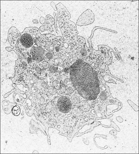
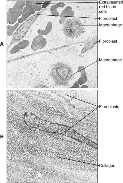
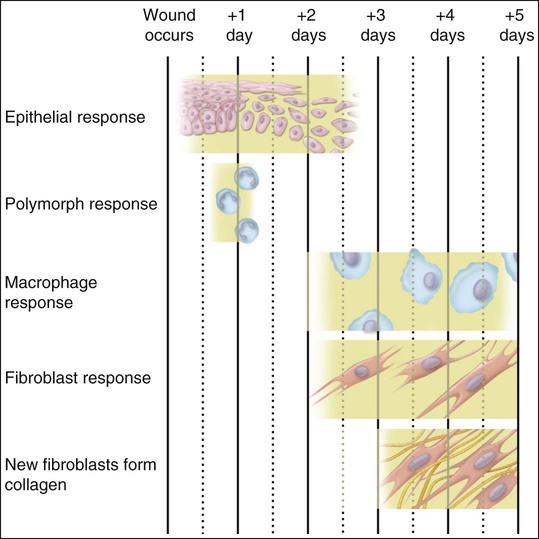
Polymorphonuclear leukocytes, mainly neutrophils, are the first inflammatory cells to invade the wound. They appear within few hours of injury and become activated in response to phagocytic stimuli or by binding of chemotactic mediators, antigen-antibody complexes to specific receptors on the cell membrane, and components of the complement system. These cells reach a maximum concentration at about 24 hours and have a short life span at the wound site before they die. Neutrophils contain various enzymes and reactive oxygen metabolites (oxygen-derived free radicals) that kill engulfed bacteria but that also can destroy damaged and normal tissue when the cells die. Neutrophils function primarily to manage bacterial invasion and hence infection, thus their absence in noninfected wounds does not hinder the repair process. Macrophages and other mononuclear leukocytes enter the wound after 24 hours and are the predominant cell type in damaged tissue at 5 days (Figure 15-1). Macrophage infiltration into the wound site is mediated by various chemotactic factors that are released by platelets in the fibrin clot, keratinocytes at the wound margins, fibroblasts, and leukocytes resulting in cellular and humoral responses and in phagocytosis of damaged tissue components and foreign material. Platelets also release many potent growth factors (transforming growth factor β [TGF-β], platelet-derived growth factor [PDGF], interleukin-1, and others), cytokines, and chemokines. These soluble mediators are critical for the next phase of wound repair involving cell recruitment and differentiation and the commencement of rebuilding damaged tissues. Macrophages are a major source of cytokines involved in lymphocyte chemotaxis and later constitute the most prominent leukocyte subset in wounds. TGF-β in particular stimulates fibroblasts to proliferate and synthesize extracellular matrix proteins. In the absence of macrophages, fewer fibroblasts are stimulated during healing, so that healing is slower.

Mast cells represent an important source of proinflammatory mediators and cytokines that promote inflammation and vascular changes.

Another interesting cytokine is osteopontin, which accumulates in calcified tissues. Osteopontin is expressed widely by a variety of inflammatory cells, including T lymphocytes and macrophages. Osteopontin is also known as early T lymphocyte activation-1 and is implicated in macrophage recruitment and activation. Locally produced osteopontin and some found in serum and tissue fluids may act as an opsonin that facilitates uptake of material, possibly including bacteria, by macrophages. Mutant mice lacking a functional osteopontin gene show an aberrant skin healing response and are more susceptible to infection. Thus by direct action and ability to stimulate fibroblasts, macrophages have a direct effect on the repair process.

Reparative Phase

Successful repair of the injured tissues requires resolution of the inflammatory reaction. As the acute inflammatory phase subsides, regeneration of the tissue begins, occurring first in the epithelium and then in the connective tissue.

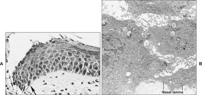
Damage to the epithelium results in mobilization and migration of epithelial cells at the wound margin. The cells lose their close attachment to each other and to the underlying connective tissue within 24 hours of wounding; this is apparent histologically as a widening of the intercellular spaces (Figure 15-2). Twenty-four to 48 hours after wounding, cell division in the basal epithelium increases a short distance behind the wound margin, and those cells immediately adjacent to the margin begin to migrate laterally beneath the clot or coagulum (Figure 15-3). As they migrate, the epithelial cells deposit basal lamina constituents that facilitate movement through the subepithelial connective tissue. Migration and subsequent adhesion of epithelial cells to the basal lamina implicates remodeling of the cytoskeleton and redistribution of integrin membrane receptors, interaction with laminin-332, and ultimately the formation of hemidesmosomes.

Initially, basal cells move, but suprabasal cells slide or roll over the basal cells subsequently. Epithelial cells continue to migrate until they reach the cells from the opposing wound margin, when contact inhibition restricts further movement. At this time an increase in cell division leads to stratification and differentiation, reestablishing a normal epithelial tissue.

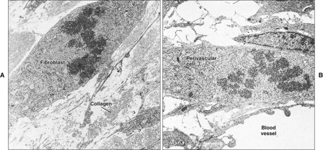
Initially, the wounded connective tissue consists of fibrin, necrotic tissue, and an acute inflammatory cell infiltrate. Fibroblasts migrate and proliferate within the healing connective tissue within 24 hours. The fibroblasts involved in wound repair derive from two sources: division of undamaged fibroblasts at the wound periphery (Figure 15-4, A) and undifferentiated connective tissue (mesenchymal) cells (Figure 15-4, B). The resulting daughter cells from both sources migrate into the wound defect to form the collagen of scar tissue (Figure 15-5). Moreover, endothelial cells proliferate and capillaries develop from preexisting vessels at the wound margin. New blood vessels play an essential role in tissue healing by participating in connective tissue formation, providing nutrients and oxygen, secreting bioactive substances (endothelial cells), and allowing for inflammatory cell migration to the site of injury. Angiogenesis is a complex event regulated by growth factors acting in synergy. Vascular endothelial growth factor, fibroblast growth factor [FGF], and TGF-β are major components in wound angiogenesis. Extracellular matrix molecules, such as fibronectin, laminin, and collagens, are also important in vessel growth by acting as a scaffold for cell migration and reservoir for growth factors.

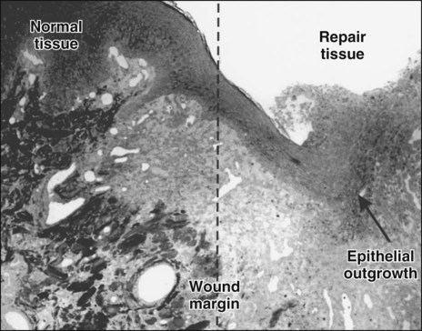
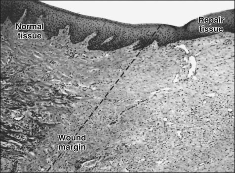
At 3 days the healing lamina propria is predominantly cellular, consisting of inflammatory cells, developing capillaries, and abundant fibroblasts among fibrin remnants and new collagen fibrils (see Figure 15-3). Between days 5 and 20 after wounding, collagen is deposited rapidly in the wound, with a corresponding increase in tissue tensile strength, although up to 150 days may be required to regain normal tissue strength (Figure 15-6). The relative proportion of cells and fibers approaches that of unwounded tissue by 20 days.

Persistence of the inflammatory response delays wound healing by generating unbalanced proteolytic activity and tissue destruction at the repair site. Infiltrating macrophages and neutrophils produce numerous proteinases, including various matrix metalloproteinases. Resident cells at the wound site, such as keratinocytes, fibroblasts, and endothelial cells, also up-regulate production of proteinases. Bacterial components and degraded tissue perpetuate the problem by sustaining continued influx of inflammatory cells.

Wound Contraction and Scarring

Scar formation is a physiologic and inevitable outcome of wound repair in mammals, the function of which is to restore tissue integrity quickly. Evidence indicates that scar formation is linked intimately to the inflammatory phase of repair. By controlling infection, the rapid initial inflammatory response allows the wound to heal quickly but ultimately results in the production of a tissue of lesser quality. Interestingly, repair in early fetal life shows no typical inflammatory phase, and healing of the skin, for instance, is scarless.

In skin the first fibroblasts that enter the wound contain abundant actin and myosin and have contractile properties, so that they often are called contractile fibroblasts or myofibroblasts. These cells have junctions with one another and with connective tissue fibrils. By contracting, myofibroblasts are able to draw the edges of the wound together, thereby reducing the surface area and facilitating healing. Myofibroblasts are believed to derive from a clonal population within the connective tissue.

Collagen that is laid down may form scar tissue and lead to rigidity and immobilization of the area, with impairment of function. Myofibroblasts and wound contraction have been described in oral mucosa, but scar tissue that is formed usually is remodeled so that most surgery within the mouth can be undertaken without fear of producing disabling scar tissue. The reason for the differences in wound healing between skin and oral mucosa is not understood, but increasing evidence indicates that fibroblasts in oral mucosa are phenotypically different from those of skin and more closely resemble fetal fibroblasts. Such differences can be seen in the synthesis of glycosaminoglycans and in the response to the cytokine TGF-β. Figure 15-7 provides a summary of this simple account of repair.

Wound Healing at the Dentogingival Junction

If gingivitis progresses to periodontitis, the junctional epithelium migrates apically and is responsible for the formation of the pocket epithelium. This process requires not only cell proliferation but also migration of the cells over the connective tissue substratum that has been modified by the inflammatory process. Recent studies have identified variable expression of integrins and other adhesion molecules at the epithelial-connective tissue interface during the inflammatory process and subsequent migration of the junctional epithelium. When healing occurs, a new structure with the same histologic characteristics as the original junctional epithelium develops from the phenotypically different oral (or gingival) epithelium. The underlying connective tissue is believed to play a significant role in determining the formation of the junctional epithelium. Connective tissue is destroyed during periodontal disease, and the junctional epithelium therefore extends until it reaches intact connective tissue that provides the signal to stop its migration, forming a long junctional epithelium. Some believe that lack of mechanical stability at the wound site favors formation of a long junctional epithelium and that following periodontal surgery, formation of a fibrin clot against the denuded root surface favors formation of a connective tissue attachment that prevents apical migration of the gingival epithelium.

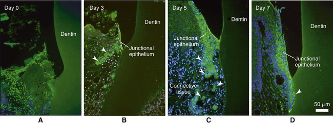
The recent discovery that odontogenic ameloblast–associated (ODAM) protein is produced by the junctional epithelium, and the fact that it is expressed early during its regeneration in animal models (Figure 15-8) implicates this molecule as a potential target for novel prevention and regenerative strategies. ODAM may indeed behave as a matricellular protein influencing both matrix and cellular events.

Only gold members can continue reading. Log In or Register to continue

Stay updated, free dental videos. Join our Telegram channel

Jan 5, 2015 | Posted by in General Dentistry | Comments Off on 15: Repair and Regeneration of Oral Tissues

VIDEdental - Online dental courses

Get VIDEdental app for watching clinical videos