Oral Soft Tissue Lesions and Minor Oral Surgery
Göran Koch and Dorte Haubek
Children and adolescents present a large variety of oral pathologic conditions, such as lesions of the oral mucosa, bone lesions, cysts, and tumors or tumor‐like lesions. Knowledge on pathologic changes of oral tissues is a prerequisite for providing correct diagnosis and adequate treatment. The present chapter deals with the most common pathologic conditions or changes of the oral mucosa in children and adolescents.
Oral surgery in children and adolescents is, with few exceptions, similar to oral surgery in adults. However, there are some specific and common surgical procedures which, due to the special condition of a growing individual, are different to procedures performed in adults. The most common of such surgical procedures will be presented in this chapter. For more extensive knowledge on oral pathology and more detailed information on pediatric oral surgery, the reader is referred to textbooks listed in section “Background literature.”
Oral mucous lesions
Apart from specific skin lesions, a number of common infectious diseases in children are also manifested as oral lesions. For example, bacteria, viruses and fungi may cause infections of the oral mucosa. During the child’s disease and the week after recovery, unnecessary dental treatment should be avoided due to potential risk of contamination.
Bacterial infections
Impetigo contagiosa
This lesion is more common in children than adults. It is caused by streptococci and staphylococci and often affects the perioral area. The infection causes inflammatory vesiculo‐bullous lesions that rupture, leaving secreting or crust‐covered lesions (Figure 15.1). The lesions will cause only a low sensation of pain. As the disease is contagious, it can be spread within the family and between playmates. In the acute stage, dental treatment should be avoided. In most cases, the lesions heal without complication after strict hygiene measures, but intake of antibiotics are often recommended.
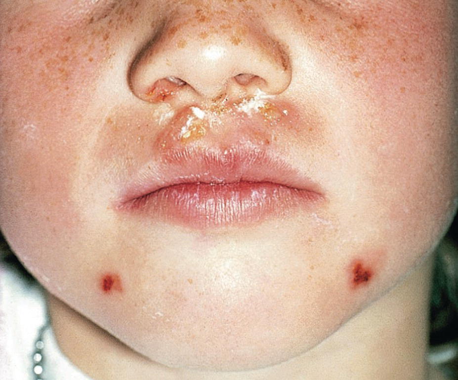
Figure 15.1 Impetigo contagiosa in a 7‐year‐old girl.
Scarlet fever
Scarlet fever is a common disease in childhood caused by beta‐hemolytic streptococci. The general symptoms are fever, tonsillitis and lymphadenitis. A papular red skin rash will develop within a few days. Besides swollen tonsils, the most characteristic oral manifestation is the gradual change of the appearance of the tongue from a “strawberry” to a “raspberry” tongue. In the initial stage of the infection the white‐coated tongue shows a scattered pattern of hyperemic fungiform papillae. Later, this coating is lost and the red edematous fungiform papillae dominate the clinical picture.
Viral infections
A number of viruses may cause infections of the oral mucosa in children. In recent years, many vaccination programs have, however, been introduced. One of these is measles, mumps and rubella (MMR) vaccine which is an immunization vaccine against measles, mumps and rubella. In some types of MMR vaccines, the protection against varicella is included (MMRV). In countries with a well‐established vaccination program, many children do not get these diseases anymore. However, brief descriptions of some of these diseases, mainly manifesting in childhood, are given below.
Rubella
Rubella is a virus infection characterized by light red macules on the skin. Occasionally, macules are also found on the soft palate, called Forschheimer’s spots (Figure 15.2).
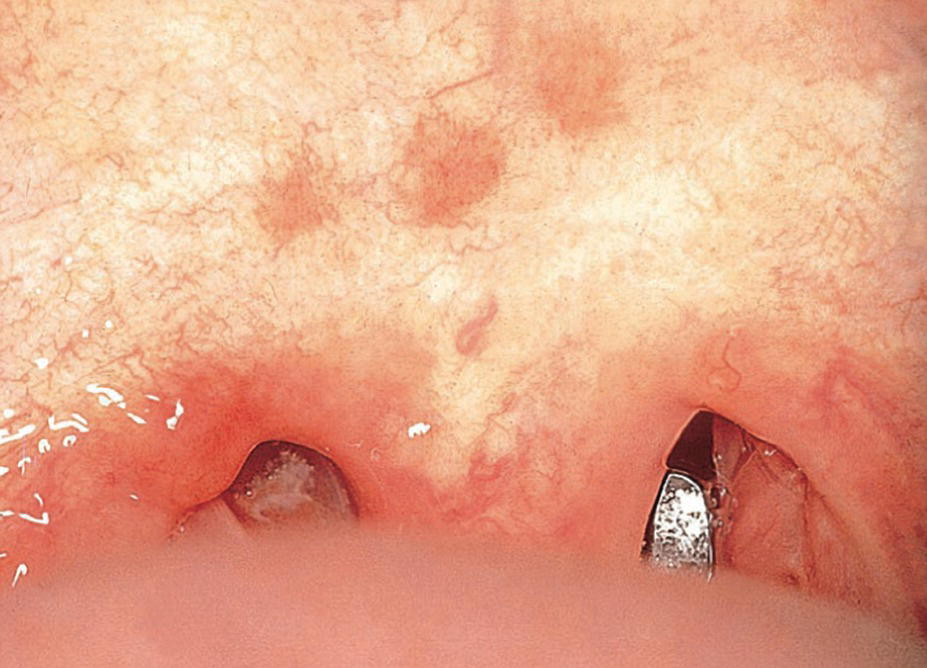
Figure 15.2 Forschheimer’s spots on the soft palate in a child with rubella.
Measles
Measles is a viral disease. After an incubation time of 10–12 days, the child will have a cough, fever and photophobia. Some days later, light red maculopapular lesions develop all over the skin. The oral manifestations, Koplik’s spots, occur on the buccal mucosa as prodromal grayish‐white macules surrounded by a slightly erythematous zone some days before eruption of the skin lesions (Figure 15.3).
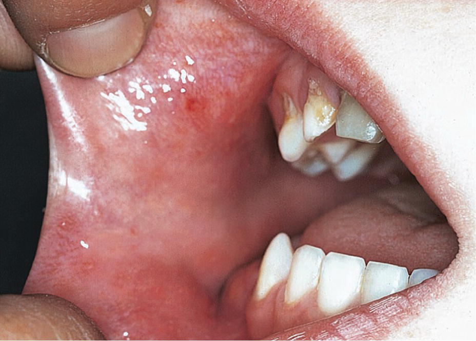
Figure 15.3 Koplik’s spots in the cheek mucosa in a child with measles.
Mumps
Mumps is a viral infection affecting the salivary glands. The general symptoms are fever and pain from the infected glands. In cases where the parotitis is unilateral, difficulties in distinguishing the condition from a swelling of odontogenic origin may arise.
Varicella (chickenpox)
Varicella is a viral disease mostly seen in children. The general symptoms are fever, pharyngitis and vesicular lesions on the skin, beginning on the trunk and then spreading all over the skin (Figure 15.4). The lesions have varying degrees of development. The oral manifestations are whitish vesicles surrounded by a red halo found mostly on the mucosa of the lips, bucca and tongue (Figure 15.5).
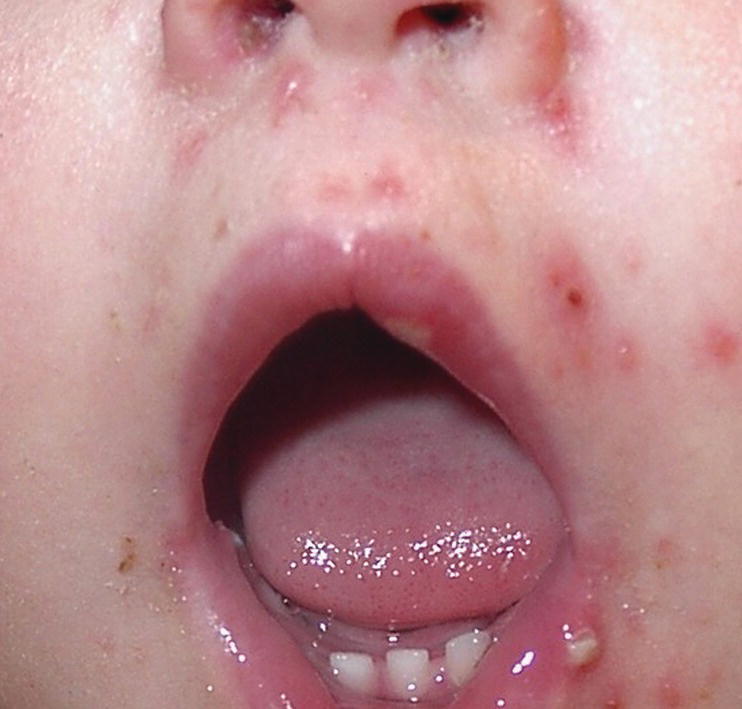
Figure 15.4 Vesicular lesions on the skin in a child with varicella (chickenpox).
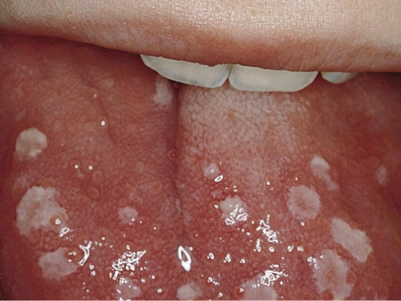
Figure 15.5 Vesicles on the mucosa of the tongue in a child with varicella.
Herpetic gingivostomatitis
Herpetic gingivostomatitis is caused by herpes simplex virus (HSV) and communicated through personal contact, e.g., transmission via the saliva of the mother. Primary oral infection occurs following the first exposure to the virus. Studies have found antibodies to HSV in 40–90% of individuals in different populations showing that HSV infection affects almost all individuals even if many of them only suffer the infection subclinically. The incidence of primary herpes simplex infection increases from the age of 6 months. It reaches its peak at age 2–5 years. Below 6 months of age, the child is usually protected by maternal antibodies. The primary herpes simplex infection is therefore mainly a disease in children. The incubation period is 3–5 days. The primary infection is often manifested as an acute herpetic gingivostomatitis where the entire gingiva is red, edematous and inflamed. The general symptoms are fever, headache, malaise and pain. After 1–2 days, small vesicles develop on the oral mucosa. Subsequently, they rupture, leaving painful ulcers with a diameter of 1–3 mm (Figure 15.6).
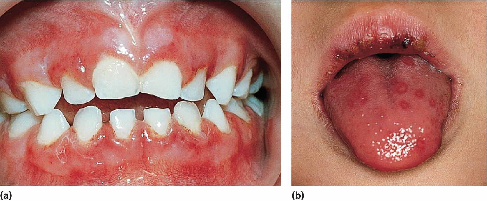
Figure 15.6 (a) Herpes simplex lesions spread over the alveolar mucosa and (b) tongue.
Normally, the primary herpes simplex infection is self‐limiting. The child will recover within a 10‐day period. Treatment is primarily supportive, e.g., painkillers for the pain and fever, fluid to maintain sufficient hydration and, if necessary, topical anesthetic ointments to facilitate eating. In severe cases, hospitalization and/or the use of antiviral agents are necessary.
It has been shown that recurrent herpes simplex infection originates from a reactivation of HSV, which remains dormant in nerve tissue between periods of excitation. Reactivation may be caused by injuries to the mucosa caused by sunlight, citrus fruits or dental treatment, etc. Recurrent herpetic infection will develop at the same location at every excitation, e.g., herpes labialis (Figure 15.7). Topical treatment with ointments containing acyclovir, available on the market, might minimize the symptoms.
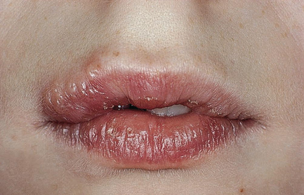
Figure 15.7 Herpes labialis.
Verruca vulgaris
Intraoral warts may present as solitary or multiple lesions and are caused by human papillomavirus. They can be associated with skin warts, and precautions to minimize risk of contamination should be considered (Figure 15.8).
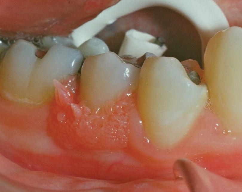
Figure 15.8 Wart.
Fungal infections
Fungal infection may cause mucosal pathology in children and adolescents.
Oral candidiasis
Oral candidiasis is caused by the fungus Candida albicans, which is normally found in the oral cavity. The fungus invades the mucosa only when there is a change in the oral environment or a general impairment of the immunologic or hormonal balance. Such changes can be brought about by the administration of antibiotics and immunosuppressive drugs. The clinical manifestations in the oral mucosa can vary. Pseudomembranous candidiasis (thrush) is the most common fungal infection in newborn children and in children with chronic disease. It is characterized by raised, pearly‐white patches (Figure 15.9) that can be rubbed off, leaving an erythematous or bleeding mucosa. The clinical manifestations can vary from acute to chronic forms. In some patients, a hypertrophic variant can be found (Figure 15.10). The treatment is medication with antifungal preparations, such as nystatin, amphotericin B or miconazole. Systemic administration or topical application by mouthrinses, sucking lozenges and gels containing these preparations will be effective in most cases.
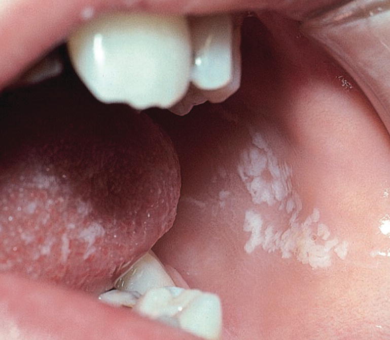
Figure 15.9 Candidiasis lesion in cheek mucosa.
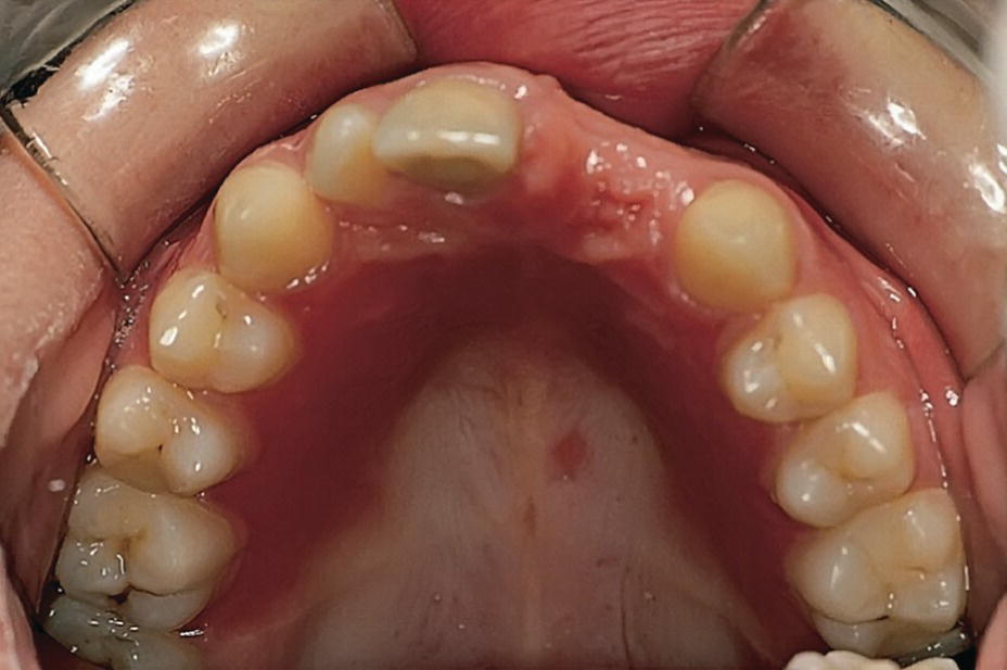
Figure 15.10 Candida albicans infection of the palatal mucosa in child with a partial denture.
Immunologically‐related and other mucous lesions
Aphthous ulcers
Recurrent aphthous ulcerations (RAU) or recurrent aphthous stomatitis (RAS) are the most common oral ulcerations seen in children. Studies indicate a prevalence of about 35% in adolescents. An altered immune defense system has been suggested as the main predisposing factor. This is confirmed by the frequent aphthous ulcers seen in children on immunosuppressive drugs and by the familiar occurrence. As a trigger factor, infection with specific oral streptococci has been suggested.
The lesions are most frequently localized to the non‐masticatory mucosa, e.g., the vestibulum and the tongue. The lesion starts as a small white papule which gradually ulcerates. The ulcers are 0.2–1 cm in diameter with the central part covered with a yellowish–gray coating, and a crateriform base with raised reddened margins. The surrounding tissue shows a light swelling. The ulcers are extremely painful and may be of varying size and number (Figure 15.11). The ulcers heal without scars in 1–2 weeks. The intervals between recurrent episodes may vary from a week to several months.
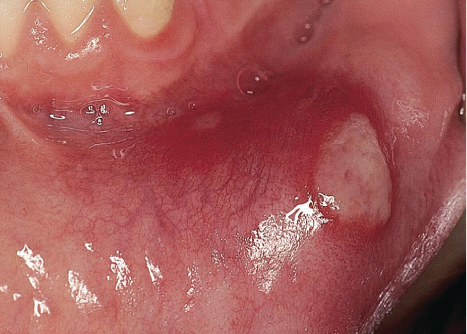
Figure 15.11 Aphthous ulcer.
Other than reassurance, treatment is often unnecessary, as the lesions usually heal within 10–14 days. However, a variety of treatment modalities has been suggested to be used in the most severe cases. In general, the treatment has followed one or more of the following approaches:
- treatment of the oral microbiota (chlorhexidine, hydrogen peroxide‐producing enzyme solutions)
- increasing the immune system (e.g., intake of Longo‐vital®)
- symptomatic treatment of pain (etching solutions, topical anesthetics).
Erythema multiforme
Erythema multiforme is a dermatologic disease, which may also develop oral lesions or in which the oral lesions are the only symptom of the disease. Erythema is not regarded as a specific disease, but as a general reaction to a series of independent precipitating factors, such as food or drug allergy, infection, radiotherapy or general disease. The onset is very rapid. In 24 hours, a child may develop extensive lesions of the skin and/or mucosa. The skin lesions are asymptomatic erythematous macules or papules, mostly affecting hands and feet. In the generalized vesiculo‐bullous erythema multiforme even the eyes and genitals are involved (Stevens‐Johnson syndrome). Initially, the oral lesion has the character of vesicles or bullae, which rapidly burst, forming ulcers (Figure 15.12a). Compared with viral lesions the ulcers are larger, deeper, and often bleed. Involvement of the lips is most common. After some days, the ulcers will crust and healing takes place within 2 weeks (Figure 15.12b,c).
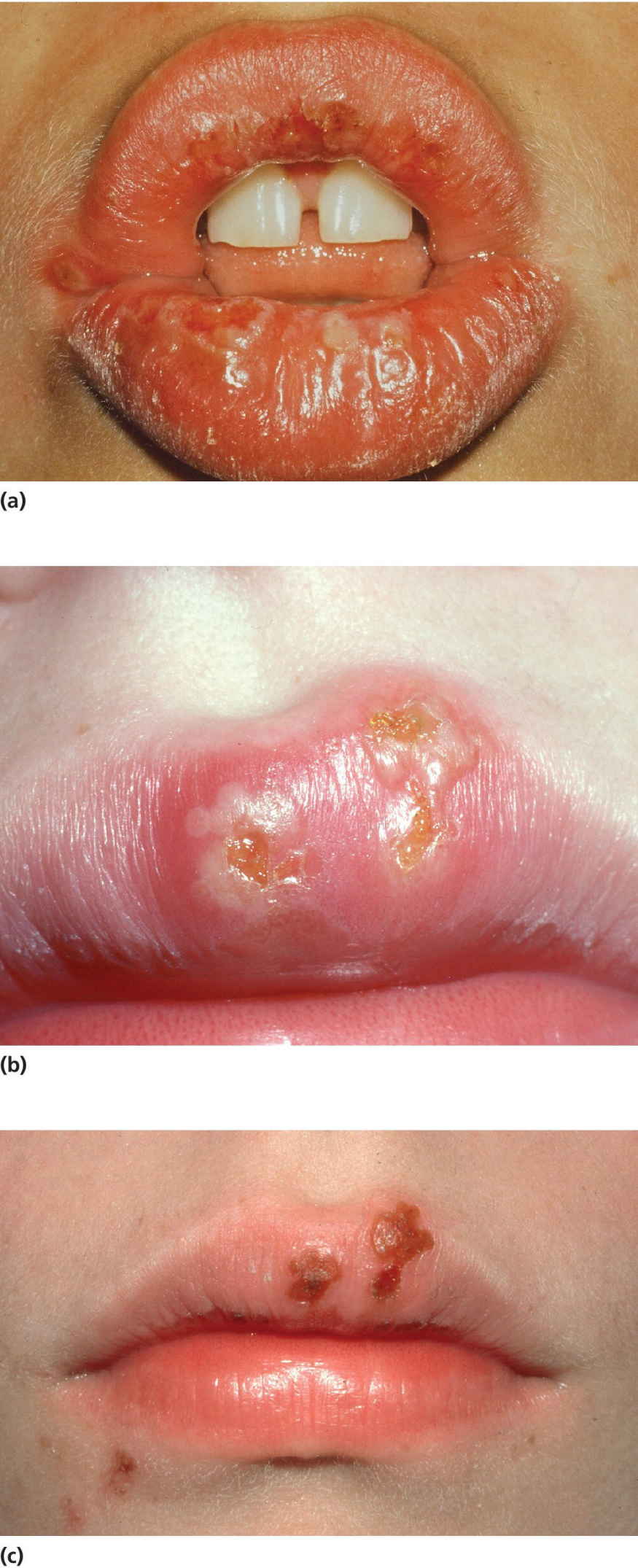
Figure 15.12 Erythema multiforme: (a) oral lesions showing vesicles and bullae which rapidly burst; (b) lesions after 2–3 days; (c) crust formation (healing) 2 days later.
In cases where the lips are severely affected the patient has great difficulties in eating and drinking. In mild cases, topical anesthetics may be the only treatment. In severe cases, systemic treatment with corticosteroids and antibiotics can be justified.
The diagnosis is based on the overall clinical assessment and examination.
Traumatic lesions
Traumatic irritation of the oral mucosa is occasionally seen in children. The most common situation is cheek, lip or tongue bites occurring accidentally or due to decreased sensation of the oral tissues usually after dental local anesthesia (Figure 15.13). The clinical appearance is considerable swelling and bleeding followed by the development of a large, whitish pseudomembranous mucous lesion. The lesion is self‐limiting and will heal in about a week.
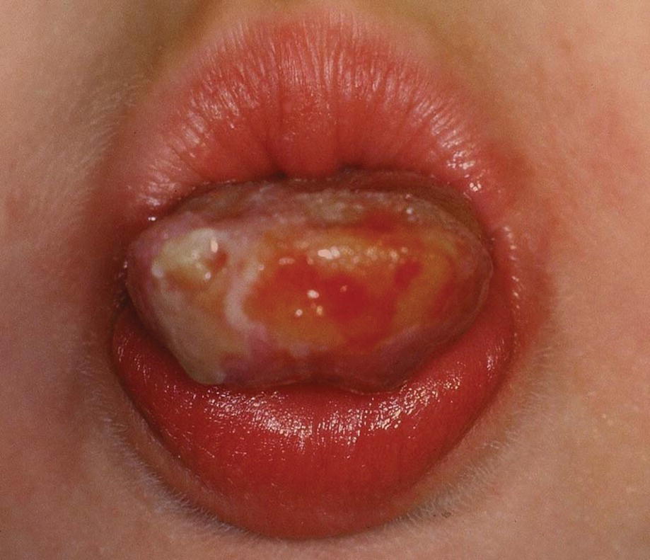
Figure 15.13 Tongue mutilation after local anesthesia.
Self‐mutilation, also called self‐harm or self‐injury, of the mucosa is most commonly seen in children with disabilities, for example, having uncontrolled tongue movements (see Chapter 24). The irritation of the mucosa is caused by the child forcing the tongue out of the mouth and thereby injuring it on the mandibular incisors. Hyperkeratotic wounds may develop. Smoothing of the incisal edges of the incisors or an acrylic splint in the mandible may help to heal the lesion. Other types of self‐harm may also include biting, hitting, or bruising oneself, or picking or pulling the oral mucosal and/or tissues. Self‐mutilation can be episodic or repetitive.
Crohn’s disease
Crohn’s disease is commonly associated with oral lesions. The lesions may also precede symptoms of bowel disease. The most common oral pathologic findings in Crohn’s disease are aphthous ulcers, areas of inflammatory hyperplasia, including mucosal fissuring and indurated “tag‐like” lesions on the retromolar area (Figure 15.14). The lesions tend to run a remitting course.
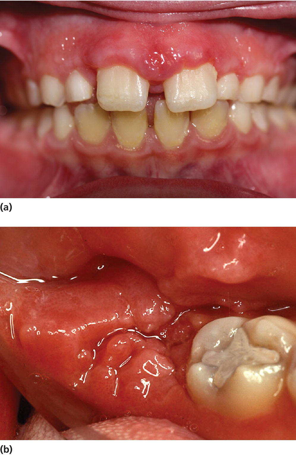
Figure 15.14 Crohn’s disease, gingival characteristics: (a) incisal region; (b) molar region.
Geographic tongue
Geographic tongue is a benign migratory glossitis occurring in children. Clinical manifestation is pinkish red, irregularly depapillated areas surrounded by a well‐defined, slightly raised whitish border. Affected areas vary from day to day, and the appearance can be described as a continuously changing map (Figure 15.15). Occasionally, other parts of the oral mucosa are affected (stomatitis migrans). A hereditary tendency is seen, but the etiology is unknown. It is normally symptomless, but some children will experience discomfort and burning sensation from the lesions, especially when eating, e.g., spicy food. In severe cases, topical anesthetics are recommended during the acute phase.
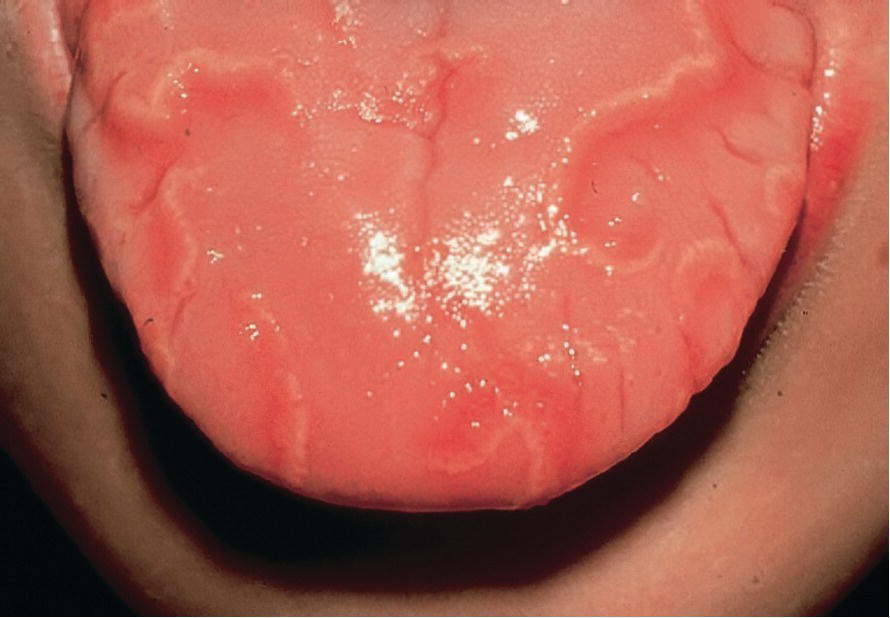
Figure 15.15 Geographic tongue is a relatively common condition found in children.
Cysts, tumors, and tumor‐like lesions
Oral cysts are often described as a cavity in the oral tissues containing fluid or gas and frequently lined with epithelium. Some of the most well‐known and frequently occurring cysts are listed in Box 15.1.

VIDEdental - Online dental courses


