Tooth Development and Disturbances in Number and Shape of Teeth
Göran Koch, Irma Thesleff, and Sven Kreiborg
Mechanisms of normal and abnormal tooth development
Teeth develop as epithelial appendages from the ectoderm covering the oral cavity. The morphologic aspects of tooth development at the microscopic level have been described in great detail long ago, and more recent research has started to uncover the molecular details underlying the morphogenesis of teeth, dental cell differentiation and extracellular matrix formation, and mineralization. The genetic regulation of normal tooth development has been elucidated by experimental studies mainly using mouse teeth as models, and studies in human molecular genetics have led to the identification of gene defects causing dental aberrations.
Principles of tooth development
Tooth development starts from placodes, local thickenings of the dental lamina forming as a stripe of oral epithelium at the sites of future dental arches. The placodes bud to the underlying mesenchyme which condenses around the bud. Rapid growth and folding morphogenesis of the epithelium leads to the cap stage of development. The shape of the tooth crown becomes established during the bell stage when the locations and heights of tooth cusps are set by additional foldings and growth of the epithelium. The odontoblasts and ameloblasts differentiate during the bell stage, and deposit the extracellular matrices of dentin and enamel, respectively. Differentiation, matrix deposition, and mineralization start from the tips of the cusps and proceed cervically, and after they reach the future cemento‐enamel junction, root development starts (Box 4.1).
Permanent (secondary) teeth develop from the successional dental lamina associated with the primary (deciduous) tooth germs, and their budding starts during the cap stage of primary tooth development. Also posterior molars form in succession from the dental lamina at the distal aspect of the previous molar. Both the primary and successional dental laminae contain stem cells which have the capacity to form teeth. Sox2, a known marker gene of stem cells, is expressed in the primary as well as successional dental lamina [1].
Bone formation is initiated in the maxilla and mandible only after the initiation and budding of primary teeth, but the subsequent development of alveolar bone from the mesenchyme surrounding the tooth germs is tightly coordinated with tooth morphogenesis. At the time of eruption, the teeth are surrounded by bone, and their eruption depends on precisely regulated bone remodeling, i.e., bone resorption between crown and the oral cavity and bone apposition at the base of the roots.
The most important mechanism regulating tooth development is the interaction between the epithelial and mesenchymal cells. During the initiation of tooth development, the epithelium has an instructive role and determines the type of the tooth to be formed as well as the odontogenic fate of the mesenchymal cells. The capacity to regulate tooth development shifts to the mesenchyme during budding and, subsequently, mesenchymal signals regulate the morphogenesis of the epithelium and the shape of the tooth. The differentiation of the odontoblasts, ameloblasts, and cementoblasts, as well as their matrix deposition, is also regulated by interactions between the different tissues. Hence, tooth development is controlled by a series of reciprocal tissue interactions. It is noteworthy that similar sequential and reciprocal cell interactions govern the development of all organs in the embryo.
Molecules regulating tooth development
The most important molecules regulating tooth development are those that participate in the signaling networks mediating epithelial–mesenchymal interactions. The actual signals are small peptides which are secreted by one cell and exert their influence on nearby cells through binding to specific receptors (Figure 4.1). This leads to changes in gene expression in the responding cell and subsequent changes in cell behavior. Extensive studies on the signaling molecules in teeth and other organs and in different animals have shown that they have been conserved to an astonishing extent during evolution. It is now known that the same signals regulate the development of practically all organs in the embryos of all species, but depending on the organ and stage of development, the signals elicit different responses.
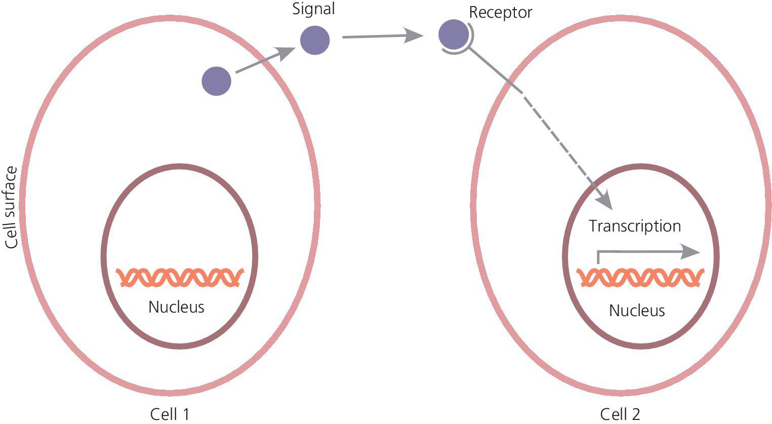
Figure 4.1 Cells communicate via soluble signal molecules regulating gene expression (transcription).
The actual signals belong to a few families, of which four have been mostly studied and they also, importantly, regulate tooth development. These are the Hedgehog, FGF (fibroblast growth factor), BMP (bone morphogenetic protein) and Wnt families. These signals are already expressed in the very early dental epithelium and regulate the initiation of tooth development. They also signal back from mesenchyme to epithelium and initiate epithelial folding and thereby shape development. During advancing development several signals are locally expressed in the dental epithelium in small clusters of cells, the dental placodes and the enamel knots, which are signaling centers regulating tooth initiation and crown shape. The placodes form at sites where the dental epithelium buds to mesenchyme. The primary enamel knot forms at the tip of the bud and induces the folding of the epithelium and onset of cap formation. In molars, secondary enamel knots appear at the sites of new cusps and produce signals, which stimulate the growth of the cusps (Figure 4.2). The signals regulate a great number of different transcription factors which determine the identities of cells and the ways in which the cells respond to signals. The central roles of several transcription factors in tooth development have been demonstrated by transgenic mouse experiments in which inhibition of their function has resulted in arrested tooth development. The chain of reciprocal interactions between the dental epithelium and mesenchyme, and the mediating signals and transcription factors, can be considered the “program of tooth development.”
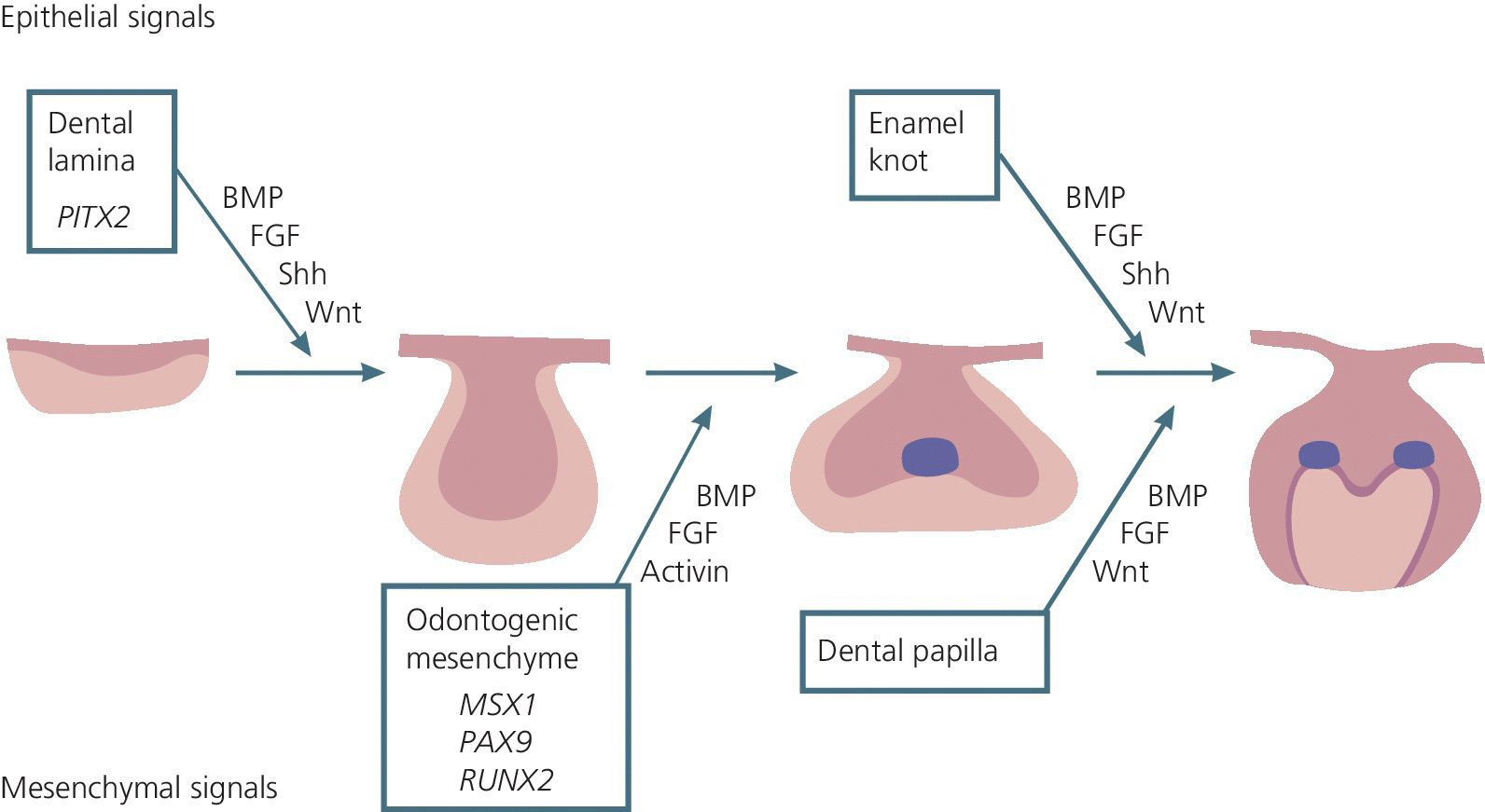
Figure 4.2 Reciprocal interactions between the epithelial and mesenchymal tissues are mediated by conserved signaling molecules (BMP = bone morphogenetic protein; FGF = fibroblast growth factor; Shh = sonic hedgehog; Wnt). Numerous transcription factors are associated with signaling, and only those in which mutations cause dental abnormalities are indicated in the boxes (PITX2, MSX1, PAX9, RUNX2).
Disturbances in tooth development
Disturbances in tooth development are seen as:
- numerical variations (missing or supernumerary teeth)
- variations in size and shape of teeth
- defects in the mineralized tissues, enamel, dentin, and cementum, which may result from defects in the composition of the respective extracellular matrices and/or their mineralization (see Chapter 20)
- problems in eruption (see Chapter 5).
It is obvious that tooth development may be disturbed at different stages of morphogenesis, and that the end result depends on the timing and the type of insult. Variations in the number, shape, and size of teeth are commonly linked. In particular, hypodontia together with small and peg‐shaped teeth are often seen in the same patient. In these cases it is apparent that there is an inhibition of the early morphogenesis, and that it leads either to a complete arrest of development or to small teeth depending on the tooth type and extent of disruption. The most commonly missing teeth are the third molars, second premolars, and maxillary lateral incisors. It is believed that they are affected because they are the last teeth to develop from the respective dental placode. It is possible that the development of some of the teeth ultimately missing is actually initiated and may even have proceeded to early stages but that their development has been arrested because the tooth germs did not reach a threshold size.
Clinical aspects
Chronology of dental development
Data on the chronology of tooth development are usually given as mean values from series of observations. Even if the chronology in tooth development has a documented stability, deviations may occur. The timing of onset to completion of mineralization/development of the primary and permanent dentition is presented in Box 4.2 and Figures 4.3 and 4.4. The mineralization of the primary teeth starts during 14–18 weeks in utero. The root formation of the primary teeth is completed between 1.5 and 3 years. The crowns are halfway mineralized at birth and become fully formed during the first year of life (Figure 4.3). Mineralization of the permanent teeth starts at birth with the first molars. The incisors and canines start their mineralization during the first year of life, the premolars and second molars between the second and third years of life, and the third molar between the eighth and eleventh years of life. However, the normal range is wide. The crowns of the permanent teeth (except third molars) are generally completed between 5 and 7 years of age. Root development takes about 6–7 years. In general, the mandibular teeth develop earlier than the maxillary teeth (Figure 4.4). A marked sex difference has been observed in tooth formation, girls being on average half a year ahead of boys.
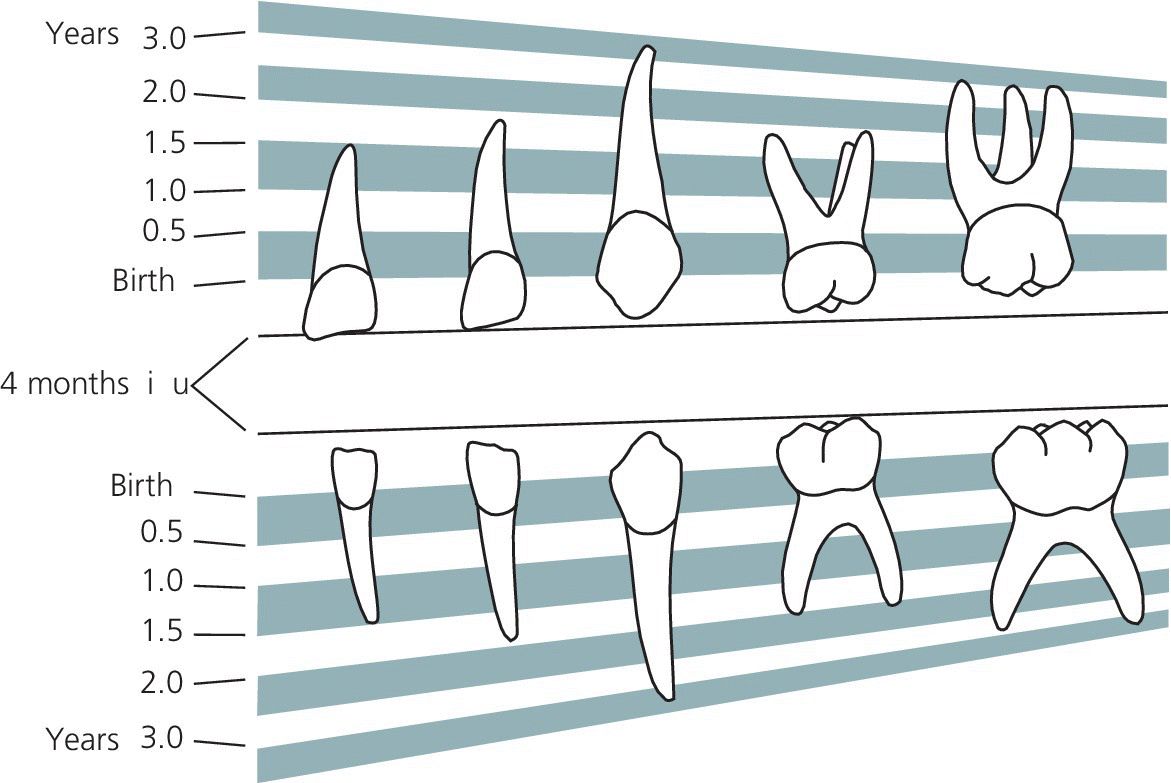
Figure 4.3 The chronology of mineralization of primary teeth.
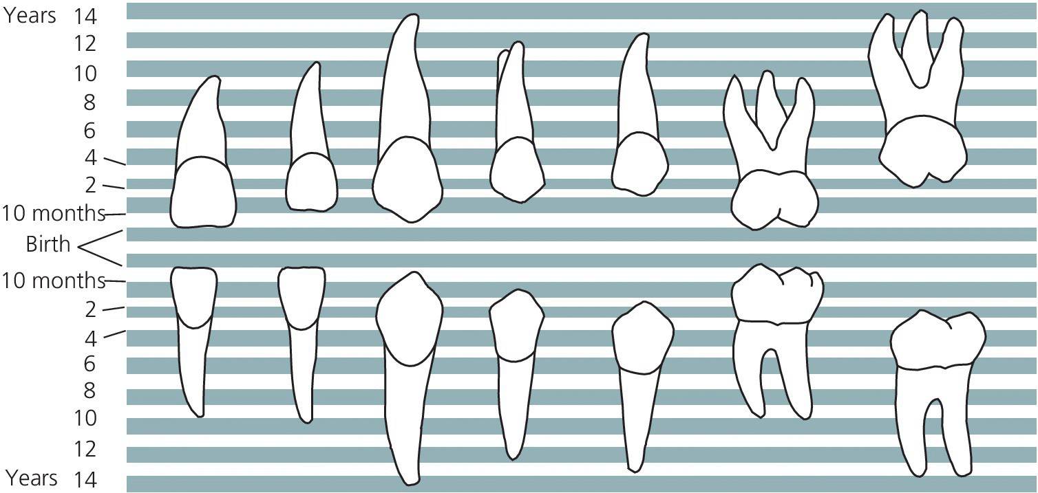
Figure 4.4 The chronology of mineralization of permanent teeth.
VIDEdental - Online dental courses


