15: Molars

Molars

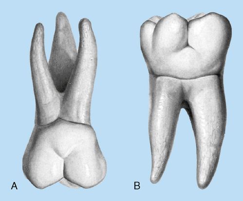
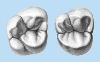
The 12 permanent molars are the largest and strongest teeth in the mouth by virtue of their crown bulk size and root anchorage in bone (Fig. 15-1). Their primary function is to grind or crush food.

The permanent molars erupt long after all the deciduous teeth have already erupted. The first permanent molars erupt distal to the primary second molars. In humans, usually the first permanent teeth to erupt are the first molars. It is only after these molars have erupted that the permanent incisors begin replacing the deciduous incisors.

The following chart shows the approximate eruption time for molars. However, much individual variation occurs. Which teeth usually erupt first, the mandibular or the maxillary?

Eruption Times of Molars

  Eruption Enamel Completion Root Completion
1st molar 6 to 7 years 4 years 9 to 10 years
2nd molar 11 to 13 years 7 to 8 years 14 to 16 years
3rd molar 17 to 22 years 12 to 16 years 18 to 25 years

image

The molars are nonsuccedaneous teeth—that is, they do not replace any primary teeth. They are referred to as accessional, or nonsuccedaneous, as opposed to succedaneous teeth, which do replace deciduous teeth.

Generally, the first molars are formed from five lobes, but some second and third molars may have only four lobes. The lobes in Fig. 15-2 are numbered in the order of their size, with number 1 being the largest.
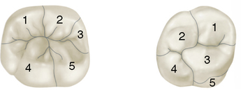
In general, each cusp of a molar is formed from its own lobe. For instance, a maxillary first molar forms from five lobes, three of which form major cusps. These major cusps are large and well developed, characteristic of, and usually present on all maxillary molars: first, second, and third. The fourth lobe on a maxillary first molar forms a minor cusp. A minor cusp has smaller proportions and less development. It is less functional than the major cusps and is not always present on second and third molars. The maxillary first molars have a fifth lobe, which develops into a supplementary cusp. A supplementary cusp is completely afunctional and is not usually present on either the second or third molars. First molars are the most highly developed and largest of the molars and are more likely to have minor and supplementary cusps in addition to their major cusp.

The maxillary molars have only three major cusps, one minor cusp, and sometimes one supplementary cusp. The mandibular molars usually have four major cusps and sometimes one minor cusp (see Figs. 5-5 and 5-6).

The minor cusps of the maxillary teeth are the distolingual cusps; those on the mandibular teeth are the distal cusps. The supplementary cusp occurs only on a maxillary first molar. However, it may not be present at all.

In review, the most developed of the molars are the first molars, whether maxillary or mandibular. The second molars usually have no supplementary cusps, and the minor cusps are even more minor in relation to the major cusps. Third molars may not develop minor cusps at all. A maxillary third molar may have only three major cusps, or a mandibular third molar may have four major cusps.

MAXILLARY MOLARS

First Molars

Evidence of calcification: Birth

Enamel completed: 4 years

Eruption: 6 to 7 years

Root completed: 9 to 10 years

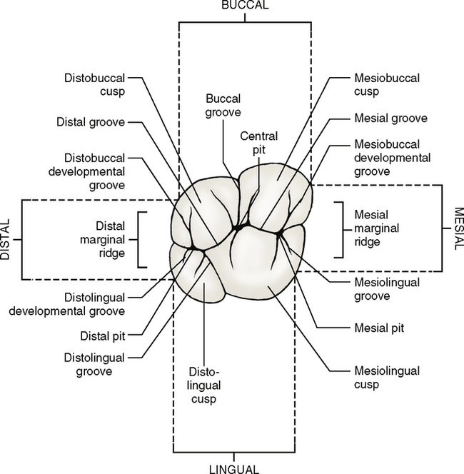
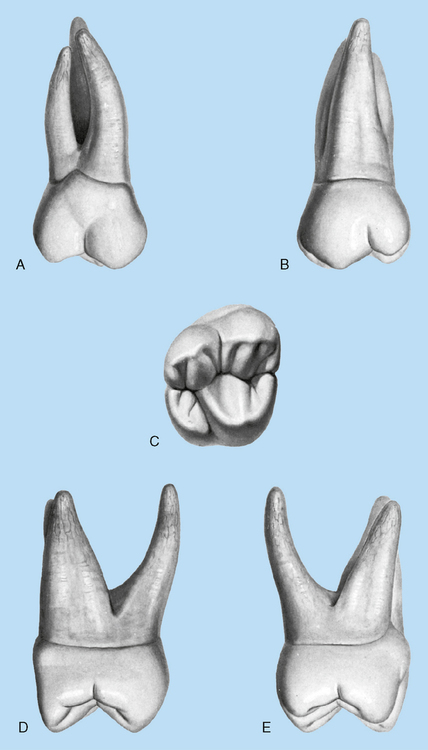
The maxillary first molars (Fig. 15-3) are normally the largest teeth in the maxillary arch. The three molars on each side are the first, second, and third maxillary molars. Each has three well-developed major cusps and one minor cusp, all of which are functional. The fifth or supplemental cusp, which is afunctional, is called the cusp or tubercle of Carabelli. This fifth cusp is usually found on all maxillary first molars.

The crown of a first molar is broad mesiodistally and buccolingually and just slightly wider buccolingually than mesiodistally. Of the four functional cusps, two are on the buccal, and two are on the lingual side.

Facial (buccal) aspect

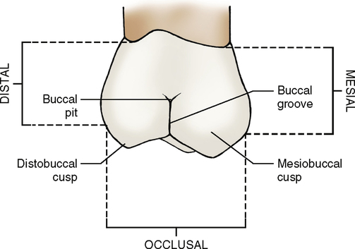
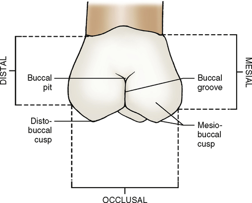
From the buccal view (Fig. 15-4; see Fig. 15-3, A), four cusps can usually be seen: mesiobuccal, distobuccal, mesiolingual, and distolingual. The two lingual cusps are not located directly behind the buccal cusps but are distal and lingual to them. Although the mesiobuccal cusp is broader than the distobuccal cusp, the latter is usually sharper and longer. The mesiobuccal cusp forms an obtuse angle (more than 90 degrees), where its mesial slope meets its distal slope at the cusp tip. The distobuccal cusp usually forms a less obtuse angle where the mesial slope meets the distal slope.

The buccal developmental groove divides the two buccal cusps. This groove runs in a line parallel to the long axis of the tooth, terminating halfway from its point of origin to the cervical line of the crown. Although not very deep at any point, at its terminal end, it splits into a buccal pit with two small grooves radiating from it. The cervical line is irregular and curved, generally toward the occlusal side at the mesial and distal ends.

The mesial outline is straight from the cervical line to the mesial contact area. Below the contact area it curves distally until it reaches the mesiobuccal cusp tip. The height of curvature is just cervical to the junction of the middle and occlusal thirds. Distally, the outline of the crown is convex, and the contact area is in the center of the middle third.

Lingual aspect

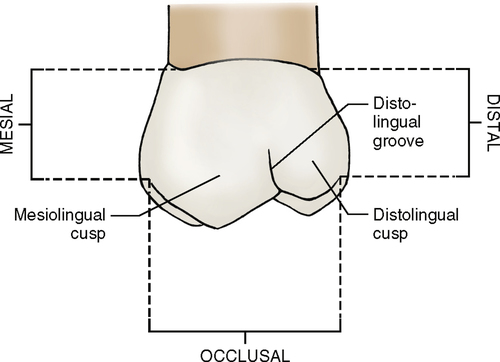
The lingual cusps alone can be seen from the lingual aspect (Fig. 15-5; see Fig. 15-3, B). The mesiolingual cusp is much longer and larger than either of the buccal cusps. Wider mesiodistally and buccolingually, the mesiofacial cusp is the next largest, although it is not as long as the distofacial cusp. The distolingual cusp is the smallest and the shortest of the functional cusps. Of course, the cusp of Carabelli is the shortest and smallest of all five cusps, but it is afunctional. In fact, the cusp of Carabelli is not a cusp at all but rather a tubercle. A fifth-cusp developmental groove, called the mesiolingual groove, separates the cusp of Carabelli from the mesiolingual cusp.

The outline of the crown is as straight mesially as it is buccally. The distal outline is more convex because of the roundness of the distolingual cusp. The lingual developmental groove extends from the center of the lingual surface occlusally, between the two lingual cusps, where it curves sharply to the distal side and becomes the distal oblique groove. These two grooves (lingual and distal oblique grooves) are sometimes considered to be one and are then referred to as the distolingual developmental groove.

The distolingual cusp of a maxillary first molar is functional even though it is small. On a first molar the distolingual cusp occupies approximately 40% of the lingual surface and the mesiolingual cusp occupies the other 60%. The distolingual cusp is progressively smaller on the maxillary second and third molars.

All three roots can be seen from the lingual aspect. On the average, the roots are about twice as long as the crown. The lingual root is usually longer than either of the two buccal roots, which are the same length.

Mesial aspect

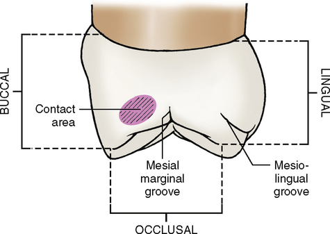
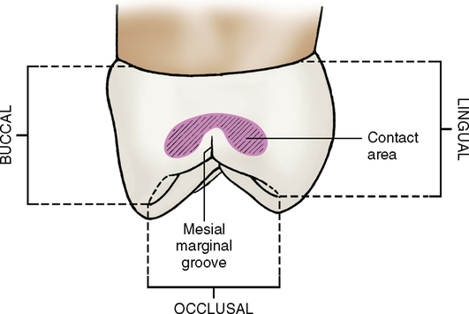
The mesial aspect (Fig. 15-6; see Fig. 15-3, D) of a max-illary first molar usually shows a clear profile of the cusp of Carabelli. The lingual crest of curvature is at the center of the middle third of the crown, the buccal crest at the cervical third. The cervical line is slightly convex mesially.

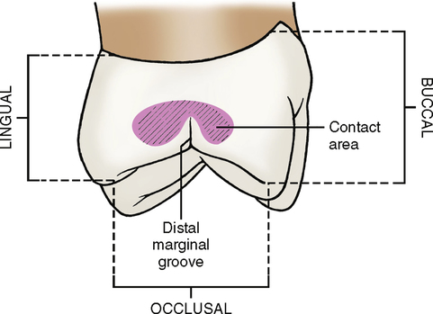
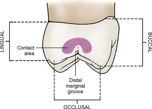
Distal aspect (Fig. 15-7; see Fig. 15-3, E)

The crown has the tendency to taper distally. The buccolingual measurement of the crown on the mesial side is greater than the same measurement distally. The distal cervical line is usually straighter and less curved than that on the mesial side. Although the distal surface of the crown is convex and smooth, a slight concavity is evident on the distal surface of the root trunk from the cervical line to the distobuccal root.

The distal marginal ridge is shorter and less prominent than the mesial marginal ridge, and more of the occlusal surface in general can be seen from the distal view. The distobuccal root is the narrowest of all three roots.

Occlusal aspect

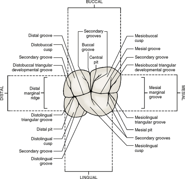
A maxillary first molar has a rhomboidal occlusal outline (Fig. 15-8; see Fig. 15-3, C). The molar crown is wider mesially than distally; it is also wider lingually than buccally. This is the only tooth that is wider lingually than buccally. Is the occlusal outline of a maxillary first molar more like a square or more like a square that has been squashed sideways? What is the difference between a rhomboidal and a square form?

Other anatomic structures

Two major fossae and two minor fossae are on maxillary first molars. The major fossae are the central fossa, which is mesial to the oblique ridge, and the distal fossa, which is distal to the oblique ridge. The minor fossae are the mesial fossa and the distal triangular fossa, both of which are located on the inside of their respective marginal ridges.

The central developmental pit lies in the central fossa. The buccal developmental groove radiates from this pit buccally between the two buccal cusps. The central developmental groove lies in a mesial direction, originating in the central developmental pit and terminating at the mesial triangular fossa. Here, it is joined by the mesiofacial and mesiolingual triangular grooves, which appear as branches of the central groove. The mesial marginal groove, a branch of the central groove, lies between these two triangular grooves and may cross the mesial marginal ridge of the crown. The mesial pit is found in the mesial triangular fossa.

Sometimes another developmental groove radiates from the central pit in a distal direction. If it crosses the oblique ridge, joining the central and distal fossae, it is called the transverse groove of the oblique ridge or the distal part of the central developmental groove.

The oblique ridge is a transverse ridge and is unique to maxillary molars. A transverse ridge is formed by two triangular ridges joining together and crossing the surface of a posterior tooth diagonally rather than straight across buccolingually. In this case the triangular ridge of the mesiolingual cusp joins the triangular ridge of the distobuccal cusp. This cannot happen unless the ridges cross the tooth transversely, as opposed to buccolingually. Therefore the oblique ridge runs from the tip of the mesiolingual cusp to the tip of the distobuccal cusp.

The distal fossa of a maxillary first molar runs parallel with and distal to the oblique ridge. This fossa is long and narrow rather than circular. In this fossa lies the distal oblique groove from which it takes its shape. The distal pit is found in the distal fossa.

The distal triangular fossa is mesial to the distal marginal ridge. The distal oblique groove terminates in the distal triangular fossa, where the distal oblique groove gives off three branches, the distofacial triangular groove, the distolingual triangular groove, and the distal marginal groove.

The primary grooves of the maxillary first molars are as follows:

As a point of clarification, it should be mentioned that triangular grooves are primary grooves that separate a marginal ridge from the triangular ridge of a cusp. These triangular grooves end in a triangular fossa. In studying the central groove, notice that the triangular groove is a continuation of the central groove. As the central groove goes toward the marginal ridge, it separates into two or three branches. The two outer Y-shaped branches curve between the marginal ridge and triangular ridges of the buccal and lingual cusps. The part of the groove that lies between the cusp ridge and marginal ridge is the triangular groove. The two triangular grooves are bisected by a third groove, which lies between them. This groove connects with the marginal ridge and is called the marginal groove.

Second Molars

Evidence of calcification: 3 years

Enamel completed: 7 to 8 years

Eruption: 11 to 13 years

Root completed: 14 to 16 years

A maxillary second molar (Fig. 15-9) supplements a first molar’s function. Generally speaking, the differences that occur between the first and second molars are even more accentuated between the first and third molars. In other words, certain characteristics in form and development occur in a first molar that occur to a lesser degree in a second molar and possibly not at all in a third molar. What are these characteristics? Following is a list of several traits that may occur, but in general they have a tendency to be more accentuated in a second molar and most accentuated in a third molar.
1. The maxillary molar crowns are shorter occlusocervically and narrower mesiodistally in the second molars than in the first molars. The third molars continue to be smaller in all crown proportions, including buccolingually.

2. The molar crowns show more supplemental (secondary) grooves and pits on the second molars than on the first. The third molars show even more supplemental grooves and accidental (tertiary) grooves and pits.

3. The oblique ridge is less prominent on the second molars and in some instances disappears almost entirely on the third molars.

4. The fifth lobe, or cusp of Carabelli, usually disappears on the second molars and occurs infrequently on the third molars.

5. The distolingual cusp is less developed on the maxillary second molars and disappears almost entirely on most maxillary third molars.

6. The occlusal outline of the second molars is less rhomboidal and more heart shaped. A third molar is even more heart shaped in occlusal outline.

7. The roots of the second molars have a tendency to lie closer together and may even be fused.

8. The mesiobuccal roots of the second and third molars have a greater tendency to curve distally in their apical third. The distobuccal root of a maxillary second molar is straighter than that of a maxillary first molar, with little or no mesial curvature. The third molar’s distobuccal root has a tendency to curve distally in its apical third.

9. The roots of the second molars are almost as long and sometimes even longer than those of the first molars. The roots of the third molars are almost always smaller than those of either the first or second molars.

10. The second molars show more variety of form than the first molars, not only in the crown but also in root development. The third molars show unlimited variety in crown and root formations and are often congenitally missing.

Lingual aspect

The lingual view (Fig. 15-11; see Fig. 15-9, B) of a maxillary second molar shows no fifth cusp (cusp of Carabelli). The distolingual cusp is smaller than that of the first molars.

Mesial aspect

The mesial view (Fig. 15-12; see also Fig. 15-9, D) shows the second molar crown to be shorter than the first molar, but its buccolingual measurement is about the same as that of a maxillary first molar. The roots are closer together.

Occlusal aspect

The occlusal outline (Fig. 15-14; see Fig. 15-9, C) of the crown of a maxillary second molar is less rhomboidal than that of a maxillary first molar. The increase in size of the mesiolingual cusp and the absence of the cusp of Carabelli makes this possible. Also the distolingual cusp is smaller.

The mesiodistal diameter of the crown is smaller, but the buccolingual diameter is about the same as that of a maxillary first molar.

The mesiobuccal and mesiolingual cusps are just as developed as in a first molar. The distobuccal cusp is just barely smaller, and the distolingual cusp is noticeably smaller.

More supplemental grooves and pits are present on a maxillary second molar than on a maxillary first molar.

Third Molars

Evidence of calcification: 7 years

Enamel completed: 12 to 16 years

Eruption: 17 to 22 years

Root completed: 18 to 25 years

A maxillary third molar varies more than any other maxillary tooth in size, shape, and relative position to the other teeth. Rarely is it as well developed as a maxillary second molar. It often appears as a developmental anomaly or does not form at all. What term is descriptive of the latter situation?

The crown of a third molar is shorter than that of a second molar, and the roots tend to fuse into one fused root. The occlusal outline of a maxillary third molar is heart shaped. The distolingual cusp is poorly developed or even absent (Fig. 15-15).

Third molars have a tendency to become impacted. If a tooth does not erupt because it is obstructed by bone or another tooth or if it is prevented from eruption because of the angle at which it is situated within the bone, it is said to be impacted. Third molars, maxillary or mandibular, have a greater tendency to be impacted than any other teeth. The impaction of third molars is to a large extent caused by an underdeveloped jaw and hence insufficient space to accommodate them. Thus they are blocked out and prevented from erupting.

Our ancestors’ need for third molars was critical. The eruption of the third molars helped push together the remaining teeth, especially necessary if a portion of a tooth or a whole tooth was lost through attrition or accident.

As civilization advanced, survival became less dependent on keeping one’s teeth. More and more people survived without third molars. Today, the congenital absence of third molars presents no problem whatsoever. Congenitally missing third molars are a modern genetic trend in humans, which is becoming more dominant. More and more people will never have one or more third molars formed, and they will experience no consequences because of it.

Roots

Maxillary molars are trifurcated and have three roots—mesiobuccal, distobuccal, and lingual—connected to a single root trunk (Figs. 15-16 and 15-17). Trifurcation gives maxillary molars sturdy anchorage against forces that would tend to displace them. The lingual root is the longest, and the distobuccal is the shortest.
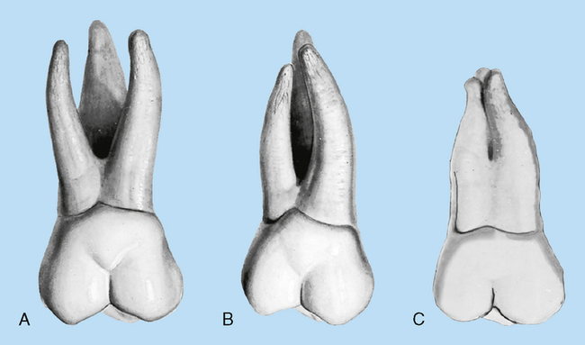
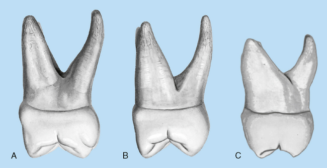
All three roots are usually visible from the buccal view (see Fig. 15-16). The two buccal roots incline distally, with the mesiobuccal root starting to curve at its middle third. The distal root is usually straighter and tends to cur/>

Only gold members can continue reading. Log In or Register to continue

Stay updated, free dental videos. Join our Telegram channel

Jan 4, 2015 | Posted by in General Dentistry | Comments Off on 15: Molars

VIDEdental - Online dental courses
Get VIDEdental app for watching clinical videos