14
Use of Digital Models/Dental Casts and their Role in Orthodontics/Maxillofacial Surgery
The dental plaster model is the physical three- dimensional (3D) representation of the dentition and oral anatomy that clinicians can hold in their hands and use to view the dental occlusion in any 3D spatial perspective. It is a necessary clinical and legal1 record for diagnostic purposes and the planning of treatment in orthodontics, prosthodontics, oromaxillofacial surgery, and cleft lip and palate surgery, where dental interceptive intervention is indicated. In addition to its role as a record for diagnosis and treatment planning, the dental plaster model is routinely used as the positive and accurate proxy for the fabrication of oral appliances and prostheses in the definitive treatment process. With the physical dental model, clinicians have been able to:
- perform measurements of intra- arch tooth size and inter- arch parameters (the overjet) using calipers;
- rearrange the positions of the teeth to simulate the prospective dental arch alignment and occlusion, as in the case of orthodontic planning (the Kesling setup2);
- simulate the expected positions of the dental arches for occlusal wafer construction immediately prior to orthognathic surgery.
From the physical measurements of the mesiodistal widths of the teeth, the Bolton analysis3 and the estimation of the degree of crowding or spacing are determined. In the Kesling setup, the physical rearrangement of the dental arch form to determine whether arch space may be gained through an extraction of teeth requires a drill – and – saw method to section parts of the plaster model and reset the plaster crowns of the teeth in a new arch arrangement. It is a time- consuming procedure and much disliked by clinicians.
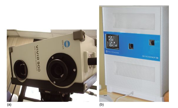
Figure 14.1 (a) The Minolta VIVID 900 surface laser scanner (Konica Minolta Sensing, Inc, Osaka, Japan) and (b) the NextEngine (Santa Monica, CA, USA) 3D scanner.

With the advancement of computer technology in the area of image acquisition, the accuracy and speed with which a physical object may be converted from a physical to a 3D digital form has improved significantly over the last 10 years. The cost of a surface laser scanner to accurately acquire the surface topology of a physical object is also no longer prohibitive. Compared with a portable industrial surface laser scanner from Konica Minolta Sensing, Inc (Osaka, Japan), the VIVID 900 model, which was priced at about US$40,000 some 5 years ago, a 3D desk-top surface laser scanner from NextEngine, Inc (Santa Monica, CA, USA) can at the time of writing be bought for under US$3,000 (Figure 14.1).
Such technologic improvements and cost reductions permit the application and adoption of modern image acquisition technology to dentistry. This has huge potential specifically in rendering the current processes in orthodontic diagnosis, the planning of treatment, and simulations more efficient and substantially more effective. Hence, 3D digital dental models could offer distinct technologic and clinical application advantages over physical plaster models.
TECHNOLOGIC ADVANTAGES OF DIGITAL DENTAL MODELS
There are several technologic advantages, described below. First, digital dental models, in that they are computer images by nature, are “unbreakable” and not subject to physical wear. In addition, they require no laboratory setup to duplicate them as the images can be replicated easily and quickly.
The storage of multiple sets of digital dental models requires little physical space and cost. They can be contained within the size of a conventional thumb- drive, and therefore free up valuable physical storage space that could be used for more productive purposes. The price of computer memory storage has never been lower; the retail price in April 2009 of a 4 gigabyte thumb – drive was approximately US$10. Since each digital dental model (via surface laser scanning) takes up between 5 and 10 megabytes of computer memory space, one could technically store 400–800 laser- scanned digital dental models in a 4 gigabyte thumb drive.
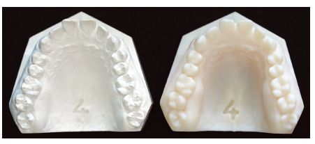
Figure 14.2 The accurate rapid- prototyped model (right) of the original dental model (left), which was created from computed tomographic scanning of the original dental model.

Accessibility to and security of an archive of digital dental models can be easily provided around the clock. The retrieval of digital data is quick through a dedicated computer server, and permissibility to view the digital dental models may be granted via secure passwords. Hence, it is impossible to “misplace” digital dental models or lose them through “theft.”
Additionally, as a form of digital data, they can be sent via high- speed broadband networks from one geographic location to another across miles in a blink of the eye in compressed and uncompressed modes. Using commercially available digital document technology through the software Adobe Acrobat 9 Pro Extended (Adobe Systems, Inc, San Jose, CA, USA), the 3D image of the digital dental model can be easily “inserted” as an image file into a portable document format (.pdf) text file, and sent to a recipient via email; the recipient can then view and interact with the 3D image of the dental model through Adobe Reader version 7.07 and higher (Adobe Systems, Inc). This technology renders visual communication and collaborative treatment between interested parties a reality at a high level of information clarity and efficiency without expensive freight charges.
Rapid prototyping reproduction from digital to physical forms can be achieved when needed. In the event of needing to convert a digital dental model into a physical hard copy, the digital image of the dental model can be easily converted to an appropriate format suitable for the rapid prototyping system to print out the 3D physical shape of the dental model (Figure 14.2).4
CLINICAL APPLICATIONS OF DIGITAL DENTAL MODELS IN ORTHODONTICS AND MAXILLOFACIAL SURGERY
Quantitative measurement of dental and oral anatomy
On acquiring the 3D image of the plaster dental model from contemporary 3D scanners, the digital image undergoes a series of “processing” steps before the scanned images can be made meaningful and useful to the clinician.
Figure 14.3 (a) The individually scanned digital maxillary and mandibular models, and (b) with the models in occlusion, in natural “white” color and texture.

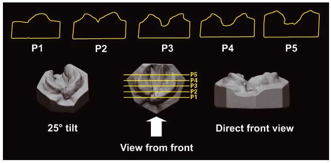
Figure 14.4 Digital model of a cleft palate that is sliced in the anteroposterior direction from planes P1 to P5. The corresponding palatal contour is viewed further above from P1 to P5.

The surface of the digital dental model is mathematically represented as a surface of points (vertices) joined together to form a network lattice of triangles. The overall surface color and texture is displayed when each triangle is computer- coded for that specific color (a color map) and texture (a texture map). Color and texture added to the surface of the image provide a realistic visual representation of the plaster dental model (Figure 14.3).
With accompanying programming for capability to make geometric measurements within a graphical user interface environment, any linear, angular, area, and volumetric measurements, or a combination of these measurements, can be taken from the surface on the 3D image of the digital dental model.
Digital models can be “sliced” in any preferred orientation plane to reveal the surface contours (Figure 14.4) of the dental and oral anatomy5 and the spatial relationship of the contours of teeth surfaces in an occlusion. From the “slices,” the digital anatomic contours can be highlighted. Linear and area measurements can be made from these two – dimensional (2D) slices. Volume measurements (Figure 14.5) can also be made from the digital anatomy based on the slice plane orientation5 and predefined anatomic reference indicators.
Figure 14.5 The boundaries of the volume o/>
Stay updated, free dental videos. Join our Telegram channel

VIDEdental - Online dental courses


