Premolars
• To identify an extracted premolar as maxillary or mandibular, first or second, right or left
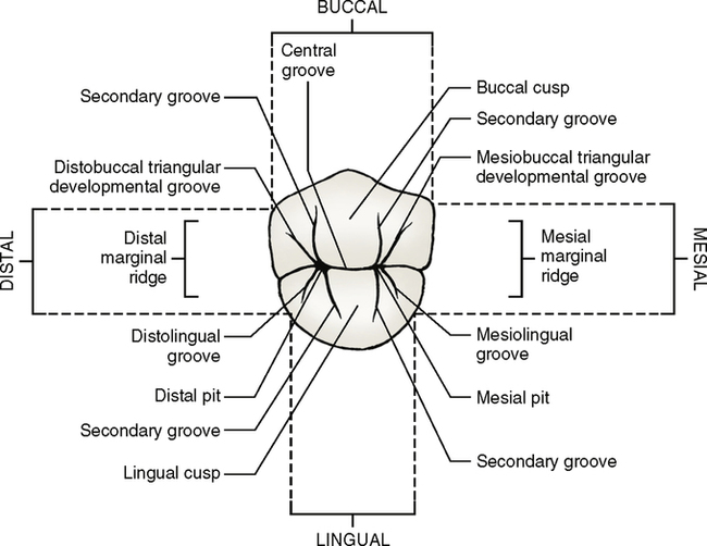
• To recognize and name the pertinent dental anatomic form of each tooth such as cusps, ridges, developmental grooves, triangular grooves, pits, and developmental depressions
• To make comparisons between maxillary and mandibular premolars
• To discuss the major differences and similarities between the maxillary first and second premolars
• To describe briefly the various occlusal forms possible for a mandibular second premolar
• To compare the mandibular first premolars with the mandibular second premolars (development, shape, and diversities of anatomic form)
• To understand how development occurs through the formation and fusion of the lobes
• To understand how the form of a tooth relates to its ultimate function
MAXILLARY PREMOLARS
First Premolars
Evidence of calcification: 1.5 years
Enamel completed: 5 to 6 years
Root completed: 12 to 13 years
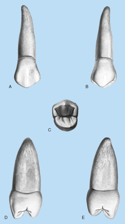
Maxillary first premolars (Fig. 14-1) have buccal and lingual cusps. The buccal cusp is usually 1 mm or more longer than the lingual cusp. These teeth are also the only premolars that normally have two roots, a buccal and lingual, although occasionally, only a single root is evident.
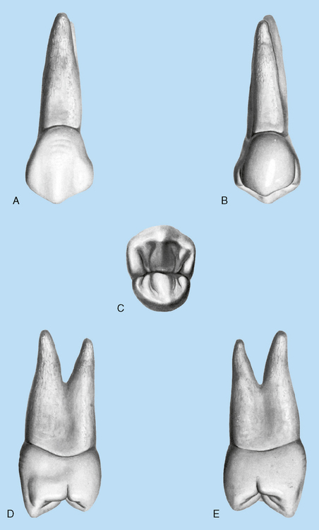
Mesial aspect
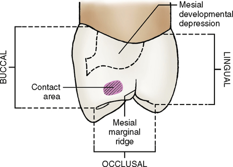
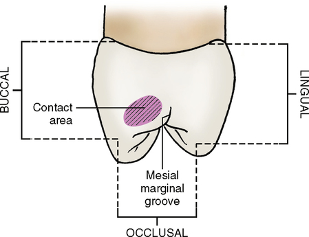
On the mesial surface (Fig. 14-4; see Fig. 14-1, C) of the crown, a groove extends from the mesial marginal ridge cervically. This groove is called the mesial marginal groove. It crosses the mesial marginal ridge and runs from the occlusal third to the middle third of the crown, lingual to the contact area. The mesial surface can also be identified by a mesial developmental depression located cervically to the mesial contact area. The concavity continues cervically from above the contact area across the cervical line, where it joins a deep developmental depression between the roots. The mesial marginal groove is not always present, but the mesial developmental depression usually is quite evident.
Distal aspect
From the distal view (Fig. 14-5; see Fig. 14-1, E) a maxillary first premolar is similar to the mesial view, except no groove usually crosses the distal marginal ridge and no developmental depression is present. Some specimens show a distinct distal marginal groove, but the mesial marginal groove is deeper and more obvious. The cervical line is less curved on the distal surface than on the mesial surface. The crown also appears more rounded and smooth. Both the buccal and lingual cusp tips are centered over the root, and this is also true from the mesial view. All maxillary premolars have their cusp tips centered over their root.
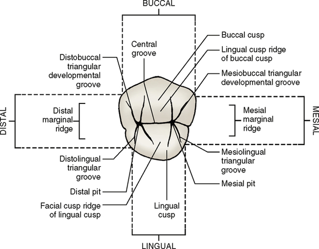
Occlusal aspect
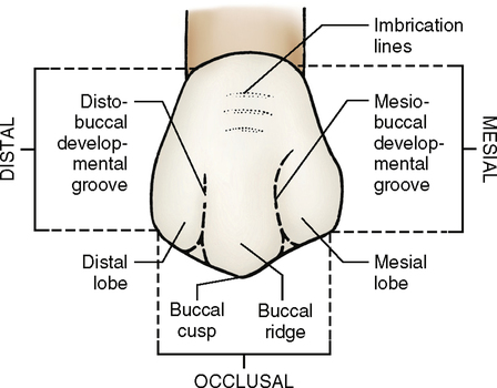
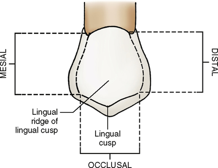
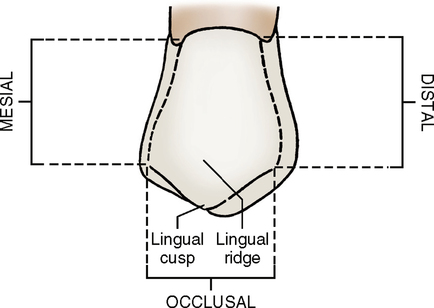
The occlusal surface (Fig. 14-6; see Fig. 14-1, C) shows two well-developed cusps. The lingual cusp is more pointed than the facial cusp, but the facial cusp is much larger and longer than the lingual. Each cusp has four ridges emanating from it, and each ridge is named according to its location: facial, lingual, distal, and mesial.
The lingual cusp ridge extends from the cusp tip lingually to the central area of the occlusal surface. Any ridge that runs from the cusp tip to the central groove of the occlusal surface is called a triangular ridge. Examples are the lingual cusp ridge of the buccal cusp and the buccal cusp ridge of the lingual cusp, which runs from the cusp tip of the lingual cusp to the central groove (Fig. 14-7).
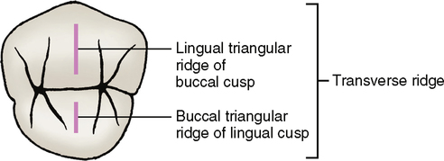
When two triangular ridges join, after traversing the tooth buccolingually, they form a transverse ridge. Thus a transverse ridge exists on the occlusal surface of a maxillary first premolar. It is formed by the union of the two triangular ridges—the lingual cusp ridge of the buccal cusp and the facial cusp ridge of the lingual cusp. See Fig. 14-7 for the transverse ridge formed by the two joining triangular ridges.
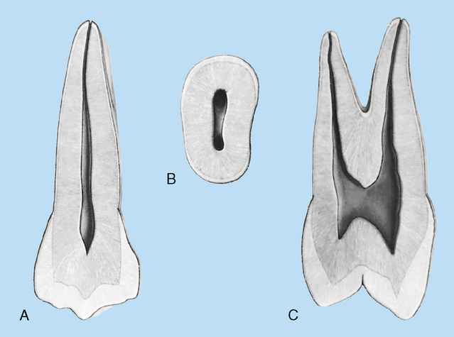
Pulp cavity
The pulp cavity (Fig. 14-8) of the maxillary first premolar has two pulp horns, one for each cusp, and two root canals, one for each root. Sometimes only one undivided root is present. When this occurs, usually two root canals are still evident, although they often combine to form one apical foramen. In some specimens with only one root, only one single root canal is present.
General characteristics
1. The posterior teeth have a greater faciolingual measurement in relation to their mesiodistal measurements.
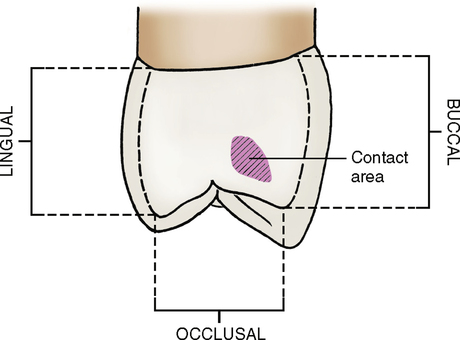
2. The mesial and distal contact areas are broader and closer to the same level on the tooth.
3. The mesial and distal curvature of the cervical line is less.
4. The crown measurements cervicoocclusally are less, giving the appearance of shorter crown length.
Second Premolars
Evidence of calcification: 2 years
Enamel completed: 6 to 7 years
Root completed: 12 to 14 years
Maxillary second premolars (Fig. 14-9) resemble the maxillary first premolars in both form and function. The crown, however, has a less angular and more rounded appearance. The second premolars also vary from the first in that they usually have only one root. How many roots do the maxillary first premolars have?
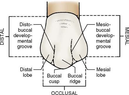
Facial (buccal) aspect
From the buccal view (Fig. 14-10; see Fig. 14-9, A), it is evident that the buccal cusp of a second premolar is not as long as that of a first premolar, and it appears less pointed. Also, a second premolar has the same general markings as the first, but they are not as well defined.
Mesial aspect
The mesial view (Fig. 14-12; see Fig. 14-9, D) shows the difference in cusp length between the maxillary first and second premolars. The buccal cusp of a second premolar is shorter than the buccal cusp of a first premolar, and the lingual cusp is almost as long; thus the buccal and lingual cusps are nearly the same length.
Occlusal aspect
The occlusal outline (Fig. 14-13; see also Fig. 14-9, C) is more rounded than that of a first premolar, and the second premolar is ovoid rather than hexagonal.
Stay updated, free dental videos. Join our Telegram channel
VIDEdental - Online dental courses


