12 The Permanent Mandibular Molars
Mandibular First Molar
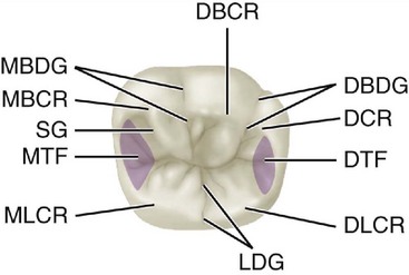
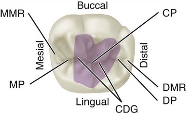
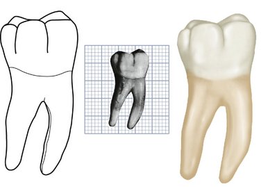
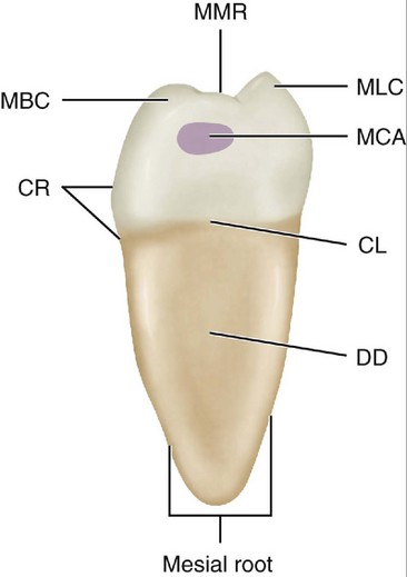
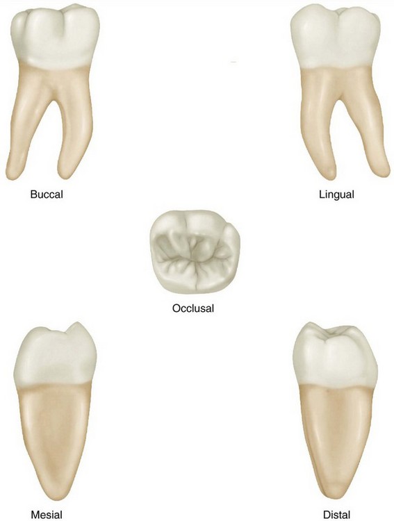
Figures 12-1 through 12-17 illustrate the mandibular first molar from all aspects. Normally, the mandibular first molar is the largest tooth in the mandibular arch. It has five well-developed cusps: two buccal, two lingual, and one distal (see Figure 12-1). It has two well-developed roots, one mesial and one distal, which are very broad buccolingually. These roots are widely separated at the apices.
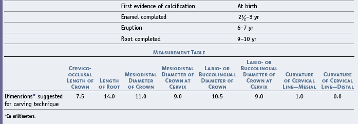
The dimension of the crown mesiodistally is greater by about 1 mm than the dimension buccolingually (Table 12-1). Although the crown is relatively short cervico-occlusally, it has mesiodistal and buccolingual measurements that provide a broad occlusal form.
The mesial root is broad and curved distally, with mesial and distal fluting that provides the anchorage of two roots (see Figure 13-22). The distal root is rounder, broad at the cervical portion, and pointed in a distal direction. The formation of these roots and their positions in the mandible serve to brace the crown of the tooth efficiently against the lines of force that might be brought to bear against it.
DETAILED DESCRIPTION OF THE MANDIBULAR FIRST MOLAR FROM ALL ASPECTS
Buccal Aspect
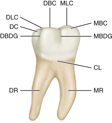
From the buccal aspect, the crown of the mandibular first molar is roughly trapezoidal, with cervical and occlusal outlines representing the uneven sides of the trapezoid. The occlusal side is the longer (see Figures 12-3, 12-4, 12-12, 12-13, and 12–14).
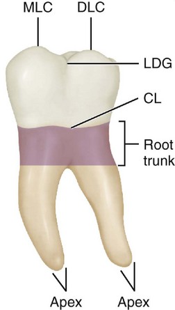
Two developmental grooves appear on the crown portion. These grooves are called the mesiobuccal developmental groove and the distobuccal developmental groove. The first-named groove acts as a line of demarcation between the mesiobuccal lobe and the distobuccal lobe. The latter groove separates the distobuccal lobe from the distal lobe (see Figures 12-2 and 12-3).
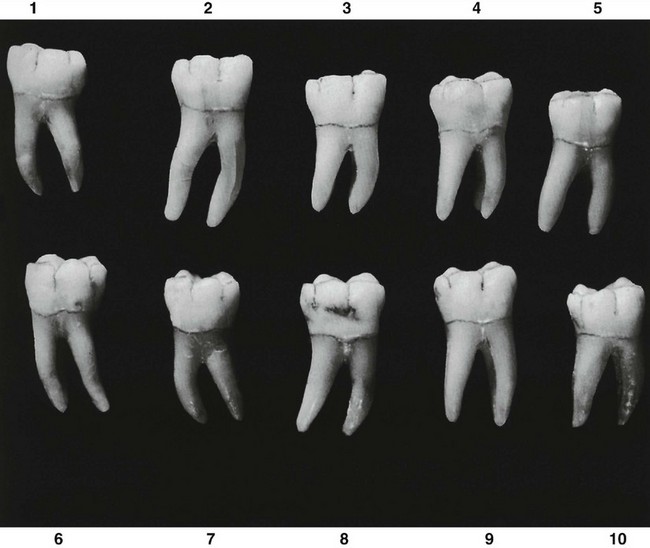
The mesiobuccal, distobuccal, and distal cusps are relatively flat. These cusp ridges show less curvature than those of any of the teeth described so far. The distal cusp, which is small, is more pointed than either of the buccal cusps. Flattened buccal cusps are typical of all mandibular molars. In most first molar specimens the buccal cusps are worn considerably, with the buccal cusp ridges almost at the same level. Before they are worn, the buccal cusps and the distal cusp have curvatures that are characteristic of each one (see Figures 12-4 and 12-14, 4).
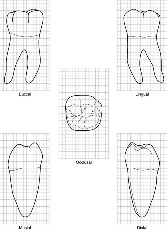
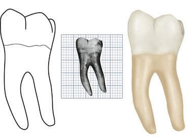
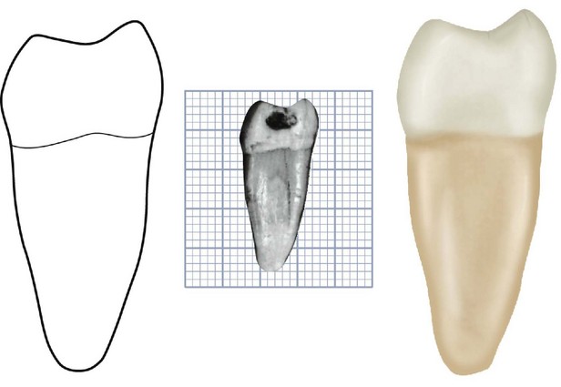
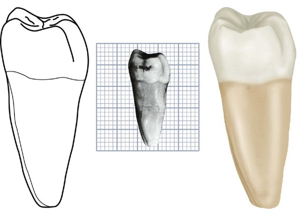
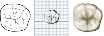
Figure 12-12 Mandibular right first molar. Graph outlines of five aspects are shown. (Grid = 1 sq mm.)
The surface of the buccal portion of the crown is smoothly convex at the cusp portions with developmental grooves between the cusps. Approximately at the level of the ends of the developmental grooves, in the middle third, a developmental depression is noticeable. It runs in a mesiodistal direction just above the cervical ridge of the buccal surface (see Figure 12-14, 6 and 8). This cervical ridge may show a smooth depression in it that progresses cervically, joining with the developmental concavity just below the cervical line, which is congruent with the root bifurcation buccally.
The roots of this tooth are, in most instances, well formed and constant in development.
The distal root is less curved than the mesial root, and its axis is in a distal direction from cervix to apex. The root may show some curvature at its apical third in either a mesial or a distal direction (see Figure 12-14, 1 and 8). The apex is usually more pointed than that of the mesial root and is located below or distal to the distal contact area of the crown. Considerable variation is evident in the comparative lengths of mesial and distal roots (see Figure 12-14).
Both roots are wider mesiodistally at the buccal areas than they are lingually. Developmental depressions are present on the mesial and distal sides of both roots, which lessens the mesiodistal measurement at those points. They are somewhat thicker at the lingual borders. This arrangement provides a secure anchorage for the mandibular first molar, preventing rotation. This I-beam principle increases the anchorage of each root (see Figure 13-22).
Lingual Aspect
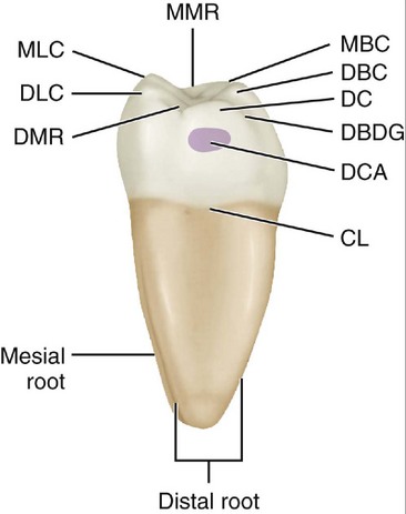
From the lingual aspect, three cusps may be seen: two lingual cusps and the lingual portion of the distal cusp (see Figures 12-5, 12-6, 12-12, and 12-13). The two lingual cusps are pointed, and the cusp ridges are high enough to hide the two buccal cusps from view. The mesiolingual cusp is the widest mesiodistally, with its cusp tip somewhat higher than the distolingual cusp. The distolingual cusp is almost as wide mesiodistally as the mesiolingual cusp. The mesiolingual and distolingual cusp ridges are inclined at angles that are similar on both lingual cusps. These cusp ridges form obtuse angles at the cusp tips of approximately 100 degrees.
The distal cusp is at a lower level than the mesiolingual cusp.
The distal outline of the crown is straight immediately above the cervical line to a point immediately below the distal contact area; this area is represented by a convex curvature that also outlines the distal surface of the distal cusp. The junction of the distolingual cusp ridge of the distolingual cusp with the distal marginal ridge is abrupt; it gives the impression of a groove at this site from the lingual aspect. Sometimes, a shallow developmental groove occurs at this point (see Figure 12-10). Part of the mesial and distal surfaces of the crown and root trunk may be seen from this aspect because the mesial and distal sides converge lingually.
The roots of the mandibular first molar appear somewhat different from the lingual aspect. They measure about 1 mm longer lingually than buccally, but the length seems more extreme (see Figures 12-6 and 12-7). This impression is derived from the fact that the cusp ridges and cervical line are at a higher level (about 1 mm). This arrangement adds a millimeter to the distance from root bifurcation to cervical line. In addition, the mesiodistal measurement of the root trunk is less toward the lingual surface than toward the buccal surface. Consequently, this slenderness lingually, in addition to the added length, makes the roots appear longer than they are from the lingual aspect (see Figure 12-9).
Mesial Aspect
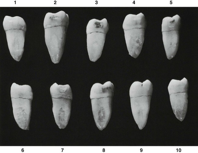
When the mandibular first molar is viewed from the mesial aspect, with the specimen held with its mesial surface at right angles to the line of vision, two cusps and one root only are to be seen: the mesiobuccal and mesiolingual cusps and the mesial root (see Figures 12-7, 12-8, 12-12, 12-13, and 12–15).
Stay updated, free dental videos. Join our Telegram channel

VIDEdental - Online dental courses


