Periodontium: Alveolar process and cementum
Learning objectives
■ Describe the nature of alveolar bone proper and supporting bone.
Key terms
Alveolar bone proper
Alveolar crest
Area of reversal
Bodily movement
Dehiscence
Extrusion
Fenestration
Haversian bone
Hyalinization
Incisor liability factor
Intrusion
Lamina dura
Leeway space
Mesial drift
Reversal line
Rotation
Sharpey fibers
Supporting bone
Tipping movement
Overview
This chapter discusses the hard tissues of the periodontium, which are cementum and alveolar bone. The alveolar process is the bony part of the maxilla and mandible that has the primary function of supporting the teeth. Alveolar bone is composed of alveolar bone proper, which is attached to the fibers embedded in the roots of the teeth. Supporting bone is the bone covering the mandible, and it serves as cortical plates that give support to the alveolar bone proper. This alveolar bone is in the process of continuous turnover, which enables the tissue to be responsive to manipulation, such as tooth movement resulting from normal physiologic function or orthodontic treatment. Cementum functions as the means of fiber attachment to the tooth roots. These fibers have the abilities to form and resorb, which are necessary for support during tooth movement. If teeth are moving in a straight line or rotating, all parts of the suspensory apparatus must change simultaneously. This phenomenon first took place during tooth eruption and continues to function for both the primary and secondary dentition. Tooth function is a prerequisite for the maintenance of the alveolar bone and cementum. Bone loss occurs during aging or periods of inactivity, resulting in possible tooth mobilization. With loss of alveolar bone, loss of periodontal fibers occurs as well. Periodontal disease can cause these conditions with possible tooth loss that could result in an edentulous jaw (Box 12-1).
Alveolar process
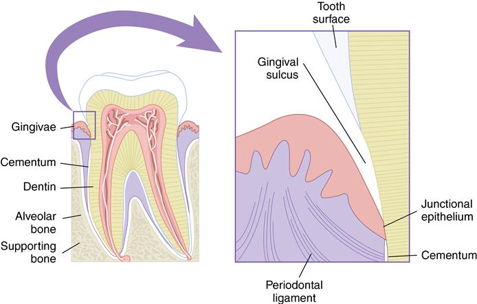
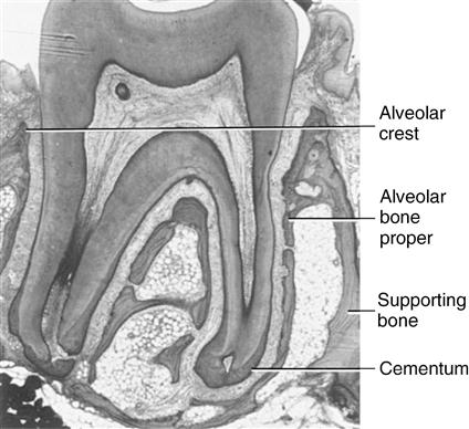
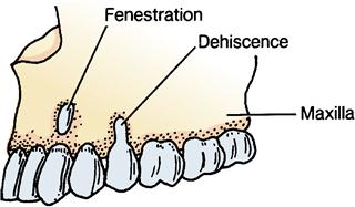
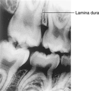
The alveolar process is the part of the maxilla and mandible that supports the roots of teeth and is composed of alveolar bone proper and supporting bone (Fig. 12-1). Alveolar bone proper is the bone lining the tooth socket. In clinical radiographic terms, it is defined as the lamina dura. Dense bone serves as the attachment bone that surrounds the roots of the teeth. Supporting bone is, as the name implies, the bone that serves as a dense cortical plate to sustain the alveolar bone proper. This cortical plate covers the surface of the maxilla and mandible and supports the alveolar bone proper. The supporting cancellous bone underlies and supports the dense cortical bone (see Fig. 12-1 and Fig. 12-2). The existence of alveolar bone is entirely dependent on the presence of teeth. Alveolar bone develops initially as a protection for the soft developing primary teeth and later, as the roots develop, as a support for the teeth. Finally, as the teeth are lost, the alveolar bone resorbs. Teeth are responsible not only for the development but also for the maintenance of the alveolar process of the mandible (see Fig. 3-21). The coronal border of the alveolar process is known as the alveolar crest (see Fig. 12-2). This crest is normally located approximately 1.2 to 1.5 mm below the dentinoenamel junction of the teeth. It is rounded on the anterior region and nearly flat in the molar area. When teeth are viewed from the buccolingual aspect, the alveolar crest may be thin or missing. The area of bone loss where an apical root penetrates the cortical bone is known as a fenestration, and bone loss in the coronal area of the root is termed dehiscence (Fig. 12-3).
 CLINICAL COMMENT
The lamina dura is an important diagnostic landmark in determining the health of the periapical tissues. Loss of density usually means infection, inflammation, and resorption of this bony socket lining.
Alveolar bone proper
The compact or dense bone that lines the tooth socket is of two types when viewed microscopically. This bone either contains perforating fibers from the periodontal ligament or is similar to compact bone found elsewhere in the body. Perforating fibers or Sharpey fibers are bundles of collagen fibers embedded in the alveolar bone proper. These fibers are at right angles or oblique to the surface of the alveolar bone proper and along the root of the tooth (see Fig. 12-1). The fiber bundles inserting in the bone are regularly spaced and appear similar to those that insert into the root surface cementum (Figs. 12-4 and 12-5). Perforating fibers are not limited to periodontal bone. They also appear anywhere in the body where ligaments or tendons attach to cartilage or bone.
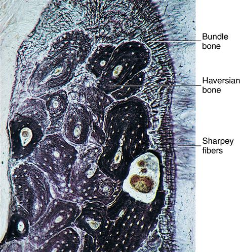
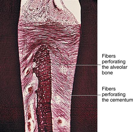
Uniformity of position of numerous fibers in cemental and bony surfaces is shown. Fiber bundles of bone are larger and less numerous than fibers entering cemental surface.
Because bone of the alveolar process is regularly penetrated by collagen fiber bundles, it can be appropriately termed bundle bone. Bundle bone, being synonymous with alveolar bone proper or lamina dura, appears more dense radiographically than the adjacent supportive bone (Fig. 12-6). This density is probably the result of the mineral content or orientation of the mineral crystals (Ca++ hydroxyapatite) surrounding the fiber bundles. Blood vessels and nerves penetrate the lamina dura through small foramina. Because the mineral density is sufficient, this bone appears opaque in radiographs (see Fig. 12-6). Tension on the perforating fibers during mastication is believed to stimulate this bone and is considered important in its maintenance.
Stay updated, free dental videos. Join our Telegram channel
VIDEdental - Online dental courses


