CHAPTER 12
ONCOCYTOMA
Oncocytes develop from the accumulation of large numbers of abnormal mitochondria within epithelial cells resulting in cytoplasmic enlargement with granularity and a low nuclear cytoplasmic ratio. Although this process can occur in many types of epithelial cells, oncocytic change within salivary glands is extremely common and seems to be an age-related phenomenon. Whether this change occurs because of an intracellular abnormality or in response to external stimuli has not been defined clearly. The term “oncocytosis” is used when oncocytic change is prominent. Oncocytosis can occur in either diffuse or nodular forms.
The 2005 World Health Organization Classification of Tumours: Pathology and Genetics of Head and Neck Tumours defines oncocytoma as a “benign tumour of salivary gland origin composed exclusively of large epithelial cells with characteristic bright eosinophilic granular cytoplasm (oncocytic cells).” Separation of nodular oncocytosis (nodular oncocytic metaplasia) from oncocytoma may be difficult; however, when an oncocytic proliferation is circumscribed, at least partially encapsulated, and large enough to be clinically detectable, it is classified best as an oncocytoma.
Oncocytomas comprise approximately 1% of all salivary gland neoplasms. They occur most commonly in the sixth through eighth decades with a mean age of 58 years. There is no sex predilection. Approximately 84% of major salivary gland oncocytomas occur in the parotid, with the remainder in the submandibular gland. Lesions developing in the minor salivary glands are uncommon, but they do occur. Parotid gland oncocytoma most often presents as a painless mass.
There are no clear etiologies for most oncocytomas. Occasional oncocytoma patients, however, have a history of prior radiation therapy or prolonged radiation exposure to the head and neck. These patients present at a younger age as compared with the nonradiation-exposed individuals.
12.3 CYTOLOGIC FEATURES WITH HISTOLOGIC CORRELATES
12.3.1 Gross Features
Oncocytomas are well circumscribed, solid, tan-brown nodules without evidence of hemorrhage or necrosis. They are encapsulated at least partially and typically measure 3–4 cm, although lesions can range up to 7 cm.
12.3.2 Cytologic Features
Fine needle aspiration biopsies of oncocytomas yield cellular specimens. The smears contain abundant large cells present as individual cells, clusters, papillary fragments, and sheets (Figure 12.1). The oncocytic cells have abundant granular cytoplasm with a low nuclear cytoplasmic ratio and distinct cytoplasmic borders (Figures 12.2–12.5). On alcohol-fixed, Papinicolaou-stained slides, the cytoplasm is blue-gray to orange-pink and granular (Figures 12.2, 12.3, and 12.5). On air-dried, Romanowsky-stained slides, the cytoplasm appears blue and without observable granules (Figure 12.4). The nuclei of oncocytic cells are round to oval, centrally placed, and may contain variably sized nucleoli (Figures 12.2–12.5). Some oncocytomas may show nuclear enlargement with vesicular chromatin. Naked nuclei representing isolated oncocytes usually are observed in the background (Figure 12.1). In tumors with a clear cell component, the tumor cells contain abundant optically clear (Papinicolaou stain) or microvacuolated (Romanowsky stain) cytoplasm. Smears from oncocytomas typically are devoid of inflammatory cells.
FIGURE 12.1. Oncocytoma. Cellular specimen containing sheets and clusters of oncocytes and scattered naked nuclei (Papanicolaou ×200).
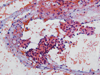
FIGURE 12.2. Oncocytoma. Oncocytes contain abundant, granular pink-red cytoplasm with uniform, round-to-oval nuclei and occasional variably prominent nucleoli (Papanicolaou ×600).
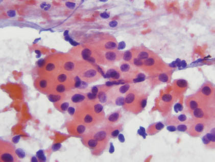
FIGURE 12.3. Oncocytoma. In this example, the oncocytes’ abundant granular cytoplasm is gray-blue, rather than pink-red, as shown in Figures 12.1 and 12.2 (Papanicolaou ×600).
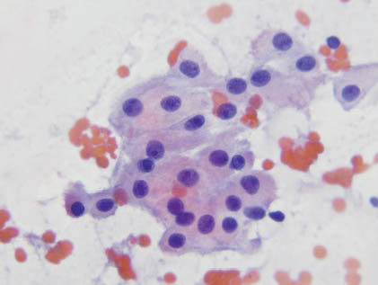
FIGURE 12.4. Oncocytoma. Oncocytes contain abundant, waxy blue cytoplasm without observable granules and uniform round-to-oval nuclei (Romanowsky ×600). Courtesy of C. Michael, University of Michigan Health System, Ann Arbor, MI.
Stay updated, free dental videos. Join our Telegram channel
VIDEdental - Online dental courses


