Incisors
• To identify the particular anatomic features of incisor teeth
• To compare maxillary central incisors with maxillary lateral incisors
• To compare maxillary incisors with their mandibular incisor counterparts
• To identify an extracted incisor
• To recognize the normal and deviated anatomic forms of incisor teeth
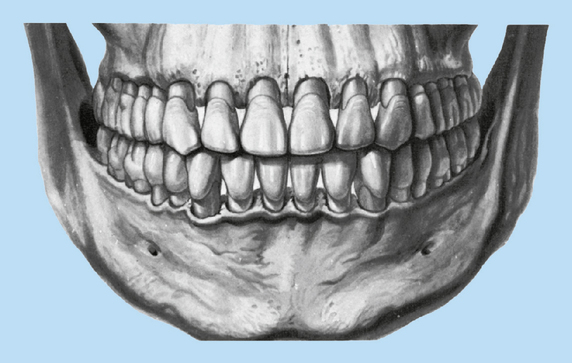
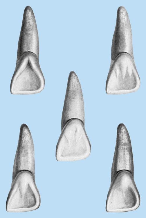
The eight permanent incisors are four maxillary (upper) and four mandibular (lower). The maxillary incisors consist of two central and two lateral incisors, as do the four mandibular incisors (Fig. 12-1).
MAXILLARY INCISORS
Central Incisors
Evidence of calcification: 3 months
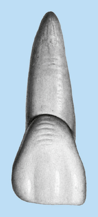
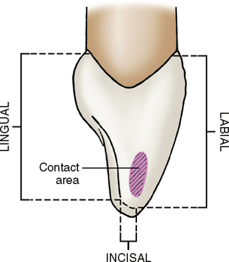
A maxillary central incisor (Fig. 12-2) is the widest mesiodistally of any of the anterior teeth. Its labial appearance is less rounded than that of a maxillary lateral incisor or canine. The crown usually looks symmetrical and normally formed, having a nearly straight incisal edge, a mesial side with a straight outline, and the distal side that is more curved. The mesioincisal angle is relatively sharp, and the distoincisal angle is much more rounded.
Labial aspect (Fig. 12-3; see also Fig. 12-8, A)
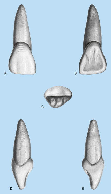

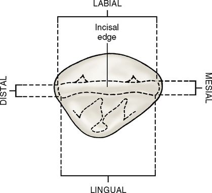
Incisal aspect (Fig. 12-7; see also Fig. 12-8, C)
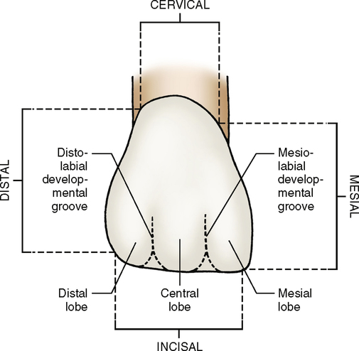
The incisal ridge tends to slope lingually as a result of the lower incisors coming more frequently into contact with the lingual edge than with the facial edge of the maxillary incisor. From an incisal view, the crown shows a triangular shape, with its apex on the lingual surface. (See Fig. 12-8 for all of the views of a maxillary right central incisor.)
Lateral Incisors
Evidence of calcification: 1 year
Labial aspect
Although the labial aspect (Fig. 12-9, A) of a maxillary lateral incisor may appear to resemble that of a central incisor, it usually has more curvature, with a rounded incisal ridge and rounded angles mesially and distally. The distal outline is always more rounded, and the height of contour is more cervical than the mesial outline.
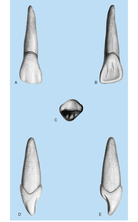
Lingual aspect
The lingual view (Fig. 12-9, B) of a lateral incisor shows more contrast than the same view of a central incisor. Mesial and distal marginal ridges are pronounced, and the cingulum is usually prominent, with a tendency toward deep developmental grooves within the lingual fossa where it joins the cingulum. The linguoincisal ridge is better developed, and the lingual fossa is more concave and circumscribed than that of the central incisor. A lingual pit is frequently present.
Incisal aspect
The incisal aspect (Fig. 12-9, C) of these teeth sometimes resembles that of the central incisors or a small canine. The cingulum and the incisal ridge, however, may be large; the labiolingual dimension may be greater than usual in comparison with the mesiodistal dimension. If these variations are present, the teeth show a strong resemblance to the canines.
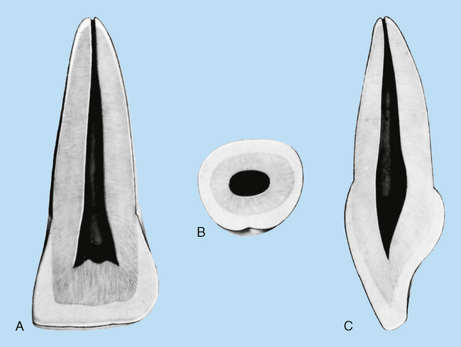
Pulp Cavity
The pulp cavity of the maxillary central incisor (Fig. 12-10) mirrors the configuration of the tooth. Only one root canal is evident, which is rather large. The pulp chamber lies in the coronal portion of the tooth and presents three sharp elongations: mesial, distal, and central pulp horns. The central pulp horn is usually shorter and more rounded than the other two.
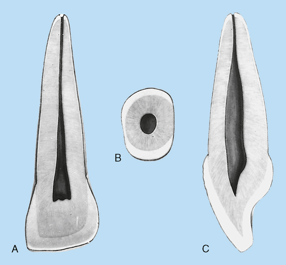
The pulp cavity of the maxillary lateral incisor is quite simple, comprising only a pulp chamber and a single pulp canal. The chamber is similar to that of the maxillary central incisor but usually does not have three sharp pulp horns. More often, the pulp chamber ends incisally as one rounded form or two less sharp pulp horns, a mesial and distal (Fig. 12-11).
Stay updated, free dental videos. Join our Telegram channel
VIDEdental - Online dental courses


