11: The Permanent Maxillary Molars

11 The Permanent Maxillary Molars

The maxillary molars differ in design from any of the teeth previously described. These teeth assist the mandibular molars in performing the major portion of the work in the mastication and comminution of food. They are the largest and strongest maxillary teeth, by virtue both of their bulk and of their anchorage in the jaws. Although the crowns on the molars may be somewhat shorter than those on the premolars, their dimensions are greater in every respect. The root portion may be no longer than that of the premolars, but instead of one root or a bifurcated root, the maxillary molar root is broader at the base in all directions and is trifurcated into three well-developed prongs that are actually three full-size roots emanating from a common broad base above the crown.

Generally speaking, the maxillary molars have large crowns with four well-formed cusps. They have three roots, two buccal and one lingual. The lingual root is the largest. The crowns have two buccal cusps and two lingual cusps. The outlines and curvatures of all the maxillary molars are similar. Developmental variations will be set forth under descriptions of the separate molars.

Before a detailed description of the maxillary first molar is begun, some statements will be made that are applicable to all first molars, mandibular and maxillary.

The permanent first molars usually appear in the oral cavity when the child is 6 years old. The mandibular molars precede the maxillary molars. The first permanent molar (maxillary or mandibular) erupts posterior to the second deciduous molar, taking up a position in contact with it. Therefore the first molar is not a succedaneous tooth because it has no predecessor. The deciduous teeth are all still in position and functioning when the first molar takes its place. Because the development of the bones of the face is downward and forward, sufficient space has been created normally at the age of 6 for the accommodation of this tooth.

The normal location of the first permanent molar is at the center of the fully developed adult jaw anteroposteriorly. As a consequence of the significance of their positions and the circumstances surrounding their eruption, the first molars may also be considered cornerstones of the dental arches. A full realization of the significance of these teeth as units in the arches and their function and positions relative to the other teeth will be gained when an opportunity comes to study the arrangement of the teeth with their occlusion and the temporomandibular articulation of the jaws. Subsequent chapters cover those phases. The mandibular molars are described in Chapter 12.

Maxillary First Molar

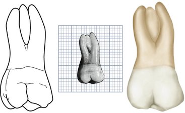
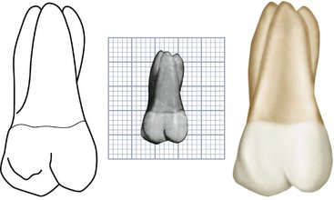
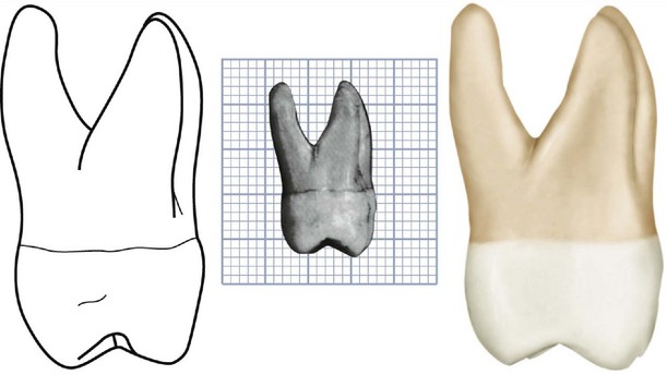
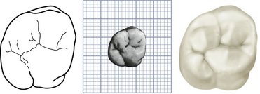
Figures 11-1 through 11-18 illustrate the maxillary first molar from all aspects. The crown of this tooth is wider buccolingually than mesiodistally. Usually the extra dimension buccolingually is about 1 mm (Table 11-1). This, however, varies in individuals (see Figure 11-17, 1, 5, 7, and 9). From the occlusal aspect, the inequality of the measurements in the two directions appears slight. Although the crown is relatively short, it is broad both mesiodistally and buccolingually, which gives the occlusal surface its generous dimensions.

The maxillary first molar is normally the largest tooth in the maxillary arch. It has four well-developed functioning cusps and one supplemental cusp of little practical use. The four large cusps of most physiological significance are the mesiobuccal, the distobuccal, the mesiolingual, and the distolingual. A supplemental cusp is called the cusp or tubercle of Carabelli. This morphological trait can take the form of a well-developed fifth cusp, or it can grade down to a series of grooves, depressions, or pits on the mesial portion of the lingual surface. This trait has been used to distinguish populations.

This supplemental cusp is found lingual to the mesiolingual cusp, which is the largest of the well-developed cusps. Usually, a developmental groove is found, leaving a record of cusp development, unless it has been erased by frictional wear. The fifth cusp or a developmental trace at its usual site serves to identify the maxillary first molar. A specimen of this tooth showing no trace of its typical characteristic would be rare.

The three roots of generous proportions are the mesiobuccal, distobuccal, and lingual. These roots are well separated and well developed, and their placement gives this tooth maximum anchorage against forces that would tend to unseat it. The roots have their greatest spread parallel to the line of greatest force brought to bear against the crown diagonally in a buccolingual direction. The lingual root is the longest root. It is tapered and smoothly rounded. The mesiobuccal root is not as long, but it is broader buccolingually and shaped (in cross section) so that its resistance to torsion is greater than that of the lingual root. The distobuccal root is the smallest of the three and smoothly rounded.

The development of maxillary first molars rarely deviates from the accepted normal. Ten specimens with uncommon variations are shown in Figure 11-18.

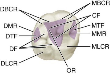
DETAILED DESCRIPTION OF THE MAXILLARY FIRST MOLAR FROM ALL ASPECTS
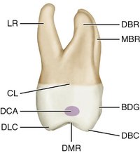
Buccal Aspect

The crown is roughly trapezoidal, with cervical and occlusal outlines representing the uneven sides (see Figures 11-4, 11-13, 11-14, and 11-15). The cervical line is the shorter of the uneven sides (see Figure 4-16, D).

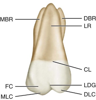
When the buccal aspect of this tooth is viewed with the line of vision at right angles to the buccal developmental groove of the crown, the distal side of the crown can be seen in perspective, which is possible because of the obtuse character of the distobuccal line angle (see Occlusal Aspect later in this chapter). Parts of four cusps are seen, the mesiobuccal, distobuccal, mesiolingual, and distolingual.

The mesiobuccal cusp is broader than the distobuccal cusp, and its mesial slope meets its distal slope at an obtuse angle. The mesial slope of the distobuccal cusp meets its distal slope at approximately a right angle. The distobuccal cusp is therefore sharper than the mesiobuccal cusp, and it is at least as long and often longer (see Figure 11-15, 4, 6, 7, 8, and 9).

The buccal developmental groove that divides the two buccal cusps is approximately equidistant between the mesiobuccal and distolingual line angles. The groove slants occlusoapically in a line of direction parallel to the long axis of the distobuccal root. It terminates at a point approximately half the distance from its origin occlusally to the cervical line of the crown. Although the groove is not deep at any point, it becomes more shallow toward its termination, gradually fading out. Lateral to its terminus is a dip in the enamel of the crown that is developmental in character and extends for some distance mesially and distally.

The cervical line of the crown does not have much curvature from mesial to distal; however, it is not as smooth and regular as that found on some of the other teeth. The line is generally convex with the convexity toward the roots.

The mesial outline of the crown from this aspect follows a nearly straight path downward and mesially, curving occlusally as it reaches the crest of contour of the mesial surface, which is the contact area. This crest is approximately two thirds the distance from cervical line to tip of mesiobuccal cusp. The mesial outline continues downward and distally and becomes congruent with the outline of the mesial slope of the mesiobuccal cusp.

The distal outline of the crown is convex; the distal surface is spheroidal. The crest of curvature on the distal side of the crown is located at a level approximately half the distance from cervical line to tip of cusp. The distal contact area is in the middle of the middle third.

Often from this aspect, a flattened area or a concave area is seen on the distal surface immediately above the distobuccal cusp at the cervical third of the crown.

All three of the roots may be seen from the buccal aspect. The axes of the roots are inclined distally. The roots are not straight, although the buccal roots show an inclination to curvature halfway between the point of bifurcation and the apices. The mesiobuccal root curves distally, starting at the middle third. Its axis usually is at right angles to the cervical line. The distal root is straighter, with its long axis at an acute angle distally with the cervical line. It has a tendency toward curvature mesially at its middle third.

The point of bifurcation of the two buccal roots is located approximately 4 mm above the cervical line. This measurement varies somewhat, of course. Nevertheless, the point is much farther removed from the cervical line than in the deciduous molars. This relation is typical when all permanent molars are compared with all deciduous molars.

A deep developmental groove buccally on the root trunk of the maxillary first molar, which starts at the bifurcation and progresses downward, becoming more shallow until it terminates in a shallow depression at the cervical line. Sometimes this depression extends slightly onto the enamel at the cervix.

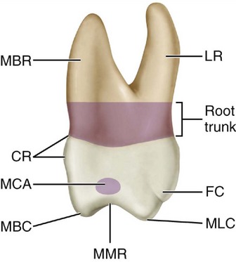
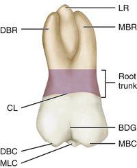
The reader must keep in mind the fact that molar roots originate as a single root on the base of the crown. They then are divided into three roots, as in the maxillary molars, or two roots, as in the mandibular molars. The common root base is called the root trunk (see Figures 11-3 and 11-8).
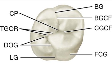
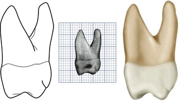
In judging the length of the roots and the direction of their axes, we must indicate the part of the root trunk that is congruent with each root as part of it, because they [i.e., the root trunk and the root] function as an entity. Usually, the lingual root is the longest, and the two buccal roots are approximately equal in length. Considerable variance is evident, although the difference is a matter of a millimeter or so only in the average first molars with normal development.

From the buccal aspect, a measurement of the roots inclusively at their greatest extremities mesiodistally is less than a calibration of the diameter of the crown mesiodistally.

No invariable rule covers the relative length of crown and root when the upper first molar is described. On the average, the roots are about twice as long as the crown.

Lingual Aspect

From the lingual aspect, the gross outline of the maxillary first molar is the reverse of that from the buccal aspect (see Figures 11-5, 11-6, 11-13, and 11-14). Photographs or drawings show this only approximately, because all teeth have breadth and thickness; consequently, the perspective of two dimensions plus the human element, which enters into the technique of posing specimens and making drawings and photographs, are bound to result in some error in graphic interpretation.

The variation between the outline of the mesial surface and that of the distal surface is apparent. Because of the roundness of the distolingual cusp, the smooth curvature of the distal outline of the crown becoming confluent with the curvature of the cusp creates an arc that is almost a semicircle. The line that describes the lingual developmental groove is also confluent with the outline of the distolingual cusp, progressing mesially and cervically and ending at a point at the approximate center of the lingual surface of the crown. A shallow depression in the surface extends from the terminus of the lingual groove to the center of the lingual surface of the lingual root at the cervical line and then continues in an apical direction on the lingual root, fading out at the middle third of the root.

The lingual cusps are the only ones to be seen from the lingual aspect. The mesiolingual cusp is much larger, and before occlusal wear, it is always the longest cusp the tooth possesses. Its mesiodistal width is about three fifths of the mesiodistal crown diameter, with the distolingual cusp making up the remaining two fifths. The angle formed by the mesial outline of the crown and the mesial slope of the mesiolingual cusp is almost 90 degrees. An obtuse angle describes the junction of the mesial and distal slopes of this cusp.

The distolingual cusp is so spheroidal and smooth that it is difficult to describe any angulation on the mesial and distal slopes.

The lingual developmental groove starts approximately in the center of the lingual surface mesiodistally, curves sharply to the distal as it crosses between the cusps, and continues on to the occlusal surface.

The fifth cusp appears attached to the mesiolingual surface of the mesiolingual cusp. It is outlined occlusally by an irregular developmental groove, which may be des/>

Stay updated, free dental videos. Join our Telegram channel

Jan 9, 2015 | Posted by in Occlusion | Comments Off on 11: The Permanent Maxillary Molars

VIDEdental - Online dental courses

Get VIDEdental app for watching clinical videos