11: Root Morphology

Root Morphology

FUNCTIONS OF ROOTS

The roots of teeth have several general functions. First, the roots offer a system of support or anchorage for the teeth. Second, the roots afford a sensory system to warn of threats to the teeth. The roots also afford external and internal reparative methods to respond to pathology, pressure, trauma or movement. Finally, the roots house the nourishment system of the tooth.

Sensory Functions

The inner part of tooth roots is composed of pulpal tissue. This tissue not only nourishes the tooth but also contains nerves that can elicit a pain response. In fact once pulp tissue nerves are stimulated the only response they can emit is a pain response.

These nerves can be stimulated in a variety of ways. The most common way is for the dentin tubules to carry a stimulus from the root surface through the dentin tubules to the nerve tissue lining the root canal. If that stimulus is dehydration, caused by air or chemicals, the response is pain. Root decay, root resorption or abrasion can allow the tubules to be opened and air, sweets, or other chemicals such as bleaching agents can cause the tubules to become dehydrated. Rubbing the root surface with an instrument, friction, or abrasion can also elicit a pain response.

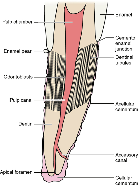
The dentin that surrounds the pulp tissue of the root is itself covered by an imperfect layer of cementum. This patchwork of cementum has bare areas where dentin is exposed. If the tooth root is exposed because of periodontal disease, recession, trauma or any form of pathology, these exposed spots of dentin can stimulate the pulpal nerves through their tubules (Fig. 11-1).

The roots are a warning system to external or internal trauma. If the pulpal tissue inside the root canal becomes inflamed, then hot or cold stimulation can cause pain. If the nerve is alive but badly inflamed, a cold stimulus such as cold water can cause a painful response. The longer the response takes to subside, the more damaged the pulpal tissue. If gases are present within the pulpal tissue, such as when the pulp is infected or necrotic, these gases can be expanded with a heated stimulus such as hot coffee, tea, food or water. Once affected by heat the gases expand, but the root canal that houses these gases remains the same size. These expanded gases then put pressure on any nerve tissue that remains vital. The expanded gases then force themselves out the root apex into the surrounding bone and periodontal ligament. This in turn causes a serious pain response that lingers sometimes with extreme intensity and duration.

Pressure and temperature responses are not elicited by pulp tissue. Instead the nerve tissue within the bone, gums and periodontal ligament elicits these responses. The root canal tissues do not have nerves that can stimulate the feelings of temperature or pressure. The root canals can only respond to pain.

Reparative and Nourishment Functions

The pulp canals house the nourishment system of the tooth. It is here within the root canal that arteries, veins, and lymph tissue nourish the tooth from the inside. These vessels enter and exit through an orifice in the apex of the root called the apical foramen. They allow nutrients and oxygen to circulate throughout the pulp chamber. They also provide a system to remove harmful products and carbon dioxide from the tooth. This is the way that the nerves and odontoblasts within the tooth are nourished and replenished.

If the flow of these vessels is restricted over a long time or totally constricted for as little as several minutes, then the nerves and other tissues inside the pulp chamber could die of anoxia (lack of oxygen).

Inside the tooth the odontoblasts allow secondary and reparative dentin to be formed in response to trauma (see Chapter 20). This process is not restricted to the root but to the entire pulp chamber.

The apical third of the root at or near its apex can continue to form cementum on the outside of the root. If this process is extreme, it is called hypercementosis, and it forms a cementoma at the apex of the root (see the section on Dental Anomalies in Chapter 7 and Root Formation in Chapter 21). A cementoma is usually associated with bone destruction and or trauma. This cementum is different from the cementum that lines the rest of the root. First, it can continue to grow and add on to itself. Second, in this process of adding on to itself, cells that form cementum are trapped in the cementum. This cementum is called cellular cementum (see Fig. 11-1).

Support Functions

The shape and length of the roots have a direct effect on how much anchorage and support they can afford. The longer and wider is the root, then the more support the tooth receives. The longer is the root, then the more firmly the root is embedded into the bone. The greater is the surface area of the root, then the more periodontal fibers can attach the root to the bone and the better it can resist displacement from the forces exerted on it. A tooth with multiple roots has its periodontal ligaments disbursed in many more different directions than a single-rooted tooth. This allows resistance to displacement from a greater variety of forces. Likewise, concavities and grooves in the root also allow for more surface area and for the periodontal ligament to attach at different angles.

Roots with triangular cross section offer resistance to lateral displacement. Curved roots afford resistance to occlusal, apical and distal forces. Multiple periodontal fiber directions in the furcation areas between roots give the tooth direct resistance to occlusal displacement.

The width, shape, length, curvature, and number of roots, concavities and direction of the periodontal fibers all affect the amount and direction of resistance a tooth can offer to withstand the forces exerted on it (Fig. 11-2).

Tooth Movement

The teeth are not embedded into the bone but rather they are supported between the root and the bone by a hammock of periodontal fibers. These fibers attach to the cementum of the root on one end and the alveolar bone on the other. This hammock of living tissue is called the periodontal ligament (PDL). It is composed of collagenous fibers of connective tissue and is capable of being tensed or compressed.

The periodontal ligament (PDL) fills the thin area of space that exists between the tooth and the bone. Pressure exerted on the tooth compresses the PDL fibers on one side and tenses them on the other. If you push on a tooth with an instrument you can see the tooth move ever so slightly. This small movement is called mobility. A slight amount of mobility is healthy and normal.

When the external force is strong enough to exert pressure on the bone it triggers a resorption process within the alveolar bone. This causes certain white blood cells (WBCs), or osteoclasts, to dissolve bone in the area of the pressure. When enough bone is resorbed the pressure ceases, the PDL regains its normal width, and the tooth moves away from the external force and into this newly remodeled area.

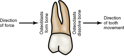
On the opposite side of the tooth, the PDL is tensed to accommodate the tooth’s movement. This tension is exerted on the bone. Osteoblastic bone-forming cells respond to the tension by forming new bone (Fig. 11-3).

In this way, responding to pressure and tension, the bone is remodeled. The net result is that the tooth moves away from the external force into a new location.

Orthodontics is accomplished by exerting a force onto a tooth. This sustained force causes the tooth to move gradually into the desired new location. Only a slight amount of movement is possible during a specific amount of time. Most orthodontic cases require almost two years to complete.

For orthodontics to succeed, the tooth must be held fixed in its new location long enough for the collagenous fibers of the PDL to remodel. The PDL fibers that were lengthened from tension must have sufficient time to relax and relieve their tension. Otherwise the residual tension in these fibers causes the tooth to move back and relapse.

If the pressure is too extreme, bone and root can be resorbed. The same cells that resorb bone can also resorb the root. However, the root does require a greater pressure than bone to be resorbed.

Even without external forces, a tooth still moves. If all of the occlusal forces are removed from a tooth and it has no occlusal antagonist, the tooth continues to erupt. A tooth moves mesially if it does not have another tooth mesial to it; this is called mesial drift (see Chapter 6). The shape of the roots and the residual tension within the PDL may play some part in causing these above phenomena.

Stay updated, free dental videos. Join our Telegram channel

Jan 4, 2015 | Posted by in General Dentistry | Comments Off on 11: Root Morphology

VIDEdental - Online dental courses

Get VIDEdental app for watching clinical videos