11: Oral Cancer: Prevention, Management, and Treatment

CHAPTER 11

Oral Cancer: Prevention, Management, and Treatment

Patricia M. Crum, Deborah A. DesRosiers, Christoph A. Ramseier and Paul E. Vankevich

CHAPTER OUTLINE

Incidence and Prevalence
Prognosis and Staging
Causation

Tobacco
Alcohol
Other Causes
Role of the Dental Team in Head and Neck Cancer

Patient Education
Patient History
Findings From the Clinical Examination: Signs and Symptoms

   Clinical Presentation

      Soft Tissue Changes
      Hard Tissue Changes
      Skin Lesions
Prevention Strategies

Barrier Protection
Smoking Cessation

   Information for the Patient About the Effects of Tobacco
   Smoking Cessation Strategies
Promoting a Healthy Lifestyle
Diagnosis of Oral Cancer

Importance of Early Diagnosis
Differential Diagnoses
Confirming the Diagnosis
Referral for Treatment
Cancer Therapies

Cancer Therapy Modalities

   Surgical Excision
   Chemotherapy
   Radiation Therapy
Adverse Oral Effects of Cancer Therapies
Planning Dental Treatment After Diagnosis of Oral Cancer

Coordinating Dental Care in Patients Before Cancer Therapy

   Urgent Intervention
   Planning for the Future
Supportive Dental Care During Cancer Therapy

   Maintaining Oral Hygiene
   Monitoring for Adverse Effects
   Symptomatic Care
Coordinating Dental Care Following Cancer Therapy

   Immediate: Hygiene and Prevention
   Long-term Follow-up: Maintenance and Surveillance
   Prosthetic Rehabilitation
Conclusion

Oral cancer can occur in any part of the oral cavity, including the lips and oropharynx. This chapter will focus on the identification, management, and treatment of oral cancer and the means of preventing it from occurring. The causes of oral cancer will be reviewed with special emphasis on tobacco use. Systemic and oral effects of tobacco use, implications of tobacco use with regard to dental treatment planning, and tobacco cessation programs are also included in this chapter.

INCIDENCE AND PREVALENCE

In the United States, approximately two thirds of oral cancers occur in the oral cavity and one third in the oropharynx. The most common type of oral and oro-pharyngeal cancer is squamous cell carcinoma, accounting for about 9 out of every 10 oral malignancies. Other malignancies of the oral cavity include melanomas, lymphomas, sarcomas, and metastases from other areas of the body.

Metastasis to the oral region from a malignant tumor elsewhere in the body is rare, but those that occur are most often found in the mandible. Figure 11-1 is a radio-graph of cancer metastasis in the mandible. The majority of cancers found to have metastasized to the jaw are adenocarcinomas. The most common primary tumor sites with metastasis to the jaws are breast, lung, and kidney.1 See Table 11-1 for the relative frequency of metastatic lesions to the jaws.

Table 11-1

Relative Frequency of Origins of Metastasic Lesions in the Jaws

  Percent
Breast 30
Lung 20
Kidney 15
Thyroid 5
Prostate 5
Colon 5
Stomach 5
Cutaneous melanoma 5

image

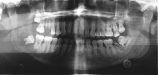
Figure 11-1 Panoramic radiograph of a 48-year-old male smoker showing metastatic lung cancer to the right mandible.

The incidence of squamous cell carcinoma in the oral cavity, including the lips, differs by anatomic sites. Some sites seem to be relatively resistant, whereas other locations are particularly susceptible. In the past, when all anatomic sites were considered, the lower lip was cited as the most susceptible site, accounting for 30% to 40% of all oral squamous cell carcinomas. Data from the National Cancer Institute SEER (Surveillance, Epidemio-logy, and End Results) Program shows that in the last 10 years the incidence of squamous cell carcinoma of the lip has significantly decreased in the United States and is no longer the most prevalent oral site.13 Squamous cell carcinoma of the lip is still much more common in males than in females and occurs most commonly in individuals between the ages of 50 and 80 years old. Most of these lesions occur on either the right or left vermilion borders, seldom at the midline. Current statistics show that the highest incidence of oral squamous cell carcinoma is found at the lateral borders and the adjacent ventral surfaces of the tongue.

Intraoral squamous carcinoma lesions frequently form a horseshoe-shaped pattern as shown in Figure 11-2. The lateral and ventral surfaces of the tongue and the floor of the mouth are the most common sites. The soft palate and the lateral posterior regions adjacent to the anterior faucial pillars are the next most frequently seen sites for squamous cell carcinoma. Other areas less frequently affected are the gingiva and alveolar ridges, and the buccal mucosa. These areas combined account for less than 10% of oral squamous cell carcinomas.1

image

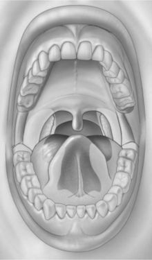
Figure 11-2 Horseshoe-shaped intraoral area most prone to the development of squamous cell carcinoma. It consists of the anterior floor of mouth, lateral borders of the tongue, tonsillar pillars, and lateral soft palate. (From Sapp JP, Eversole LR, Wysocki GP: Contemporary oral and maxillofacial pathology, ed 2, St Louis, 2004, Mosby.)

The incidence of oral cancer has remained at approximately 3% of all cancers in the United States. Approximately 30,000 new cases of oral and oropharyngeal cancer are diagnosed annually in men and women in the United States. About 95% of all oral cancers occur in persons over the age of 40.15 In the last decade, the ratio of male to female has been slightly less than 2 to 1. In the 1950s, this ratio was 6:1; this shift has been attributed to an increase in smoking by women.1,4

The total number of deaths annually attributed to oral and oropharyngeal cancer is as high as 7500 in the United States.3 Deaths caused by oral cancer represent approximately 2% of the total deaths caused by cancer in men and 1% in women. Although survival rates for most cancers have increased during the past several decades, there has been no such trend in the survival of patients with oral cancer. The overall 5-year survival rate of all patients with oral cancers has remained at about 50% over many decades. Approximately two thirds of all oral cancers are in the advanced stages (III or IV) at the time of diagnosis, decreasing the 5-year survival rate to 30%. Because 80% of early stage oral cancer (I or II) is localized, the cancer can be cured with appropriate treatment.2,5,6 Black males have the highest mortality from oral cancer when compared with other men of the same age.2,3,5 Genetics, education, socioeconomic status, and access to health care also seem to play a role in survival rates. The primary reason for the poor survival rate of individuals with oral cancer appears to be the result of the advanced stage of the cancer typically present at the initial diagnosis (See the What’s the Evidence? box on p. 276).13,6 The mortality statistics underline the importance of early detection and treatment for improving survival and the critical role played by the dental team.1,2,5,7

PROGNOSIS AND STAGING

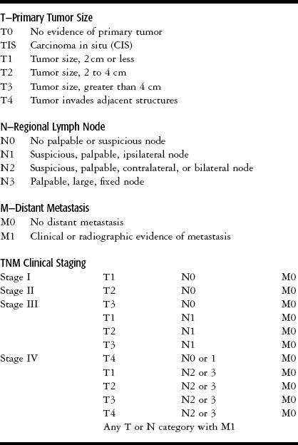
As noted above, the prognosis for patients with oral cancer depends on the clinical stage of the tumor at the time of diagnosis. Staging is based on clinical findings: How large is the lesion? Has it spread to the lymph nodes? Has it metastasized? The abbreviations used in staging systems are TNM (tumor, nodes, and metastasis). See Table 11-2 for the TNM definitions and staging groups for the oral cavity. Staging is used in planning the treatment protocol for the cancer and in determining prognosis. For example, a patient with a stage I cancer might have a tumor of 2 cm or less with no palpable lymph nodes and no distant metastases. A patient with a cancer larger than 4 cm, palpable bilateral nodes, and with or without metastasis would be classified as a stage IV. As seen in Table 11-2, there can be many combinations that fall into the advanced stages III and IV. Oral cancers are initially staged based on clinical examination. The initial stage may change after findings from additional tests, imaging studies, and biopsy procedures. As a result, oral cancers that are first considered to be at stage I or II may be reclassified as stage III or IV if further information is obtained, showing, for example, tumor invasion into deeper structures, spread into lymph nodes, or distant metastases.1,2,6

Table 11-2

TNM Definitions for Malignant Tumors of the Oral Cavity

image

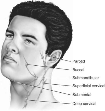
As the tumor grows, it may invade adjacent structures, such as the carotid artery sheath or neurovascular bundles. Squamous cell carcinomas of the oral cavity are known to spread by invading the lymphatic vessels. Once inside the lymphatic vessels, the tumor cells lodge and continue to proliferate in the regional lymph nodes. The lymph nodes most commonly invaded by intraoral squamous cell carcinoma are the submandibular and the superficial and deep cervical nodes. Squamous cell carcinomas of the lower lip metastasize first to the submental nodes and then may spread to the submandibular and the cervical lymph nodes. Tumor cells that spread beyond the regional lymph nodes of the head and neck often metastasize to the lungs, liver, and bone.1,2,6 Figure 11-3 provides a diagram of lymphatic drainage from oral cancers.

image

Figure 11-3 Lymphatic drainage pathways and major lymph nodes of the head and neck. (From Sapp JP, Eversole LR, Wysocki GP: Contemporary oral and maxillofacial pathology, ed 2, St Louis, 2004, Mosby.)

CAUSATION

The actual cause of oral cancers is not known. A number of causative factors have been speculated to contribute to the development of oral cancer (See the What’s the Evidence? box on p. 276). These factors include tobacco use, alcohol consumption, exposure to carcinogens and ultraviolet light (sun exposure), and gene mutation. Preexisting conditions, such as chronic candidiasis, viral infection (herpes simplex virus [HSV-1], human papillomavirus [HPV], human immunodeficiency virus [HIV], Epstein-Barr virus [EBV]), immunosuppression, and chronic irritation have also been implicated as risk factors.1,2,6 Poor dental health and poor nutritional status are often co-morbid factors in patients with oral cancer, but this as a causative factor has not been confirmed.2,4,6 Of all factors, tobacco use is regarded as the most important and is believed to contribute to the majority of oral cancers.4,6


What’s the Evidence?

What Is the Evidence That Oral Cancer Survival Rates Are Related to Early Diagnosis or Race?

Evaluation of data collected by the U.S. National Cancer Institute’s Survival, Epidemiology, and End Results (SEER) Program showed that the stage of oral cancer at diagnosis (localized, regional, or distant) is related to 5-year survival rates (1986-1991).1 Individuals diagnosed with localized cancer were found to have the highest 5-year survival rate, 81%, whereas those with regional cancer had a 42% survival rate, and those with distant metastasis only an 18% survival rate. Although no major differences were found between males and females, there were significant differences based on race. African Americans were more likely to be diagnosed with more advanced stages of disease. Thirteen percent of the cancers in African Americans had distant metastases, whereas only 7% of cancers in whites had metastasized. Racial differences in survival were also evident in survival rates. The 5-year survival rate for localized cancers in white males was 80%, whereas African-American males with localized cancer had a lower survival rate of 60%. Regional and distant cancers also showed a similar gap in survival rates, with African Americans, in particular African-American males, having the lowest survival rates.

Similar results linking early diagnosis and race to oral cancer survival have also been found at the U.S. state and medical facility levels. Information from the North Carolina Central Cancer Registry from 1987 to 1990 showed that survival was related to the stage at which the cancer was detected.2 Seventy-two percent of individuals with localized cancer had an 18-month survival rate, whereas only 28% of those with a metastasized cancer survived at least 18 months. Racial differences were evident for those with localized cancer; African Americans had a significantly lower survival rate than whites. The 18-month survival rate for whites with localized cancer was 73%, whereas African Americans had a survival rate of only 61%. The authors concluded that the stage at diagnosis alone does not explain the difference in survival rates between African Americans and whites. They suggest that the difference may be explained by such factors as social support and variation in treatment. They also emphasize the importance of oral cancer screenings and early diagnosis because oral cancer is most curable when detected early.

A 10-year study at The University of Texas M.D. Anderson Cancer Center found the stage of oral cancer at diagnosis to be associated with survival rates.3 Among 909 patients, the 5-year survival rates were 81% for those with local disease, 42% for those with regional disease, and 21% for those with distant metastases. After accounting for stage of disease, race was a significant predictor of survival. For whites, the 5-year survival rates for local, regional, and distant stages of disease were 82%, 44%, and 23%, respectively. African Americans had significantly lower 5-year survival rates of 56%, 27%, and 14%, respectively.

Another large longitudinal study, completed in Brazil, reached similar conclusions.4 Over a period of 28 years, 4527 patients with oral cancer were followed. In this sample, gender, race, and the stage of cancer at diagnosis were highly associated with survival rate. Males were diagnosed with tumors at more advanced stages and, in addition, even more pronounced differences among races were found. Blacks had more advanced tumors. Seventy-six percent of the nonwhites (black and Asian) were diagnosed with cancer at the most severe stage (IV), whereas less than half (48%) of the whites had stage IV cancer at time of diagnosis.


1. Swango, PA. Cancers of the oral cavity & pharynx in the United States: An epidemiologic overview. J Public Health Dent. 1996; 56:309–318.

2. Caplan, DJ, Herz-Picciotto, I. Racial differences in survival of oral and pharyngeal cancer patients in North Carolina. J Public Health Dent. 1998; 58:36–43.

3. Moore, RJ, Doherty, DA, Do, KA, et al. Racial disparity in survival of patients with squamous cell carcinoma of the oral cavity and pharynx. Ethnicity & Health. 2001; 6:165–177.

4. Franco, EL, Dib, LL, Pinto, DS, et al. Race and gender influences on the survival of patients with mouth cancer. J Clin Epidemiol. 1993; 46:37–46.

Tobacco

The plant alkaloid nicotine, found in all forms of tobacco, is the psychoactive and addictive chemical agent, which in small quantities can have a profound effect on human physiology and psychology. This naturally occurring, plant-produced substance has many local and systemic toxic and physiologic effects (Table 11-3). When a small dose is delivered to the brain, nicotine can stimulate, relax, or induce a state of euphoria in the user. This psychoactive pleasurable perception is the primary reason for continuation and habituation of tobacco use. The longer an individual uses tobacco, the greater is the risk of developing dependence as a result of the effects of nicotine on the brain.

Table 11-3

Pathophysiologic Effects of Nicotine

Effect on Cells and Cellular Functions Effect on Oral Environment
Inhibition of fibroblast function Poor wound healing
Increased collagenase function Increased periodontal disease
Increased inflammatory response Vasoconstriction
Impaired cellular immunity (chemotaxic function of macrophages and polymorphonuclear leukocytes [PMNs]) Increased risk of infection
Impaired humoral immunity (decreased salivary and serum antibodies) Quantitative and qualitative decrease in salivary secretions


What’s the Evidence?

What Are the Risk Factors for Oral Cancer?

The most important risk factor for oral cancer is tobacco smoking. Among the multiple elevated risks associated with tobacco use, the substance has been shown to be the primary cause of oral cancers.15 Many investigations have also provided conclusive evidence that alcohol use can increase the risk of oral cancer.14,6 Numerous studies on the association of tobacco and alcohol have shown that this combination synergistically further increases the risk of oral cancer compared with the risk of each factor alone.13,5,711 Oral cancer risk increased more than thirty-fivefold among those who consumed two or more packs of cigarettes and more than four alcoholic drinks per day. Combining tobacco use and excessive alcohol use has been estimated to account for 75% of all oral cancers in the United States, 80% among men only.1 Additionally, it has been shown that using tobacco and alcohol together contributes to the risk of oral cancer recurrence.12

Among men who were diagnosed with oral cancer between 1986 and 1989, 93% of the cancers in African Americans and 86% in white Americans were attributed to consuming more than one alcoholic drink per day and/or smoking at least one pack of cigarettes per day.14 In another study, after accounting for the effect of smoking, heavy drinking was associated with a seventeenfold increased risk among African Americans and a ninefold increased risk among white Americans.13 The higher level of alcohol and tobacco use among African Americans accounted for most of the higher oral cancer incidence among that group. It has been suggested that in the absence of alcohol and tobacco, the rates of oral cancer between races and genders would be nearly equal.

Studies have shown that consumption of certain foods is associated with a decreased risk of oral cancer. A panel from the World Cancer Research Fund in association with the American Institute for Cancer Research concluded that there is convincing evidence that diets high in vegetables and fruits decrease the risk of oral cancer.15 Many studies have found that fruits1620 and, in particular, citrus fruits3,20 are protective against oral cancer. Studies have also found that vegetable consumption1720 in particular, carrots,3,1719,21 helps reduce the risk of oral cancer.

In addition to tobacco use, excessive exposure to the sun is a risk factor for lip cancer.22,23 Ultraviolet light damages the epidermal cell DNA, causing mutation of tumor suppressor cells, and weakening the immune system, which is then unable to destroy abnormal cells.2427 Cancer occurs more frequently on the lower lip, in males, in individuals over 50 years in age, in those with fair skin, and among those in occupations involving extensive exposure to the sunlight.23,2840 Associations between lip cancer and chronic exposure to solar radiation have been studied in populations in Scotland,41 Sweden,42 Finland,23,4345 The Netherlands,46 France,47 Italy,30 Greece,35 Canada28,48 and the United States.29,40,49 Individuals with light skin have a much higher incidence of lip cancer than individuals with darker skin.23,30,50 It is believed that the additional melanin protects the lip against the damaging effects of the sun.51,52

Researchers have hypothesized that human papilloma-virus (HPV) may be a risk factor for oral cancer. Many observations of oral cancer have occurred after a diagnosis of cervical cancer caused by HPV.5355 An increasing volume of evidence shows that some oral cancers contain HPV DNA.53,5658 A study of data from the U.S. Surveillance, Epidemiology, and End Results Program (SEER), showed that individuals with HPV-associated anogenital carcinoma had a higher risk of having oral cancer develop than individuals without HPV-associated anogenital carcinoma.59 A meta-analysis that included 4680 oral mucosa samples from 94 studies concluded that the likelihood of detecting HPV in cancerous oral tissue was 47%, which is significantly higher than the 10% likelihood of detecting HPV in normal oral tissue.60 Yet a recent systematic review, which included 60 studies in 26 countries, found fewer links between HPV and oral cancer. HPV was present in 24% of intraoral cancer cases and 36% of oropharyngeal cancer cases.61 Although some evidence suggests that HPV may contribute to the development of oral cancer, this issue is still under debate. In a review of the current literature, Ha and Califano conclude that the presence of HPV contributes to a small subset of oral cancer cases, but that the presence of HPV is not necessary for the development of oral cancer.62

SUGGESTED READINGS

1. Blot, WJ, McLaughlin, JK, Winn, DM, et al. Smoking and drinking in relation to oral and pharyngeal cancer. Cancer Res. 1988; 48(11):3282–3287.

2. Franceschi, S, Talamini, R, Barra, S, et al. Smoking and drinking in relation to cancers of the oral cavity, pharynx, larynx, and esophagus in northern Italy. Cancer Res. 1990; 50(20):6502–6507.

3. Franco, EL, Kowalski, LP, Oliveira, BV, et al. Risk factors for oral cancer in Brazil: a case-control study. Intl J Cancer. 1989; 43(6):992–1000.

4. Merletti, F, Boffetta, P, Ciccone, G, et al. Role of tobacco and alcoholic beverages in the etiology of cancer of the oral cavity/oropharynx in Torino, Italy. Cancer Res. 1989; 49(17):4919–4924.

5. U. S. Department of Health and Human Services: The health consequences of smoking: a report of the surgeon general, Atlanta, 2004, U. S. Department of Health and Human Services, Centers for Disease Control and Prevention, National Center for Chronic Disease Prevention and Health Promotion, Office on Smoking and Health.

6. Elwood, JM, Pearson, JC, Skippen, DH, et al. Alcohol, smoking, social and occupational factors in the aetiology of cancer of the oral cavity, pharynx and larynx. Intl J Cancer. 1984; 34(5):603–612.

7. Graham, S, Dayal, H, Rohrer, T, et al. Dentition, diet, tobacco, and alcohol in the epidemiology of oral cancer. J Natl Cancer Inst. 1977; 59(6):1611–1618.

8. La, Vecchia C, Tavani, A, Franceschi, S, et al. Epidemiology and prevention of oral cancer. Oral Oncol. 1997; 33(5):302–312.

9. Marshall, JR, Graham, S, Haughey, BP, et al. Smoking, alcohol, dentition and diet in the epidemiology of oral cancer part B, oral oncology. Eur J Cancer. 1992; 28B(1):9–15.

10. Mashberg, A, Boffetta, P, Winkelman, R, et al. Tobacco smoking, alcohol drinking, and cancer of the oral cavity and oropharynx among U. S. veterans. Cancer. 1993; 72(4):1369–1375.

11. Rothman, K, Keller, A. The effect of joint exposure to alcohol and tobacco on risk of cancer of the mouth and pharynx. J Chronic Dis. 1972; 25(12):711–716.

12. Day, GL, Blot, WJ, Shore, RE, et al. Second cancers following oral and pharyngeal cancers: role of tobacco and alcohol. J Natl Cancer Inst. 1994; 86(2):131–137.

13. Day, GL, Blot, WJ, Austin, DF, et al. Racial differences in risk of oral and pharyngeal cancer: alcohol, tobacco, and other determinants. J Natl Cancer Inst. 1993; 85(6):465–473.

14. Brown, LM, Hoover, RN, Greenberg, RS, et al. Are racial differences in squamous cell esophageal cancer explained by alcohol and tobacco use? J Natl Cancer Inst. 1994; 86(17):1340–1345.

15. Food nutrition and the prevention of cancer: a global perspective/World Cancer Research Fund in association with American Institute for Cancer Research, Washington, DC, 1997, American Institute for Cancer Research.

16. McLaughlin, JK, Gridley, G, Block, G, et al. Dietary factors in oral and pharyngeal cancer. J Natl Cancer Inst. 1988; 80(15):1237–1243.

17. La, Vecchia C, Negri, E, D’Avanzo, B, et al. Dietary indicators of oral and pharyngeal cancer. Intl J Epidemiol. 1991; 20(1):39–44.

18. Franceschi, S, Barra, S, La, Vecchia C, et al. Risk factors for cancer of the tongue and the mouth. A case-control study from northern Italy. Cancer. 1992; 70(9):2227–2233.

19. Winn, DM, Ziegler, RG, Pickle, LW, et al. Diet in the etiology of oral and pharyngeal cancer among women from the southern United States. Cancer Res. 1984; 44(3):1216–1222.

20. Zheng, W, Blot, WJ, Shu, XO, et al. Risk factors for oral and pharyngeal cancer in Shanghai, with emphasis on diet. Cancer Epidemiol Biomarkers Prev. 1992; 1(6):441–448.

21. Franceschi, S, Bidoli, E, Baron, AE, et al. Nutrition and cancer of the oral cavity and pharynx in north-east Italy. Intl J Cancer. 1991; 47(1):20–25.

22. Keller, AZ. Cellular types, survival, race, nativity, occupations, habits and associated diseases in the pathogenesis of lip cancers. Am J Epidemiol. 1970; 91(5):486–499.

23. Lindqvist, C. Risk factors in lip cancer: a questionnaire survey. Am J Epidemiol. 1979; 109(5):521–530.

24. Buzzell, RA. Carcinogenesis of cutaneous malignancies. Dermatol Surg. 1996; 22(3):209–215.

25. Granstein, RD. Evidence that sunscreens prevent UV radiation-induced immunosuppression in humans. Sunscreens have their day in the sun. Arch Dermatol. 1995; 131(10):1201–1204.

26. Kripke, ML. Immunologic mechanisms in UV radiation carcinogenesis. Adv Cancer Res. 1981; 34:69–106.

27. Rees, J. Genetic alterations in non-melanoma skin cancer. J Investig Dermatol. 1994; 103(6):747–750.

28. Spitzer, WO, Hill, GB, Chambers, LW, et al. The occupation of fishing as a risk factor in cancer of the lip. N Engl J Med. 1975; 293(9):419–424.

29. Preston-Martin, S, Henderson, BE, Pike, MC. Descriptive epidemiology of cancers of the upper respiratory tract in Los Angeles. Cancer. 1982; 49(10):2201–2207.

30. Dardanoni, L, Gafa, L, Paterno, R, et al. A case-control study on lip cancer risk factors in Ragusa (Sicily). Intl J Cancer. 1984; 34(3):335–337.

31. Wiklund, K, Holm, LE. Trends in cancer risks among Swedish agricultural workers. J Natl Cancer Inst. 1986; 77(3):657–664.

32. Krolls, SO, Hoffman, S. Squamous cell carcinoma of the oral soft tissues: a statistical analysis of 14,253 cases by age, sex, and race of patientsl. J Am Dent Assoc. 1976; 92(3):571–574.

33. Fahmy, MS, Sadeghi, A, Behmard, S. Epidemiologic study of oral cancer in Fars Province, Iran. Community Dent Oral Epidemiol. 1983; 11(1):50–58.

34. Chen, J, Katz, RV, Krutchkoff, DJ, et al. Lip cancer. Incidence trends in Connecticut, 1935-1985. Cancer. 1992; 70(8):2025–2030.

35. Antoniades, DZ, Styanidis, K, Papanayotou, P, et al. Squamous cell carcinoma of the lips in a northern Greek population. Evaluation of prognostic factors on 5-year survival rate I, Part B, Oral Oncology. Eur J Cancer. 1995; 31B(5):333–339.

36. Muir, C, Weiland, L. Upper aerodigestive tract cancers. Cancer. 1995; 75(1 Suppl):147–153.

37. Ostman, J, Anneroth, G, Gustafsson, H, et al. Malignant oral tumours in Sweden 1960-1989 an epidemiological study, part B, oral oncology. Eur J Cancer. 1995; 31B(2):106–112.

38. Faye-Lund, H, Abdelnoor, M. Prognostic factors of survival in a cohort of head and neck cancer patients in Oslo, Part B, oral oncology. Eur J Cancer. 1996; 32B(2):83–90.

39. King, GN, Healy, CM, Glover, MT, et al. Increased prevalence of dysplastic and malignant lip lesions in renal-transplant recipients. N Engl J Med. 1995; 332(16):1052–1057.

40. Pogoda, JM, Preston-Martin, S. Solar radiation, lip protection, and lip cancer risk in Los Angeles County women (California, United States). Cancer Causes Control. 1996; 7(4):458–463.

41. Macfarlane, GJ, Boyle, P, Evstifeeva, T, et al. Epidemiological aspects of lip cancer in Scotland. Community Dent Oral Epidemiol. 1993; 21(5):279–282.

42. Wiklund, K, Dich, J. Cancer risks among male farmers in Sweden. Eur J Cancer Prev. 1995; 4(1):81–90.

43. Lindqvist, C, Teppo, L. Epidemiological evaluation of sunlight as a risk factor of lip cancer. Br J Cancer. 1978; 37(6):983–989.

44. Pukkala, E, Soderholm, AL, Lindqvist, C. Cancers of the lip and oropharynx in different social and occupational groups in Finland, part B, oral oncology. Eur J Cancer. 1994; 30B(3):209–215.

45. Pukkala, E, Notkola, V. Cancer incidence among Finnish farmers, 1979-1993. Cancer Causes Control. 1997; 8(1):25–33.

46. Schouten, LJ, Meijer, H, Huveneers, JA, et al. Urban-rural differences in cancer incidence in The Netherlands: 1989-1991. Int J Epidemiol. 1996; 25(4):729–736.

47. Haguenoer, JM, Cordier, S, Morel, C, et al. Occupational risk factors for upper respiratory tract and upper digestive tract cancers. Br J Ind Med. 1990; 47(6):380–383.

48. Gallagher, RP, Threlfall, WJ, Jeffries, E, et al. Cancer and aplastic anemia in British Columbia farmers. J Natl Cancer Inst. 1984; 72(6):1311–1315.

49. Lyon, JL, Gardner, JW, Klauber, MR, et al. Low cancer incidence and mortality in Utah. Cancer. 1977; 39(6):2608–2618.

50. Onuigbo, WI. Lip lesions in Nigerian Igbos. Intl J Oral Surg. 1978; 7(2):73–75.

51. Douglass, CW, Gammon, MD. Reassessing the epidemiology of lip cancer. Oral Surg Oral Med Oral Pathol. 1984; 57(6):631–642.

52. Johnson, NW, Warnakulasuriya, KA. Epidemiology and aetiology of oral cancer in the United Kingdom. Community Dent Health 10 Suppl. 1993; 1:13–29.

53. Franceschi, S, Munoz, N, Bosch, XF, et al. Human papillomavirus and cancers of the upper aerodigestive tract: a review of epidemiological and experimental evidence. Cancer Epidemiol Biomarkers Prev. 1996; 5(7):567–575.

54. Yeudall, WA, Paterson, IC, Patel, V. Presence of human papillomavirus sequences in tumour-derived human oral keratinocytes expressing mutant p53, part B, oral oncology. Eur J Cancer. 1995; 31B(2):136–143.

55. Yeudall, WA, Campo, MS. Human papillomavirus DNA in biopsies of oral tissues. J Gen Virology. 1991; 72(Pt 1):173–176.

56. Smith, EM, Ritchie, JM, Summersgill, KF, et al. Human papillomavirus in oral exfoliated cells and risk of head and neck cancer. J Natl Cancer Inst. 2004; 96(6):449–455.

57. Franceschi, S, Munoz, N, Snijders, PJ. How strong and how wide is the link between HPV and oropharyngeal cancer? Lancet. 2000; 356(9233):871–872.

58. Steenbergen, RD, Hermsen, MA, Walboomers, JM, et al. Integrated human papillomavirus type 16 and loss of heterozygosity at 11q22 and 18q21 in an oral carcinoma and its derivative cell line. Cancer Res. 1995; 55(22):5465–5471.

59. Frisch, M, Biggar, RJ. Aetiological parallel between tonsillar and anogenital squamous-cell carcinomas. Lancet. 1999; 354(9188):1442–1443.

60. Miller, CS, Johnstone, BM. Human papillomavirus as a risk factor for oral squamous cell carcinoma: a meta-analysis, 1982-1997. Oral Surg Oral Med Oral Pathol Oral Radiol Endod. 2001; 91(6):622–635.

61. Kreimer, AR, Clifford, GM, Boyle, P, et al. Human papillomavirus types in head and neck squamous cell carcinomas worldwide: a systematic review. Cancer Epidemiol Biomarkers Prev. 2005; 14(2):467–475.

62. Ha, PK, Califano, JA. The role of human papillomavirus in oral carcinogenesis. Crit Rev Oral Biol Med. 2004; 15(4):188–196.

Initiation in the use of tobacco and progression to nicotine addiction is influenced by sociocultural, psychological, physiologic, and genetic factors.8 Chronic tobacco use is characterized by psychological (habituation, behavioral) and pharmacologic (addiction, chemical dependency) factors. Because tobacco use may decrease appetite and increase the user’s basal metabolic rate, some smokers may use tobacco as a weight management strategy. Both male and female smokers may experience a weight gain of 5 to 10 lb upon smoking cessation, a frequently cited justification for not quitting.9 The health risks of continued smoking are far greater than the health benefit of avoiding a 5 to 10 lb weight gain,10 but unfortunately in the mind of the continuing tobacco user, the psychosocial reinforcement associated with preventing weight gain and the concurrent psychological pleasure of tobacco use may outweigh the obvious health benefits of smoking cessation.

Tobacco can cause oral cancer in multiple ways. There are 43 identified carcinogens in tobacco smoke and 28 in smokeless tobacco.11,12 The effects of these agents at the cellular and DNA levels are profound and continue to be better understood. Chemical components in tobacco products, and heat generated by smoked tobacco act as direct physical irritants to the oral tissue. Some forms of smokeless tobacco contain a gritty additive that, by intent, abrades and creates microscopic lacerations in the mucosa, thereby increasing the absorption of the nicotine and carcinogens. Chronic irritation to oral soft tissue may precede the formation of premalignant or malignant epithelial lesions in susceptible individuals. Perhaps more importantly, the microulceration and chronic inflammation facilitate the ingress of tobacco constituents and provide the carcinogens with increased cellular contact.13

In the United States, tobacco use, though declining, is still pervasive, resulting in significant morbidity and mortality to both users and nonusers.14 Worldwide, tobacco use continues to increase. Cigarette smoking is a major risk factor for heart disease, various forms of cancer (oral and systemic), stroke, and chronic obstructive pulmonary disease. It is estimated that one in five deaths is related to smoking and that one in four smokers will die prematurely of a tobacco-related disease, losing on average 15 years of life.8 Women who smoke are at risk for the same adverse effects as men and, in addition, a pregnant woman who smokes also puts her unborn child at risk for complications, such as premature birth and low birth weight. Tobacco smoking causes 30% of all cancer deaths in the United States.14 Tobacco smoking has been cited as the leading preventable cause of mortality and morbidity worldwide.15 The morbidity and mortality from cigarette smoking are related to the total years of smoking, the number of cigarettes per day, and the depth of inhalation. The use of nonfiltered cigarettes and mentholated cigarettes is also related to increased morbidity.16 Examples of tobacco products for smoking are seen in Figure 11-4.

image

Figure 11-4 Types of tobacco products: pipe, cigarettes, cigar.

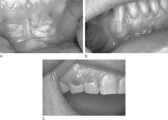
Cigar and pipe smoking are associated with the same health concerns as cigarette smoking, including systemic and upper aerodigestive tract diseases, the risk of nicotine addiction, and the creation of indoor air pollution.17 Cigar and pipe smoking are considered at least as great a risk factor for the development of oral cancer as cigarette smoking.4,17 The pipe stem (smoke delivery end) delivers smoke that has both thermal effects and hot gasses produced during combustion. These act synergistically with the toxic components in tobacco smoke to enhance the risk of carcinogenesis in chronically exposed tissues, such as lips and oral soft palate. The habit of holding the lighted end of the cigarette inside the mouth, called “reverse smoking,” is also associated with a significantly higher risk of oral cancer.2,4 Oral effects of pipe smoking are shown in Figure 11-5.

image

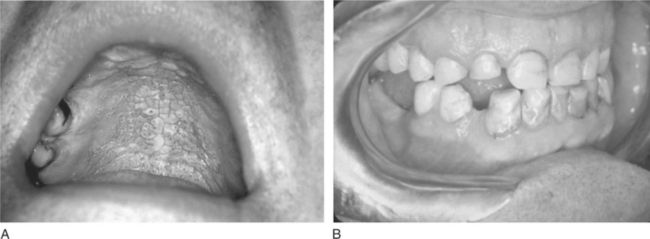
Figure 11-5 Pipe effects on oral tissue. A, Nicotine stomatitis. B, Attrition of both canines and the right maxillary lateral incisor from habitual pipe smoking in a 60-year-old male.

Currently, there are as many as 46 million smokers in the United States14 and an unknown yet important number of individuals who are indirectly exposed to passive or second-hand smoke. The second-hand smoke from cigarettes, cigars, and pipes is air pollution that poses health risks to nonsmokers.13The oral use of smokeless tobacco (commonly called spit, chew, or snuff) is not a safe alternative to smoking.6,13,18 Compelling scientific evidence documents the fact that smokeless tobacco is also dangerous, and can lead to nicotine addiction and a number of noncancerous oral pathologic conditions demonstrated in Figure 11-6. Types of smokeless tobacco to be chewed are shredded loose-leaf, pressed brick or plug, or twist made of dried ropelike strands. Snuff is a powdered or finely cut cured tobacco, which is available as a wet or dry product to be used topically in the mouth or nose. An example of smokeless tobacco is shown in Figure 11-7. All forms contain nitrosamines and other potentially carcinogenic substances. The risk of developing oral cancer increases with long-term use of smokeless tobacco.

image

Figure 11-6 Changes found in the area where the smokeless tobacco is held. A, Snuff dipper’s pouch hyperkeratosis. B, Gingival recession of left mandibular canine. C, Dental caries.

image

Figure 11-7 Smokeless tobacco product.

Alcohol

Although not considered to be an oral carcinogen itself, some research suggests that alcohol consumption may add to the risk of oral cancer.1,2,4,6,19 Alcohol consumption potentiates the oral carcinogenic effect of tobacco. It has been hypothesized that alcohol’s drying effect may result in mucosa that is more susceptible to carcinogens. Ethanol may act as a solvent for tobacco carcinogens or a promoter for chemical carcinogenesis. Alcoholic beverages contain carcinogens, such as nitrosamines and polycyclic hydrocarbons.2 It is estimated that smoking tobacco and drinking alcohol combine to account for approximately three fourths of all oral and oropharyngeal cancers in the United States.2,5,20,21 (See also Chapter 12 for additional information on the adverse effects, pathophysiology, and addictive qualities of alcohol.)

Other Causes

In addition to tobacco and alcohol, a number of other factors may increase the risk of developing oral cancer. Chronic candidiasis has been suggested as a predisposing factor for oral squamous cell carcinoma.1,2,4 Studies have shown that the HPV is found in up to 50% of oral squamous cell carcinoma lesions.4,22 Patients with prolonged exposure to direct sunlight and lack of appropriate sun protection barriers are at greater risk for having cancers of the head and neck develop (basal cell carcinoma, squamous cell carcinoma, or malignant melanoma). A compromised immune system may also render patients at an increased risk for having cancer develop. Patients with acquired immunodeficiency syndrome (AIDS), bone marrow and kidney transplant recipients, and patients who have had total-body radiation and high-dose chemotherapy are also at a higher risk of having squamous cell carcinoma develop. Oral submucosal fibrosis and chronic forms of oral lichen planus may predispose the oral mucosa to having squamous cell carcinoma develop.1 In some cultures, chewing betel (areca) nut is common and appears to be related to the increased incidence of oral cancer.1,6 Although chronic irritation is not generally believed to initiate oral cancer, it is not uncommon to find a cancer at a site of irritation. It is therefore important to have the patient return so that any oral lesions can be reevaluated, even those apparently related to a dental prosthesis.1,4

Current cancer research evaluates ways in which inherited genes may influence the risks, classification, prognosis, and treatment of head and head cancer. Microassay methodology now allows the expression of thousands of genes to be studied simultaneously. The researcher can provide gene expression profiling (GEP) for a single patient’s cancer. Comparisons of gene expression patterns between patients with the same type of cancer may allow differentiation of tumors with a poor prognosis from others with better prospective outcomes. The future is expected to open new avenues for cancer prevention and treatment.2,4,23,24


Dental Team Focus

Cancer and the Oral Health Team

Early in the patient’s dental care, awareness about oral cancer must be established. The oral health team can participate in the process of educating patients about the ways in which tobacco, alcohol, and sun exposure may cause cancer. Team members can instruct the patient in smoking cessation strategies and the use of sunscreen in the prevention of cancer and about the warning signs for early detection of cancerous lesions.

The patient who is undergoing radiation or chemotherapy for cancer may have oral complications resulting from the treatment. The oral health team can assess and instruct the patient in strategies for maintaining oral hygiene and obtaining symptomatic relief.

The administrative assistant may prepare referral letters, schedule patient visits, submit and receive laboratory reports, and manage other communication between the patient, dentist, and specialist.

ROLE OF THE DENTAL TEAM IN HEAD AND NECK CANCER

Patient Education

The dental team plays a crucial role in educating patients about oral cancers and the associated risk factors (see the Dental Team Focus box).5,7,21,25 Because many individuals may be more likely to visit the dentist than the physician for periodic or episodic visits, the dental team has a special responsibility to identify potentially malignant oral lesions and provide critical oral health information to the patient. Patients need to know if they are at risk and what they can do for the prevention of oral cancer. Excellent pamphlets are available to use as teaching tools and may be kept in the waiting area of a dental office. Specific prevention and tobacco cessation strategies are discussed later in this chapter.

Patient History

Interviewing patients to identify risk factors for oral cancer is an important part of taking the dental and medical history.5,21,25 Although gender, race, and age are easy data to gather, many people underreport their use of alcohol and tobacco. Because patients with a history of smoking or drinking are at greater risk for oral cancer, special attention should be given to obtaining accurate information on these habits. “Do you smoke?” is a simple question that may elicit an answer of “No,” but asking the same patient, “Have you ever smoked or used any type of tobacco?” may reveal a more accurate history. Further questions are needed to determine the extent of usage, including amount, length of time, and type of tobacco used since a patient may have smoked more in the past or may have recently quit smoking. These factors can be incorporated into a “pack-year” smoking history, used by some providers. Some sample questions concerning smoking history and oral changes are found in Box 11-1.


BOX 11-1   Sample Questions for Screening Oral Cancer and Assessing Risks

• Are you a smoker? Have you ever used any type of tobacco? What type (cigarette, cigar, pipe, chew, snuff)? How often? What was the most you ever smoked (packs per day)? When did you start smoking? When did you quit? Have you ever tried to quit? Would you like to quit?
• Do you consume alcoholic beverages? How often do you consume alcoholic beverages? When was the last time? What do you drink? How many glasses/bottles did you drink? What size is the bottle/can of beer (12 oz, 16 oz. or 32 oz)? Have you ever tried to quit? Would you like to quit or cut down on your consumption?
• Do you have bad breath (halitosis)? Do you notice strange tastes in your mouth? Do you have any painful areas in your mouth? Do you have any problems eating? Do you have any trouble swallowing? Do you have a sore throat or a hoarse voice?
• Have you noticed a change in the fit of your denture or partial? Have you noticed a change in your bite or the look of your teeth? Have you noticed any lumps, bumps, or lesions in or around your mouth, your face, or neck? Do you have any sores that will not heal? Have you lost weight without dieting?

Findings from the Clinical Examination: Signs and Symptoms

The head and neck cancer examination must be part of every complete examination and should be repeated at each maintenance visit, whether the patient is seen by the dentist or the dental hygienist. (See Chapter 1 for more information.) The dental team should inform the patient that the purpose of this examination is to identify any change that may be a sign of oral cancer.5 The clinician may explain the process during the examination, discussing examples of signs or symptoms that may or may not raise a concern. This is also a good opportunity to involve the patient in his or her own oral self-assessment and encourage the individual to carefully monitor lesions that are deemed benign but that need periodic follow-up. This may also provide an excellent opportunity to educate the patient about the risks of oral cancer and any recommended prevention strategies, particularly if the patient is using tobacco.

The oral cancer screening examination includes a thorough systematic visual inspection and palpation of extraoral and intraoral structures. This can be accomplished effectively in minutes and requires only adequate lighting, a dental mouth mirror, and a gauze square. If the same sequence is followed each time an exam is performed, it is unlikely that even a small lesion will be overlooked. See Box 11-2 for steps of an oral cancer screening examination. The practitioner should document any lesion’s appearance on a drawing as in Figure 11-8, or with a photograph or digital image. The size of the lesion may be measured using a small ruler or even a periodontal probe. Examples of suitable dental instruments used to measure oral lesions are seen in Figure 11-9.


BOX 11-2   Steps of an Oral Cancer Screening Examination

Step 1 Question the patient to identify any new or ongoing risk factors for cancer.

Step 2 Observe the face and neck for symmetry. Examine the skin for changes in pigmentation and texture, looking for any lesions or areas of sun damage. Palpate lesions for induration or raised borders.

Step 3 Palpate the neck muscles and lymph nodes for size, mobility, and consistency. Palpate the muscles of mastication and have the patient open and close while palpating the temporomandibular joint (TMJ) condyle. Note any areas that are painful and any changes in size, mobility, or consistency.

Step 4 Using both visual and tactile techniques, examine the upper lip then the lower lip, then proceed to the right and left buccal mucosa and buccal gingiva. Observe the flow of saliva from the parotid glands. Have the patient open wide and observe the hard and soft palates. Depress the tongue and observe the uvula and tonsillar pillars. Note any areas of unusual surface texture or color, or swelling or asymmetry of hard or soft tissue.

Step 5 Ask the patient to stick out the tongue, and grasp the tip with a piece of gauze. Gently pull and roll the tongue to the right then left so you can visualize the dorsal, lateral, and ventral surfaces. Using the mirror to retract the tongue and reflect the light, visualize the lingual gingiva and floor of the mouth. Palpate the sides of the tongue and the submandibular glands. Observe the flow of saliva from the submandibular glands.

Step 6 Document your findings in the patient’s record. Any abnormal findings should be described by appearance, location, size, and consistency. A periodontal probe can be used to measure a lesion by height and width. A diagram is also helpful for pinpointing the location of suspicious lesions. A photograph or a digital image taken with a small ruler is an excellent means to document a lesion’s size and appearance.

image

Figure 11-8

Only gold members can continue reading. Log In or Register to continue

Stay updated, free dental videos. Join our Telegram channel

Jan 5, 2015 | Posted by in General Dentistry | Comments Off on 11: Oral Cancer: Prevention, Management, and Treatment

VIDEdental - Online dental courses

Get VIDEdental app for watching clinical videos