10: Problem-Solving Clinical Techniques in Enlarging and Shaping the Root Canal

Chapter 10

Problem-Solving Clinical Techniques in Enlarging and Shaping the Root Canal

Problem-Solving List

Problem-solving issues and challenges in enlarging and shaping the root canal system addressed in this chapter are:

Prevention of Procedural Errors in Canal Enlarging and Shaping Using Traditional Techniques and Stainless Steel or Nickel-Titanium Instruments

    Loss of working length
    Deviations from the canal anatomy
    Inadequate or inappropriate enlarging and shaping that hinder the provision of quality treatment

       Enlarging and shaping beyond the canal terminus
       Excessive removal of root dentin
       Failure to properly enlarge and shape the canal
Prevention and Management of Procedural Errors in Canal Enlarging Using Contemporary Techniques and Rotary Nickel-Titanium Instruments

    Importance of the crown-down technique

“In attempting to assign the success or failure of operations upon diseased teeth to their proper causes, factors of the greatest importance are frequently left out of account, and the results ascribed to some agent which may have been entirely indifferent. One of these factors, which forms the very foundation of successful root-treatment, is the manner in which the mechanical cleansing of the canal is carried out.”< ?xml:namespace prefix = "mbp" />24

R.H. Hofheinz, 1892

Problem-solving principles in enlarging and shaping the root canal system are predicated on the guidelines presented in Chapters 8 and 9: access to the canal and establishing a proper working length. Application of these concepts will minimize or prevent problems that may be encountered in enlarging and shaping the root canal system. Even though the principles of canal enlarging imply that cleaning is being done, true cleaning occurs through the use of irrigants, important enough to be addressed in a separate chapter (see Chapter 11). But while addressing the principles of enlarging and shaping, be cognizant that they go hand in hand with using irrigants and methods of debris and irritant removal that cannot be achieved with instruments alone. This concept has been firmly established in the endodontic literature.10,14,41,43,44

Application of traditional techniques in canal enlarging and shaping using stainless steel root canal instruments has received volumes of attention historically.35,50 Likewise, there are abundant articles and textbooks detailing these traditional approaches to enlarging and shaping.37,45 The global population of dental clinicians has either adopted or is moving forward with contemporary techniques using newer instruments and philosophies of canal treatment,26,37 so minimal time will be spent on problem solving the traditional approaches to treatment. However, the reader should not feel that these principles are being neglected. These concepts and their solutions have served the clinician well for decades and are just as important today for anyone who picks up a stainless steel file and inserts it into the root canal at any time during treatment. Even with the advent of rotary nickel-titanium (NiTi) instruments and their global adoption for routine root canal procedures, in almost all cases, the use of stainless steel instruments is still indicated at some time during the procedures (e.g., pathfinding, initial penetration, severely curved canals, etc).

Prevention of Procedural Errors in Canal Enlarging and Shaping Using Traditional Techniques and Stainless Steel or Nickel-Titanium Instruments

The safe and efficacious practice of endodontics, specifically root canal procedures, is based on both biological and clinical parameters, with prevention being the operative word. This discussion precludes a focus on management of problems. Most if not all can be prevented with a knowledgeable and thoughtful approach to providing care at all times. In the discussion of endodontic outcomes, only too often when cases fail, they are labeled as “endodontic failures,” or the “persistence of what is generically termed endodontic disease.”17,18 In actuality, clinician failure is responsible for the vast majority of cases in which there is persistent pathosis, newly developed inflammatory processes, or patient symptoms. Clinician failure constitutes not practicing in a preventive manner, not applying sound principles of diagnosis, and not adhering to appropriate treatment planning and treatment. A procedural error in itself may not be the cause of the failure but rather serve as an impediment to positive outcomes.33 Sadly, this scenario may not be identified by the clinician, and in fact the clinician only too often informs the patient the treatment did not work and recommends extraction in favor of a fixed prosthesis (primarily implant). This message is for all who choose to do endodontic procedures: heed the signals, assess what has caused the problems, and change clinical practice procedures to prevent these unfortunate occurrences. Moreover, recommend these cases of clinician failure be sent to a specialist for revision considerations (see Chapter 14).

Loss of Working Length

Loss of working length during cleaning and shaping is a common and frustrating procedural error. The problem is often only noted on the master-cone radiograph or when the master apical file (MAF) is short of the intended or initial working length. Even worse, it is often not noted until the canals are filled and a crown is placed. Too many times it is never noted (Fig. 10-1). Assuming that a clean, dry canal with proper shape has been developed, reestablishing canal length becomes time consuming, tedious, and often hopeless. Loss of working length may be secondary to other procedural errors (e.g., canal blockages, ledges, perforations, fractured instruments) which occur during the canal enlarging and shaping process and can be identified early in the procedures (Fig. 10-2).21,27,29 In most instances, however, the loss of working length is due to the packing of dentin chips in the apical third of the canal. The occurrence of this problem, with some subtle exceptions, is almost exclusive to the use of hand stainless steel instruments in the root canal. The main shortcomings that promote loss of working length are listed below, and logic would support preventing the problem by eliminating these shortcomings.

Failure to irrigate frequently and copiously with a tissue-dissolving irrigant (sodium hypochlorite [NaOCl])
Failure to recapitulate (Fig. 10-3) (periodically passing a small file to the desired working length to ensure debris is not being packed); in some circles, referred to as apical clearing, defined as removal of debris from the apical portion of the preparation but not going past the canal constriction52,54
Failure to radiographically verify the working length during the enlarging process if necessary
Malpositioned instrument stops (Fig. 10-4, A)
Failure to record and regularly use stable reference points (see Fig. 10-4, B)
Skipping instrument sizes, especially in curved canals
Fracturing an instrument without realizing it has occurred
Aggressive use of instruments in small, tight, and curved canals
image

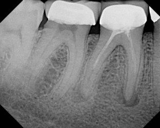
FIGURE 10-1 A 39-year-old presented with pain to biting on the mandibular first molar. He related that he could not understand why he was having pain; root canal treatment and a crown had been completed only 2 weeks prior to this painful episode. Note the incomplete root canal treatment with possible ledges in the mesial canals.

image

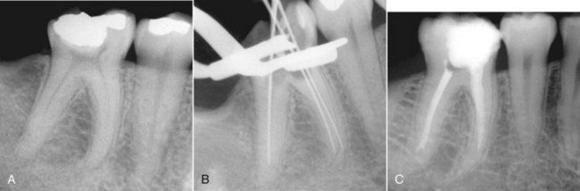
FIGURE 10-2 A, Preoperative radiograph showing significant curvature of the mesial canals, which should warn the clinician that ledging is more likely to occur if instrumentation is not performed cautiously. B, Master apical file radiograph shows that canals were instrumented larger than appropriate for size and curvature of mesial roots. Notice the loss of canal length and ledge formation. C, Mesial canals are fully ledged, and distal canal is partially ledged.

image

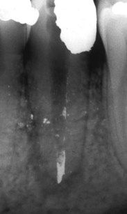
FIGURE 10-3 Packed amalgam particles in the apical extent of the canal block the apical 3 mm of the root canal.

image

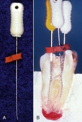
FIGURE 10-4 A, Improperly placed stop on a file can significantly change the accuracy of the working length and maintenance of canal length during enlarging and shaping. B, Cross-section of extracted tooth with files and stops in place. Stops are positioned at a proper angle with respect to the shaft of the file and the reference point on the tooth surface.

Each one of these errors is easily avoided with (1) attention to detail during instrument application and (2) generous use of irrigants during enlarging and shaping.

If nickel-titanium (NiTi) rotary instruments are used properly during canal enlarging and shaping, the same shortcomings may enter into the process, but loss of working length occurs infrequently. The major cause of loss of working length with NiTi rotary files appears to be excessive use of these instruments, retaining them in the canal for longer than 1 to 3 seconds while they are achieving their goals apically. It is not the packing of chips that occurs, but rather there is a tendency of these instruments to move laterally in the canal, especially in larger sizes and when placed apically over and over during the enlarging and shaping process.

Should loss of working length occur that is due to packed dentinal and tissue debris (Fig. 10-5, A) the clinician should choose a small but stiff instrument (e.g., No. 15 K-file) to bore through the obstruction. If the obstruction is caused by particles of temporary or permanent restorative materials, an instrument smaller than the last instrument placed into the canal system is used (e.g., No. 8, 10, or 15 K-file or C+ file [Dentsply Maillefer, Ballaigues, Switzerland]). A 45-degree curve is placed at the apical 3 to 4 mm of the instrument (see Fig. 10-5, B), and the file is inserted into the canal and slowly rotated circumferentially to detect a catch (the space between particles and canal wall; see Fig. 10-5, C).31 Once the catch is felt, the file is carefully rotated in a stem-winding fashion along with a slight in-and-out motion until the tip of the instrument bypasses the obstruction and negotiates the canal to length. Placing a relatively straight instrument into a milieu of metallic filings or dentin debris may push the particles more apically into the canal or periapical tissues, making them irretrievable. All recovery procedures should be performed with a lubricant in the canal.

image

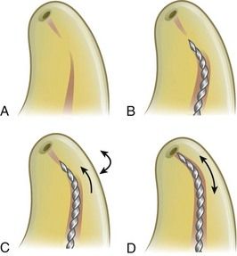
FIGURE 10-5 A, Diagram showing dentinal chips and tissue debris packed in the apical portion of the canal. B, A stiff file is curved in the apical 1 to 3 mm at a 30- to 45-degree bend and placed to the level of the blockage. C, The file is rotated slowly until it catches in the debris then advanced slowly in millimeter strokes to remove the blocking debris. D, Once the catch is felt, the file is carefully rotated in a stem-winding fashion along with a slight in-and-out motion until the tip of the instrument bypasses the obstruction and negotiates the canal to length.

Once the instrument begins to advance further into the canal, or if it expediently reaches the estimated working length, a radiograph is obtained to verify the position of the file. The instrument should not be removed until it can be used in small-amplitude strokes, moving circumferentially to dislodge the packed debris. When sufficient space has been developed through or along the side of the blockage, a smaller-sized Hedström file can be placed to length. Moving this instrument on the outstroke will eliminate the debris. To bore through dense blockage with dentinal chips, chelating agents such as RC-Prep, REDTAC, or liquid ethylenediamine tetraacetic acid (EDTA; see Chapter 11) may be used to soften the plug to facilitate penetration. If the plug has not been penetrated, the file may be creating a false canal in the dentinal wall.

Whenever there is failure to penetrate or bypass blockage, complete enlarging and shaping at a new working length coronal to the blockage (Fig. 10-6) are recommended.33 Variations in obturation techniques such as diffusion, thermoplasticized gutta-percha, warm vertical compaction, or core-carrier procedures can be used to enhance penetration of the gutta-percha and sealer around or through the blockage (see Chapter 12). Periodic reassessment is necessary, and if further treatment is deemed appropriate, surgical intervention to correct the problem may be warranted (Fig. 10-7).9 If surgery is not feasible, intentional replantation15,57 or extraction and replacement with an artificial prosthesis such as an implant may be indicated if symptoms persist.

image

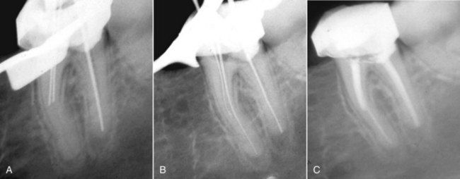
FIGURE 10-6 A, Note the block (most likely a ledge) in the mesial canals of a mandibular molar. B, After placement of a 45-degree bend, one of the canals was penetrated and negotiated to the working length. The other canal could not be penetrated, or possibly it joined the patent canal. C, Obturation with a softened gutta-percha and sealer technique.

image

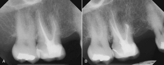
FIGURE 10-7 A, Maxillary molar with a separated instrument segment around the curve in the mesial buccal canal. The tooth was symptomatic. B, Following apical surgery and a root-end filling in the mesial buccal canal; 3-month reevaluation.

Many authors and clinicians would also list separation or breakage of intracanal instruments, especially in curved canals, as a major cause of canal blockage or loss of working length (Fig. 10-8).28 This problem has been addressed in Chapters 5 and 14. However, during the application of NiTi rotary instruments, there are unique circumstances that tend to predispose to instrument separation and canal blockage (Fig. 10-9). In many respects, the concern for instrument breakage has prevented many clinicians from adopting the use of rotary NiTi instruments. This is unfortunate because the minimal potential for instrument separation does not have to be a deterrent to usage if some basic principles are followed. First, these instruments are always to be used in a crown-down technique after a tapered pathway for the instruments has been developed. The pathway will ensure ease of movement with minimal pressure, binding, and torque on the instrument during canal penetration. The pathway can be created with stainless steel hand K-files (No. 06 to 20) intended for this purpose4 or with NiTi rotary PathFiles (Dentsply Maillefer, Ballaigues, Switzerland). The pathway that is developed favors variably tapered NiTi instruments and small constant-tapered instruments (0.04 to 0.06, Nos. 15 and 20). When time is taken to develop the pathway, additional important information is identified by the clinician, such as canal curves, the joining of canals, rapid deviations, and multiple canals. In light of these findings, most causes for loss of working length can be prevented.

image

FIGURE 10-8 Broken instrument segment around the curve at the junction of the mesial buccal and distal canals—a common canal blockage.

image

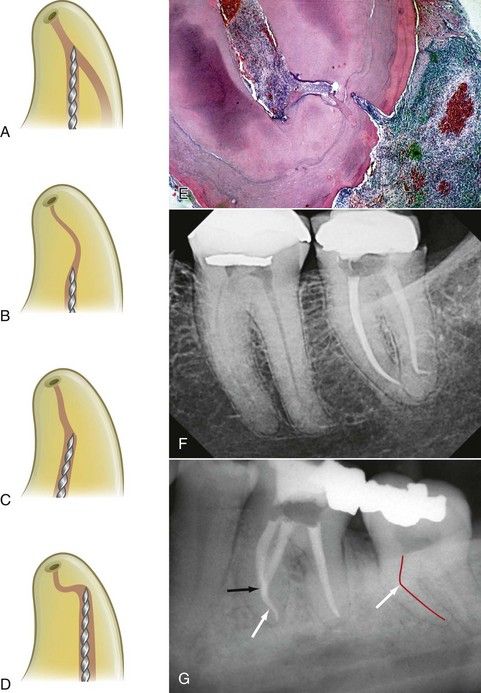
FIGURE 10-9 Diagrammatic representations of the four most common reasons for breakage of any endodontic intracanal instrument: canal convergence (A), S-shaped canals with multiple curves (B), abrupt canal deviations (C), and severe and rapid apical curvatures (D). These anatomic complexities are especially important in the application of NiTi rotary instruments. E, Histologic evidence for t/>

Only gold members can continue reading. Log In or Register to continue

Stay updated, free dental videos. Join our Telegram channel

Jan 2, 2015 | Posted by in Endodontics | Comments Off on 10: Problem-Solving Clinical Techniques in Enlarging and Shaping the Root Canal

VIDEdental - Online dental courses

Get VIDEdental app for watching clinical videos