Cementum
Learning objectives
■ Describe the development of cementum and its function on the surface of the root.
■ Discuss the aging of cementum, the formation of cementicles, and the repair of cementum.
Key terms
Cellular-acellular cementum
Cementoid layer
Intermediate cementum
Overlap, meet, and gap
Overview
Cementum, which is the focus of this chapter, has two major functions. It seals the tubules of root dentin and serves as an attachment for periodontal fibers to keep the tooth in its socket. Cementum has the ability to reverse root resorption by means of deposition as it forms a smooth patch on the cemental surface.
Two types of hard tissue cover tooth roots. The first, called intermediate cementum, is a homogenous layer originating from inner epithelial root sheath cells. The second, called cellular-acellular cementum, is a thicker deposit of a bonelike substance produced by cementoblasts that differentiate from the periodontal ligament fibroblasts. The latter is laid down in increments, usually an acellular layer followed by a cellular layer. Cementum simulates bone by displaying cells within lacunae and cell processes within canaliculi. Cementum also exhibits incremental lines but does not have a vascular and neural supply that is characteristic of bone. As a result, the cementum has unique characteristics, such as lack of neural sensitivity and a greater ability than bone to resist resorption. Both are important clinical features. Aging cementum exhibits a rough and irregular surface caused by resorption of the cemental surface. This cementum also is associated with free, attached, or embedded cementicles. These oval to round stones are similar to the denticles in pulp. They are calcified bodies that may be embedded, attached to cementum, or free in the periodontal ligament.
Role of cementum on the root surface
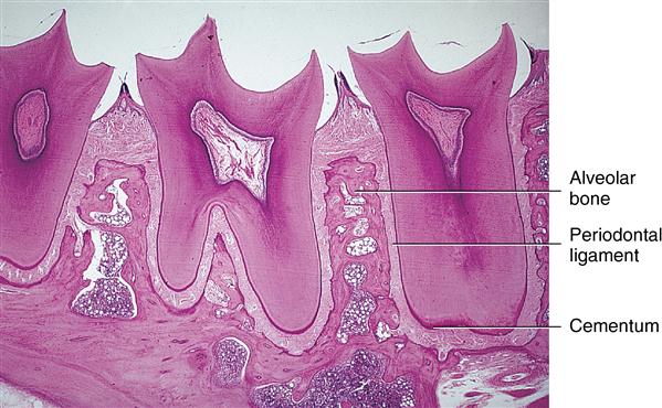
The hard tissue that covers the entire root surface is very thin but manages to carry out two important functions. First, it seals the surface of the root dentin and covers the ends of the open dental tubules. Second, perforating fibers of the periodontal ligament become embedded in the cementum. These fibers function as an attachment for the periodontal ligament fibers to the tooth root and aid in maintaining the tooth in its socket. This chapter discusses sealing of the root surface (Fig. 10-1), and Chapter 12 discusses the attachment fibers.
 CLINICAL COMMENT
Cementum can aid in maintaining the teeth in functional occlusion if it is deposited at the apical aspect of the root, especially in patients with chronic bruxism. Because cementum is the slowest growing tissue compared with the other periodontal tissues, it will be the last tissue to be added to the root surface after occlusal loss.
Development of cementum
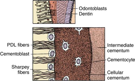
The first cementum deposited on the root’s surface is called intermediate cementum and is formed by the inner epithelial root sheath cells that formed during root dentin formation. This deposition occurs before the root sheath cell layer disintegrates (Fig. 10-2). Intermediate cementum is situated between the granular dentin layer of Tomes and the secondary cementum that is formed by the cementoblasts. These cementoblasts arise from the dental follicle (sac). The thin layer of intermediate cementum is approximately 10 μm thick. After being deposited, this layer mineralizes to a greater extent than the adjacent dentin or the cellular-acellular cementum. Under proper magnification, a thin line of radiopacity is seen covering the root.
Intermediate cementum on the right overlying dentin cellular cementum in the center of the field and periodontal attachment fibers (PDL) on the left. (From Avery JK: Oral development and histology, ed 3, Stuttgart, 2002, Thieme Medical.)
The cellular-acellular cementum is a specialized hard tissue covering the root surfaces of teeth (see Fig. 10-2 and Fig. 10-3). The initial thin layer of this cementum is acellular and is deposited on intermediate cementum. Subsequent layers alternate between cellular and acellular. Thus cementum is deposited incrementally. Both grossly and histologically, this cementum resembles bone because it is a hard tissue with cells contained in lacunae that exhibit canaliculi (see Fig. 10-2). However, unlike bone, cementum does not contain blood vessels, nerves, or haversian or Volkmann canals, which are the nutrient canals containing blood vessels and nerves in bone (see Fig. 10-3).
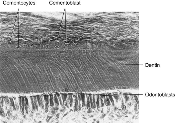
Some cementoblasts become enmeshed in cementum matrix and develop into cementocytes living in lacunae. (From Avery JK: Oral development and histology, ed 3, Stuttgart, 2002, Thieme Medical.)
Cementum is limited to the roots of teeth. In 60% of cases, cementum is formed on the cervical enamel for a short distance; in 30% it stops at the cervical line just meeting the enamel; and in 10% a small gap exists between them. This order of frequency is known as overlap, meet, and gap (OMG) (Fig. 10-4 and Table 10-1).
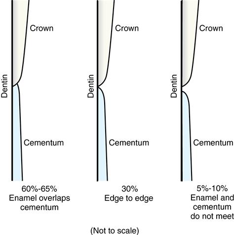
Overlap, meet, and gap (OMG) describe the order of frequency of these conditions. (Modified from Daniel SJ, Harfst SA: Mosby’s dental hygiene: concepts, cases, and competencies, 2004 update, St Louis, 2004, Mosby, p. 288.)
Table 10.1
Relationship of cementum to enamel at the cementoenamel junction
| Relationship of how enamel and cementum meet during development | Percentage of cases |
| Cementum overlaps enamel | 60 |
| Cementum meets enamel | 30 |
| Small gap exists between cementum and enamel | 10 |
Intermediate cementum
Intermediate cementum is a thin, noncellular, amorphous layer of hard tissue approximately 10 μm thick. It is deposited by the inner layer of the epithelial cells of the root sheath. Deposition occurs immediately before the epithelial root cells disintegrate as a sheet and migrate away from the root into the periodontal tissue (see Fig. 10-2). Most authors have used the term intermediate cementum, although some prefer cementoid layer. The latter term is confusing, because the initial layer of cementum is called cementoid, like osteoid in bone.
Intermediate cementum is the first layer of hard tissue deposited, and it seals the tubules of dentin. Because of its epithelial origin, intermediate cementum is composed of enamelin-like protein rather than collagen, which is the protein typical of cellular or secondary cementum. Intermediate cementum is completely formed before deposition of the secondary cementum begins. As an amorphous, noncellular layer, it is s/>
Stay updated, free dental videos. Join our Telegram channel
VIDEdental - Online dental courses


