1 Histology of the Pulp
From a functional point of view, pulp essentially exists to allow the constant formation of new dentin to compensate for natural occlusal abrasion. Therefore, the morphology of pulpal tissue changes throughout life and it should be viewed as a dynamic organ.
Histologically, pulp consists of specialized, loosely woven connective tissue, which is responsible for formation of new dentin at the periphery of the pulp chamber. Odontoblasts, which line the wall of the entire pulp chamber, lay down the so-called “reparative dentin” in response to external stimuli (e.g., bacterial invasion into dentin tubuli).
The cells in the pulpal connective tissue consist primarily of undifferentiated fibroblast-like cells (pulpoblasts). Following external stimulation, these cells can differentiate into odontoblasts or into fibroblasts that produce fibrodentin.
It is possible to differentiate between coronal pulp and root canal (radicular) pulp. Coronal pulp consists of a loose network of connective tissue in which blood vessels and nerve fibers are embedded. These vessels and nerve fibers reach the coronal pulp via the apical foramen and the root canal itself. Within the pulp chamber, these entities branch to form rich vascular and nerve plexuses in the subodontoblastic layer. In addition to the loose connective tissue, the central part of the root pulp contains collagen fibers subjacent to the odontoblastic layer, and the blood vessels and nerve fibers course through this fibrous matrix. At the apical foramen, the pulpal tissue blends with the periodontal tissues.
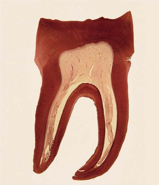
1.1 The pulp of a mandibular molar
The coronal pulp fills the wide pulp chamber. The various zones of dentin, predentin, the odontoblastic layer, and the cell-poor and cell-rich layers can be seen.
The pulpal tissue itself consists of loose connective tissue. Individual vascular elements are seen in both transverse and longitudinal sections (courtesy of Professor Dr. W. Ketterel, Mainz, Germany).
Embryonic Development of the Pulp
The pulp develops from the mesenchymal cells of the cranial ectomesenchyme, which develops from the cranial neural crest. Invagination of the superficial ectoderm of the oral cavity is followed by formation of the dental primordia, in which the individual tooth buds develop. A complex and multifaceted induction cascade between the ectoderm and mesenchyme leads to the concentration of mesenchymal cells immediately subjacent to the ectoderm of each tooth bud.
The enamel organ develops from the ectoderm surrounding condensed mesenchymal cells. Additional signaling molecules released from the invaginated epithelium into the mesectoderm stimulate the differentiation of mesenchymal preodontoblasts at the ectodermal–mesenchymal border; these cells then begin to secrete pre-dentin.
As the mineralization of dentin begins, cellular processes begin to form at the apical ends of the preodontoblastic cells. These so-called odontoblastic processes continue to produce predentin, therefore ensuring that the dentin layer becomes ever thicker. Vascular elements proliferate at the apical aspect of the tooth bud and extend into the forming dental pulp.


