Development and structure of cells and tissues
Learning objectives
■ Describe the cell and how it divides.
■ Discuss the origin of tissue and the ovarian cycle and the development of the embryonic disk.
Key terms
Absorption
Agranulocytes
Anaphase
Angioblasts
Angiogenic clusters
Appositional
Assimilation
Astral rays/asters
Basophils
Blastocyst
Cartilage
Cell cycle
Centrioles
Centromere
Cerebellum
Cerebral hemispheres
Chondroblasts
Chromatids
Chromosomes
Conductivity
Cytoplasm
Cytosol
Deoxyribonucleic acid (DNA)
Dermatome
Dermis
Ectodermal
Elastic or fibrous cartilage
Embryonic disk
Embryonic period
Endochondral bone development
Endodermal
Endometrium
Endoplasmic reticulum (ER)
Eosinophils
Epidermis
Epiphyseal line
Epithelium
Equatorial plate
Erythrocytes
Excretion
Fetal period
Fibroblasts
Fluid
Foramen ovale
Forebrain, midbrain, and hindbrain
Frontal, temporal, and occipital lobes
G1 phase, G2 phase
Gastrointestinal tract
Gene expression
Genetic mechanisms
Golgi apparatus, complex
Granulocytes
Growth
Growth factors
Hemoglobin
Hyaline cartilage
Implantation
Induction
Intercalated disks
Intercellular material
Interstitial
Interstitial growth
Intramembranous bone formation
Irritability
Leukocytes
Lymphatic system
Lymphocytes
Lysosomes
Melanocytes
Mesenchymal cells
Mesodermal
Metaphase
Metaphysis
Microtubules
Mitochondria
Monocytes
Morphogens
Morula
Myotome
Neural plate
Neural tube
Neuroblasts
Neurons
Neutrophils
Nuclear envelope
Nuclear pores
Nucleolus
Nucleus
Organizer
Osteoblasts
Plasma
Plasma membrane
Pons
Proliferative period
Prophase
Reproduction
Respiration
Ribonucleic acid (RNA)
S phase
Sclerotome
Smooth muscle cells
Somites
Spindle fibers
Striated voluntary muscles
Telophase
Umbilical system
Visceral mesoderm
Vitelline vascular system
Zygote
Overview
The smallest unit of structure in the human body is the cell, composed of a nucleus and cytoplasm. The nucleus contains deoxyribonucleic acid (DNA) and ribonucleic acid (RNA), the fundamental structures of life. The cytoplasm functions in absorption and cell duplication, in which organelles perform specific actions. The cell cycle is the time required for the DNA to duplicate before mitosis. This chapter discusses the four stages of mitosis: prophase, metaphase, anaphase, and telophase. Also described are the three periods of prenatal development: proliferative, embryonic, and fetal. The fertilization of the ovum in the distal uterine tube, zygote migration, and the zygote’s implantation in the uterine wall are discussed. In addition, the origin of human tissues—ectoderm, mesoderm, and endoderm—is presented, followed by the differentiation of tissue types, such as those of ectodermal origin, epithelium and skin with its derivatives, and the central and peripheral nervous systems. This chapter also delineates development of the mesodermal components involving connective tissues of the body, such as fibrous tissue, three types of cartilage, two types of bone, three kinds of muscles, and the cardiovascular system. The reader will better comprehend the origin, development, organization, and structure of the various cells and tissues of the human body.
Cell structure and function
The human body is composed of cells, intercellular substance (the products of these cells), and fluid that bathes these tissues. Cells are the smallest living units capable of independent existence. They carry out the vital processes of absorption, assimilation, respiration, irritability, conductivity, growth, reproduction, and excretion. Cells vary in size, shape, structure, and function. Regardless of function, each cell has a number of characteristics in common with other cells, such as cytoplasm and a nucleus, which contains a nucleolus. However, some cell characteristics are related to function. A cell on the surface of the skin, for example, serves best as a thin, flattened disk, whereas a respiratory cell functions best as a cuboidal or columnar cell to facilitate adsorption with mobile cilia to move fluid from the lung to the oropharynx. Surrounding each cell is the intercellular material that provides the cell with nutrition, takes up waste products, and provides the body with form. It may be as soft as loose connective tissue or as hard as bone cartilage or teeth. Fluid, the third component of the body, is the blood and lymph that travel throughout the body in vessels or the tissue fluid that bathes each cell and fiber of the body.
Cell nucleus
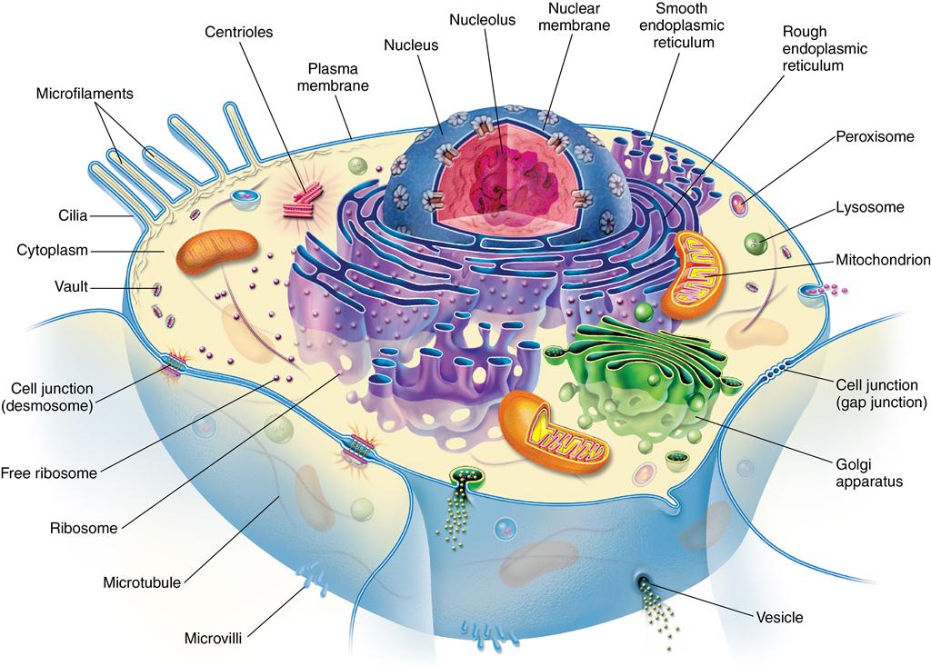
A nucleus is found in all cells except mature red blood cells and blood platelets. The nucleus is usually round to ovoid, depending on the cell’s shape. Ordinarily a cell has a single nucleus; however, it may be binucleate, as are cardiac muscle cells or parenchymal liver cells, or multinucleate, as are osteoclasts and skeletal muscle cells. The nucleus is important in the production of DNA and RNA. DNA contains the genetic information in the cell, and RNA carries information from the DNA to sites of actual protein synthesis, which are located in the cell cytoplasm. The nucleus is bound by a membrane, the nuclear envelope, which has openings at the nuclear pores. This envelope is composed of two phospholipid layers similar to the plasma membrane of the cell. The pores are associated with the endoplasmic reticulum (ER) that forms at the end of each cell division. The nucleus contains from one to four nucleoli, which are round, dense bodies constituting the RNA contained in the nucleus. Nucleoli have no limiting membrane (Fig. 1-1).

Cells communicate with each other to regulate organization, growth, and development. (From McCance KL, Huether SE: Pathophysiology: the biologic basis for disease in adults in children, ed 6, St. Louis, 2010, Mosby.)
Cell cytoplasm
Cytoplasm contains structures necessary for adsorption and for creation of cell products. The cytosol is the part of the cytoplasm that contains the organelles and solutes. The cytosol uses the raw materials brought into the cell to produce energy. It also functions in the excretion of waste products. These functions are carried out by the ER-parallel membrane-bound cavities in the cytoplasm that contain newly acquired and synthesized protein. Two types of ER, smooth surfaced and granular or rough surfaced, can be found in the same cell. Rough-surfaced ER is caused by ribosomes on the surface of the reticulum and is the site at which protein production is initiated. Proteins are vital to the cell’s metabolic processes, and each type of protein is composed of a number of different amino acids linked in a specific sequence. Amino acids form protein-containing groups, which, in turn, form acids or bases.
Ribosomes are particles that translate genetic codes for proteins and activate mechanisms for their production. They can be found as separate particles in the cytoplasm, clustered as polyribosomes, or attached to the ER membranes. Ribosomes are nonspecific as to what type of protein they synthesize. The type is dependent on the messenger RNA (mRNA), which carries the message directly from the DNA of the nucleus to the RNA in the ER. This molecule attaches to the ribosomes and gives orders about the formation of the amino acids.
The ER transports substances in the cytoplasm. The ER is connected to the Golgi apparatus via small vesicles. The Golgi apparatus or complex is critical for posttranslational modifications which help sort, condense, package, and deliver proteins arriving from the ER. The Golgi apparatus is composed of cisternae (flat plates) or saccules, small vesicles, and large vacuoles. From here the secretory vesicles move or flow to the cell surface, where they fuse with the cell membrane and the plasmalemma and release their contents by exocytosis.
Lysosomes are small, membrane-bound bodies that contain a variety of acid hydrolase and digestive enzymes to help break down substances both inside and outside the cell. They are in all cells except red blood cells but are prominent in macrophages and leukocytes. Peroxisomes, another intracellular organelle, are also important for breaking down of fatty acids.
Mitochondria are membrane-bound organelles that lie free in the cytoplasm and are present in all cells. They are important in generating energy, are a major source of adenosine triphosphate (ATP), and therefore are the site of many metabolic reactions. These organelles appear as spheres, rods, ovoids, or threadlike bodies. Usually the inner layer of their trilaminar bounding membrane inflects to form transverse-appearing plates, the cristae (see Fig. 1-1). Mitochondria lie adjacent to areas that require their energy production. They also have the ability to store ionic calcium and to release it when needed by the cell for various reactions, including signal transduction.
Microtubules are small tubular structures in the cytoplasm that are composed of the protein tubulin. These structures may appear as singles, as doublets, or as triplets. They probably function as structural and force-generating elements and relate to cilia (motile cell processes) and to centrioles in relation to mitosis. They have cytoskeletal functions in maintaining cell shape. Centrioles are short cylinders appearing near the nucleus. Their walls are composed of nine triplets of microtubules. Centrioles are microtubule-generating centers and are important in mitosis, self-replicating before mitosis begins.
Surrounding the cell is the plasma membrane or plasmalemma, which envelops the cell and provides a selective barrier that regulates transport of substances into and out of the cell. All membranes are composed mainly of lipids and proteins with a small amount of carbohydrates. The plasma membrane receives signals from hormones, growth factors, and neurotransmitters by having them bind to receptors located on the surface and within the plasma membrane, eventually activating a second messenger (e.g., cyclic adenosine monophosphate [cAMP]) that signals intracellular organelles or the nucleus/nucleolus to modify cell activity such as increasing the production of a protein. Also included in the plasma membrane are many kinds of ion channels that can activate many different cell functions. In addition, cells contain proteins, lipids, or fatty substances that provide energy in the cell and are important components of cell membranes and permeability. Carbohydrates are also important in cells as the most available energy component in the body. These carbohydrates may exist as polysaccharide-protein complexes, glycoprotein complexes, glycoproteins, and glycolipids. Carbohydrate compounds are important in cell function and for development of cell products, such as supportive tissues and body lubricants.
Genetic mechanisms help a cell to develop and maintain a high degree of order. This ability is dependent on the genetic information that is expressed within the cell. The basic genetic processes in the cell are RNA and protein synthesis, DNA repair, and replication and genetic recombination. These processes produce the proteins and nucleic acids of a cell. These genetic events are relatively simple compared with other cell processes.
Cell division
Cell cycle
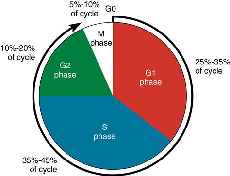
Cell division is a continuous series of discrete steps by which the cell component divides. This function is related to the need for growth or replacement of tissues and is partly dependent on the length of the cell’s life. Continually renewing cells line the gastrointestinal tract and compose the epidermis and the bone marrow. A second type of cell is part of an expanding population—the cells of the kidney, liver, and some glands. The third type of cell does not undergo cell division or DNA synthesis. An example is the neurons of the adult nervous system. For a somatic cell to undergo cell division, it must pass through a cell cycle, which ensures time for DNA genetic material in the daughter cells to duplicate that of the parent cell. However, in a sex cell, an ovum or spermatozoon, the process of meiosis occurs, in which a reduction division of chromosomes in the daughter cell takes place. The result is that half as many chromosomes are in the daughter cell as are in the parent cell. Through meiosis, after fertilization of the ovum by the male sperm, the original (diploid) number of chromosomes is regained. The duration of the cell cycle in somatic cells is now known (Fig. 1-2). After mitosis, the cells enter the reduplication or G1 phase of the interphase, the initial resting stage. This is followed by the S phase, in which DNA synthesis is completed. Next, the cell enters the G2 phase or quiescent phase of the post-DNA duplication and proceeds into the mitotic stages of prophase, metaphase, anaphase, and telophase (Fig. 1-3). The cell then reenters and remains in the interphase stage until duplication resumes the mitotic process of developing two daughter cells identical to the parent cells.
Stay updated, free dental videos. Join our Telegram channel

VIDEdental - Online dental courses


