Implant dentistry is highly successful, and a predictable treatment when performed properly by a trained and skilled team of dentists. On the other hand, the treatment of peri-implantitis has been very limited and largely disappointing. Most attempted treatments for peri-implantitis of implant infection fail. We’ll now take a look at what we think works, what may work, and what treatment do not work and should be avoided.
While the causes of peri-implantitis can be identified in most cases, effective treatment options are less obvious. From early days of dental implants, clinicians have tried a number of different treatments, with mostly disappointing results. This is perhaps due to the difficulty in decontamination of the implant surface from plaque’s toxins and the unpredictability in re-establishment of bone and soft tissue healing on that same surface.
Several methods of treating peri-implantitis have been described in the literature. Most of these methods focus on removal of the contaminating agent from the implant surface, but they remain highly unpredictable and their long-term success is unknown. These treatments include:
•Administration of systemic antibiotics alone
•Mechanical debridement with or without systemic antibiotic treatment (8)
•Mechanical debridement with or without localized drug delivery and chlorhexidine oral rinses (9)
•Mechanical debridement combined with LASER decontamination (6)
•Surgical debridement, and more recently…
•Surgical debridement with guided bone regeneration (GBR) for reparation of bony and soft-tissue defects (10)
Because nonsurgical treatment approaches have been ineffective in management of peri-implantitis, additional surgical interventions have been used including:
•Mechanical/ultrasonic debridement with localized drug delivery; i.e., antimicrobial minocycline spheres (Arestin®)
•Laser treatment with and without flap access
•Open flap debridement
•Open flap debridement with guided bone regeneration
Froum et al. reported an encouraging results in 51 cases of peri-implantitis using a regenerative approach including surface decontamination, use of enamel matrix derivative, a combination of platelet-derived growth factor with bovine bone or mineralized freeze-dried bone, and coverage with a collagen membrane or a subepithelial connective tissue graft. He demonstrated 3 mm gain of bone level over a period of 3 to 7.5 years. However, there was no histology to determine the nature of the newly formed tissue (13)
There is limited literature and research on effective treatment of peri-implantitis. Most articles are case reports on a specific technique that was attempted on a patient with variable results. There are very few studies on large patient samples that can provide us with meaningful treatment options or results. Hence, the outcome of treatments such as bone graft, tissue graft, or decontamination techniques (i.e. Laser, surgical decontamination, antibacterial) performed on the failing implants are largely unknown and should be approached cautiously. Even, if a treatment has appeared to work initially, its long-term prognosis and outcome remains questionable.
Before investing more time and money on such treatments, it is critical for the treating dentists to obtain all necessary diagnostic information, manage contributing factors, select treatments with the best long-term results, and provide patients with realistic expectations.
With such limited literature and inconsistent results from reported procedures, we decided to analyze what we have learned about the treatment of failing implants for patients with this frustrating problem. We first reviewed patients that we have seen with peri-implantitis, bone loss, and infection and identified contributing factors, when possible. We then looked at what treatments have been done on those patients and their clinical outcomes. Finally, we reviewed the literature and case reports to learn about the experience of other clinicians on different treatment modalities.
Before investing more time and money on treatments, it is critical for the treating dentists to obtain all necessary diagnostic information, manage contributing factors, select treatments with the best long-term results, and provide patients with realistic expectations.
And here is what we learned:
1.There was no consensus on which treatment modalities are consistently effective in managing failing dental implants with peri-implantitis.
2.The current classifications on different types of peri-implantitis, for the most part, are descriptive of the extent of the disease, but never have been validated regarding treatment efforts. There is also no universal or standard classification that clinicians can use for early detection and intervention. (2)
3.There were no prospective studies on the efficacy of various treatment options on failing implants.
4.Non-surgical treatment of peri-implantitis is highly unpredictable (3, 4, 5).
5.Beneficial effects of laser therapy on peri-implantitis have been shown, but its efficacy for long-term success is unknown (6,7).
6.Most procedures showed disappointing results and unpredictable outcomes.
In any disease process, treatment selection should be based on proper diagnosis, biological factors, and the probability of a long-term cure. This principle along with our observations led us to the recommendations in this chapter. First, we created a classification for various types of peri-implantitis and degree of bone and soft tissue loss. Then for each type, we listed what we know works, what we think might work, and what does not work and should be avoided.
Regardless of the type of complication, a patient with a painful dental implant should first see a specialist with expertise in management of dental implant complications as soon as possible.
There are two dental specialties that are formally trained in surgical principles of implant dentistry: oral surgeons and periodontists. However, they may not necessarily be experienced in managing dental implant complications. Therefore, patients should ask the oral surgeon or periodontist if they have specific experience in managing dental implant complications and whether they use current methods and techniques in their treatments.
When seeking the right specialist, patients should inquire:
1.Availability of CBCT (cone beam CT scan) imaging for proper diagnosis.
2.Whether the surgeon uses special tools and techniques for atraumatic removal of dental implants.
3.Experience and skill in bone and soft tissue regeneration.
4.Utilization of Platelet Derived Growth Factor (PDGF) or other biologic agents for enhancement of healing.
5.Experience in managing dental implant complications and the type of patients they have treated.
6.Evidence-based treatments that provide the best outcome with long-term results.
Before engaging in any treatment, patients should consult with a surgeon and a restorative dentist. The team dentists should obtain detailed past dental and medical history, perform a careful clinical examination, and obtain all necessary diagnostic information including CT scan (CBCT), dental X-rays, photos, and models. Equally important is discussion of patients’ emotional state and what they expect from treatment.
Treatment options include both surgical and non-surgical methods. The team should consider all historical and existing factors along with diagnostic studies to design a treatment plan that best meets the patient’s needs and goals. In some circumstances, it may also be helpful to obtain information on the initial treatments rendered by the original clinicians.
With good data and full understanding of the circumstances that led to peri-implantitis, the team can now make recommendations on optimal treatment.
Let’s take a look at treatment options for each type of peri-implantitis and other inflammatory-related complications: what works, what may work, and what absolutely does not work and should be avoided!
If you are experiencing…
•Mildly painful gum tissues
•Mild swelling of gums
•Mild bleeding of gums
•No bone or gum tissue loss
You have >> Peri-Implant Mucositis
This is the mildest form of inflammatory condition affecting dental implants with mild pain and redness of the gum tissue (Fig 16).
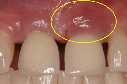
Fig 16 Peri-implant mucositis in a patient with upper incisor dental implant. The area appears red and more swollen.
What treatment works:
•Professional cleaning by your dentist every 3-4 months
•Better home care with effective brushing and flossing
•Use of other plaque control tools such as super floss, soft-picks, rubber tip stimulator, and proxabrush
•Application of chlorhexidine (peridex) to the gum tissue around the implant and its crown
•Identify and correct possible contributing factors such as fit of the components, crown shape and contours, and accessibility for effective plaque removal.
With early intervention and elimination of contributing factors, most cases of peri-implant mucositis can be effectively controlled and treated.
If you are experiencing…
•Moderate to significant painful gum tissue
•Moderate bleeding from gums
•Increasing red and swollen gums
•No bone or gum tissue loss
You have >> Peri-Implantitis Type 1
This is a more advanced form of peri-implant mucositis characterized by increased pain, bleeding, and swelling around the gum tissues (Fig 17).
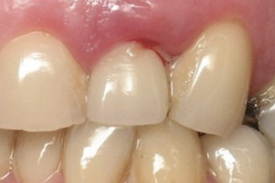
Fig 17 Peri-implantitis type 1 is characterized by increased redness, bleeding and swelling of gum tissue, but no loss of bone or soft tissue
What treatment works:
•Deep cleaning by your dentist
•Better home care with effective brushing and flossing
•Use of other plaque control tools such as super floss, soft-picks, rubber tip stimulator, and proxabrush
•Application of chlorhexidine (peridex) to the gum tissue and around the implant and its crown
•Debridement along with antibiotics, either in the form of pills or localized drug delivery; i.e., antimicrobial minocycline spheres (Arestin®)
•Identify and correct possible causes as soon as possible: proper access for plaque removal, ill-fitting crown, residual cement, smoking, etc.
 |
video Hygiene Instructions for Dental Implants |
If you are experiencing…
•Pain, redness, or bleeding of gum tissue
•1-4 mm of bone loss on one surface of the implant
•No loss of gum tissue
•Possible gray color of implant surface visible through the gum tissue
You have >> Peri-Implantitis Type 2a
In this type, bone is partially missing on one surface, but the gum tissue remains at a normal level. It is similar to peri-implantitis type 1, except that now there is loss of bone and possible grayish appearance of gum tissue in thin tissue biotypes (Fig 18).
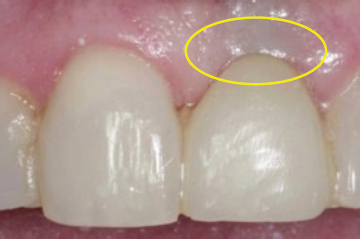
Fig 18 Peri-implantitis type 2a occurs when there is 1-4 mm loss of bone on the implant, but the gum tissue remains intact. The gum tissue may appear darker as the color of the implant becomes visible due to lack of bone coverage.
What treatments work:
•See a surgeon for a consultation. Your dentist alone cannot address this problem.
•Antibiotic therapy to control inflammation or infection
•Removal of dental implant using atraumatic technique
•Immediate bone graft to restore missing bone if appropriate based on the quality and health of the surrounding tissues and type of bony defect
•If bone graft is not appropriate at the time of implant removal, then reevaluate in 3 months with CBCT (cone beam CT scan) and determine the best bone grafting approach for the type of bony defect
•Application of platelet derived growth factor to implant removal site to enhance tissue recovery and healing
•Replace with a new dental implant in 4-6 months using “Implant-Perfect” methodology (see the glossary section)
What treatments MAY work:
If the width of the exposed implant surface is 1-3 mm AND there is minimal inflammation, AND there is no evidence of infection AND the gum tissue is relatively thick and healthy, THEN…
•The crown may be removed, and a cover screw placed on the implant—Allow 6-8 weeks for gum tissue healing
•If the gum tissue is very thin or poor quality, the surgeon can attempt to improve its quality by a connective tissue gingival graft. This will help achieve a better result after bone grafting. In this case, allow 3 months of gum graft healing before bone grafting.
•The surgeon reflects the gum tissue and uncovers the exposed part of the implant
•The exposed implant surface is then detoxified using antibacterial agents or laser
•Bone grafting is attempted over the exposed implant surface
•Bone grafting may be complemented with growth factors from rhBMP2 or PDGF for enhanced healing
•A slow-resorbing or non-resorbing occlusive membrane is placed over the bone graft
•The implant and grafted bone is covered with soft tissue completely and allowed to heal for at least 6 months
•The implant should be re-evaluated using CBCT and clinical examination to assess the degree of bone regeneration over the implant and attachment level
•If successful, uncover the implant platform, place healing abutment, and prepare for a new crown
NOTE: THE SURGICAL RESULTS MAY VARY. THERE IS NO EVIDENCE-BASED SUPPORT FOR THIS TREATMENT AND LONG-TERM SUCCESS MAY RANGE FROM NONE TO 50% DEPENDING ON BIOLOGICAL FACTORS AND THE PATIENT’S HEALING POTENTIAL. THE SURGEON AND PATIENT MUST CONSIDER THE TIME, COST, AND BENEFIT / RISK OF SUCH TREATMENTS BEFORE PROCEEDING.
Stay updated, free dental videos. Join our Telegram channel
VIDEdental - Online dental courses


