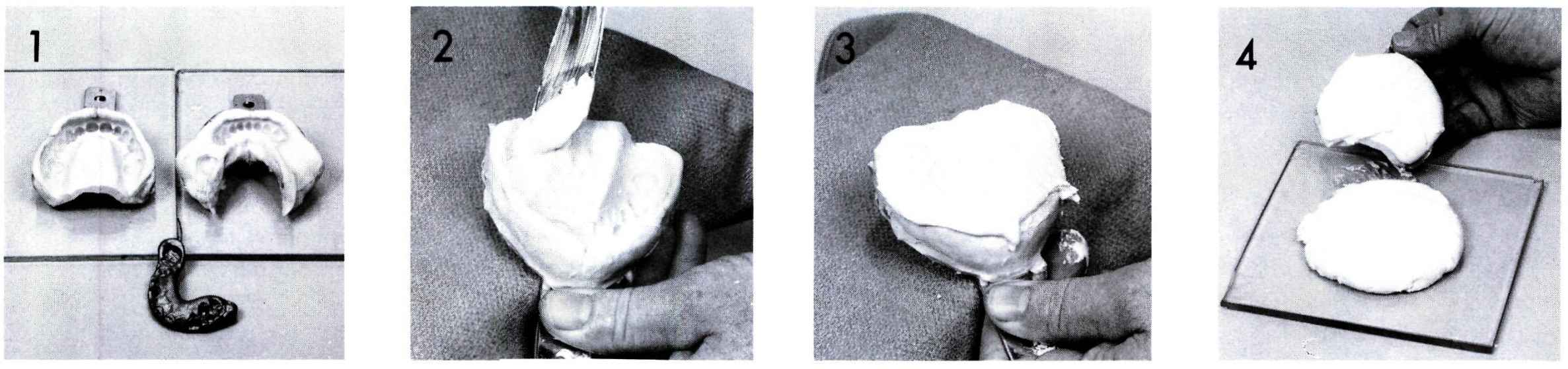
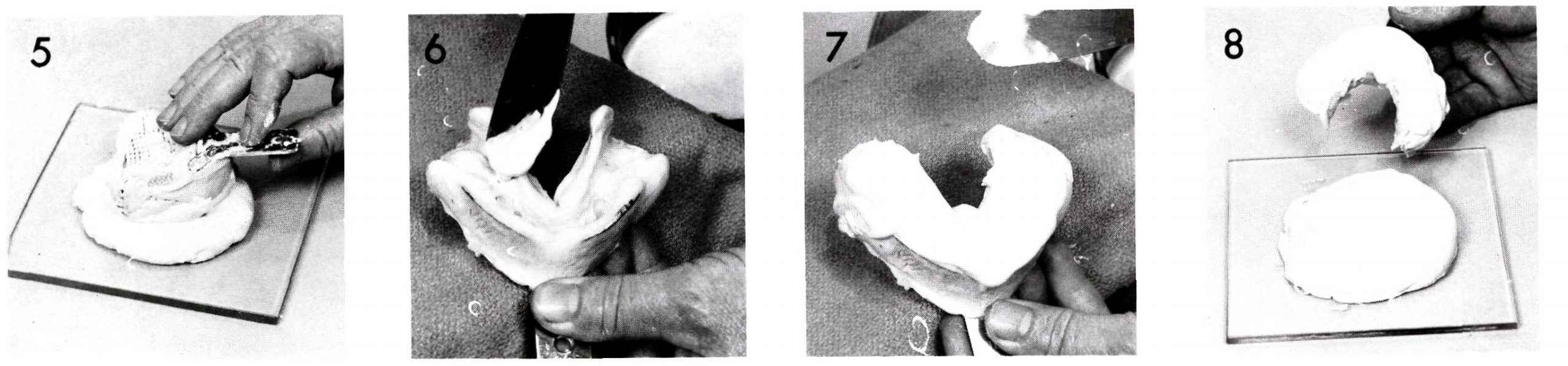
Figures 6 through 9 The same procedures are followed for the mandibular impression. Do not press the impression into the stone which has been placed on the glass slab. This prevents excess stone from flowing in the tongue space.
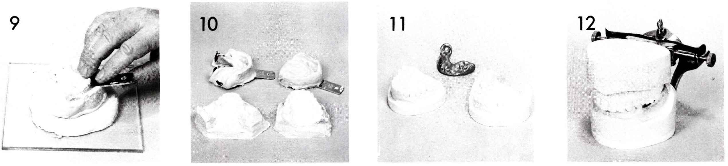
Figure 10 After the stone has set, the impressions are removed from their casts.
Figure 11 The casts are trimmed and are now ready, by means of the interocclusal jaw relation record, to be mounted.
Figure 12 If upon examination there were no occlusal discrepancies, the casts may be mounted on a simple hinge articulator. The selection of an articulator on which to mount diagnostic casts is determined by the desires of the dentist who usually bases this decision on the complexity of the restorative problem. A face-bow transfer will be supplied by the dentist if a more complex articulator is to be used.
At this stage, the dentist should indicate if any teeth associated with the removable partial denture need cast restorations or crowns. The design and contours of such restorations can be developed to enhance the appearance and function of the removable appliance.
NON-TECHNICAL ASPECTS OF PLANNING FOR REMOVABLE PARTIAL DENTURES
Planning a removable partial denture is a relatively complex procedure. The dentist must assess a number of factors present in each individual patient. The number and location of the remaining natural teeth is one factor; the teeth in the opposing arch must be considered as well as those in the arch needing the removable partial denture. Extrusion of teeth opposite an edentulous space always occurs in time and can complicate dental restorations from simple fillings to complex removable partial dentures.
The caries experience of the patient is also a consideration. Extreme caries susceptibility may influence a dentist to alter his approach to an otherwise simple restorative problem.
Periodontal disease is of importance in planning for removable partial dentures and is usually the most common complicating factor. Often these factors dictate extensive preparation including treatment of periodontal disease and restorations in conjunction with such treatment. Often the abutment teeth for removable partial dentures may be splinted together for additional strength through the use of cast crowns.
The procedures described in the balance of this section assume that the dentist has accomplished all his prior treatment objectives and is now ready to make a removable appliance.
Figure 13 This is a Jelenko surveyor, one of the more widely used surveyors.
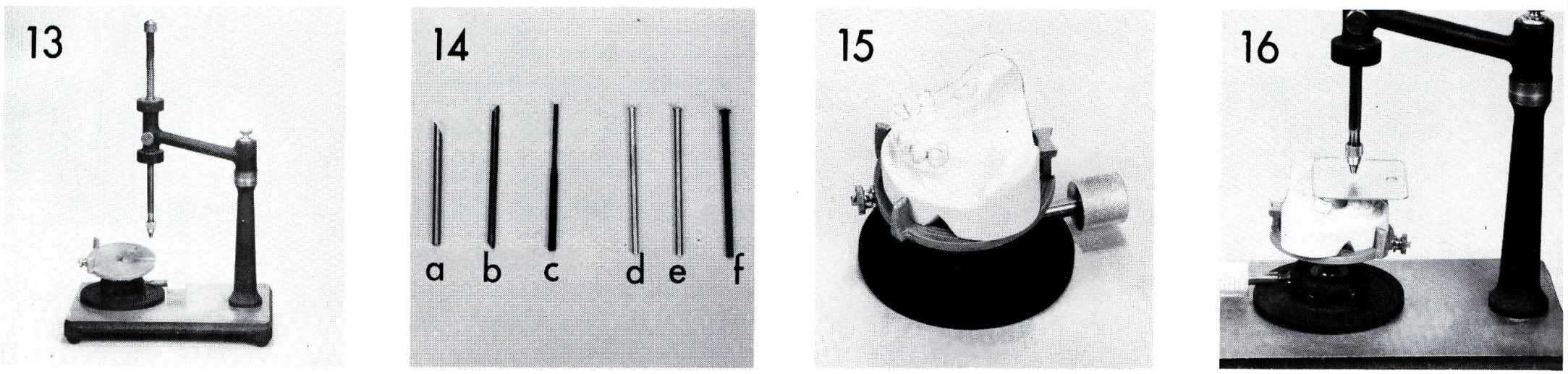
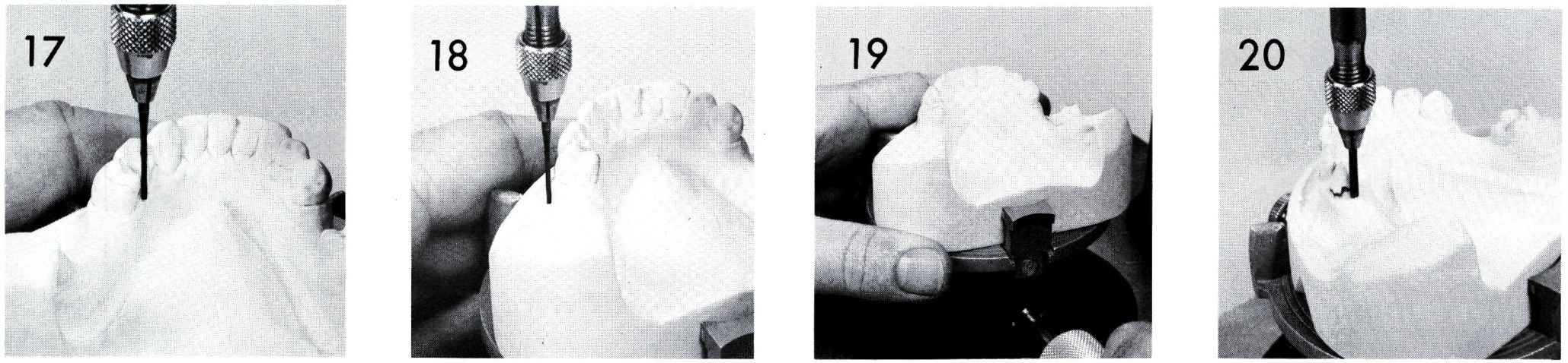
Figure 14 These are the tools which are used in a surveyor:
a is a dental bur which has been beveled and is used as a wax carver.
b is a carbon marker.
c is an analyzing rod which is used to determine undercut areas prior to scribing the height of contour with the carbon marker.
d, e and f are Ney undercut gauges: d is a .010 gauge,
e is a .020 gauge,
and f is a .030 gauge.
Figure 15 The preliminary cast is placed on the tilt-top surveying table.
Figure 16 The cast is positioned so that the occlusal plane is perpendicular to the vertical rod on the surveyor and is parallel to the surveyor base.
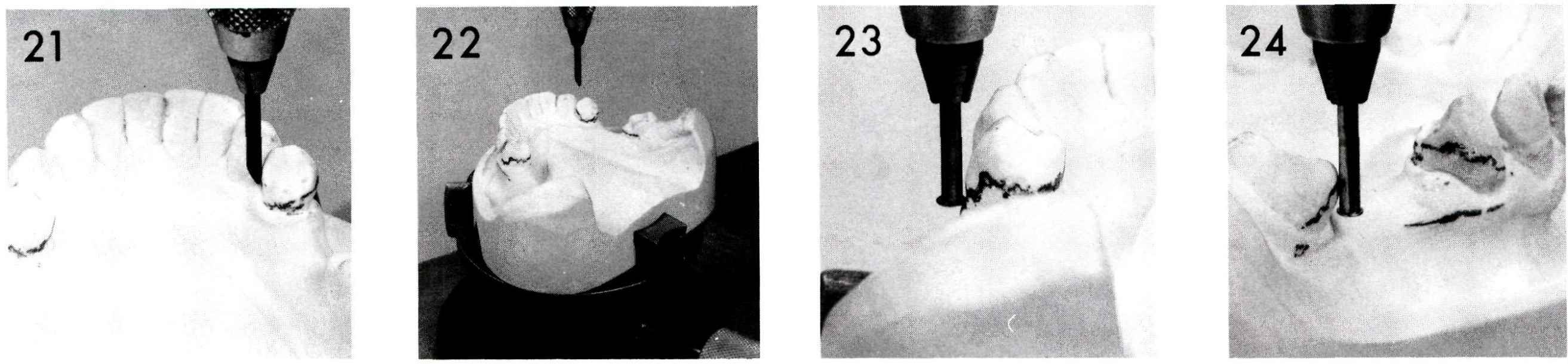
Figure 17 The analyzing rod is placed in the chuck on the surveyor and is used to check the contours of all teeth which may be involved in the partial denture.
Figure 18 Soft tissue undercuts which may influence the design of the partial denture are also checked.
Figure 19 The cast may be tipped slightly to improve undercut areas. It must be remembered that any undercut utilized for retention must be present when the occlusal plane is perpendicular to the vertical rod of the surveyor. Tilting is not often necessary.
Stay updated, free dental videos. Join our Telegram channel
VIDEdental - Online dental courses


