Chapter 6 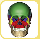 The Skull
1 Introduction
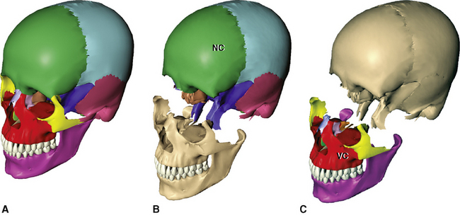
The skeleton of the head is a complex articulation of many bones and teeth, which are collectively referred to as the skull or cranium. On the basis of function the skull may be conveniently divided into two main areas: the (1) neurocranium and (2) the facial skeleton (Figure 6-1).
2 Views
FRONTAL VIEW
Bones
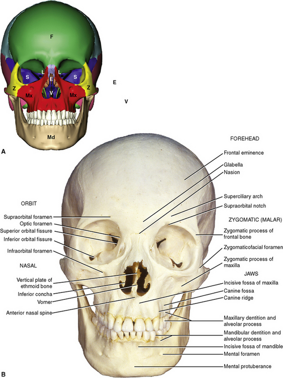
The bones evident in the anterior view (Figure 6-2, A) are the right and left maxillae, nasal bones, the right and left zygomatic bones, the right and left lacrimal bones, the right and left inferior conchae, the ethmoid bone, the vomer, the sphenoid bone, the frontal bone, and the mandible.
Features
Forehead Region
The forehead region is formed mainly by a portion of the frontal bone (Figure 6-2, B). The following features are found within the region of the forehead.
Orbital Area
The orbits contain the eyeballs and the extraocular muscles (see Figures 6-2 and 6-3). Several bones contribute to the margins (rims) and to the inner walls of the bony orbit.
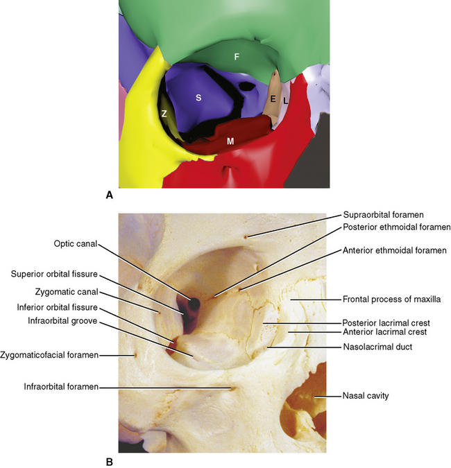
Orbital Openings
Nasal Region
The Jaws
LATERAL VIEW
Bones
The bones seen from the lateral view are the frontal bone (single), parietal bones (paired), temporal bones (paired), occipital bone (single), greater wings of the sphenoid bone (paired processes of a single bone), zygomatic bones (paired), maxillae (paired), nasal bones (paired), lacrimal bones (paired), and the mandible (single). (Figure 6-4, A).
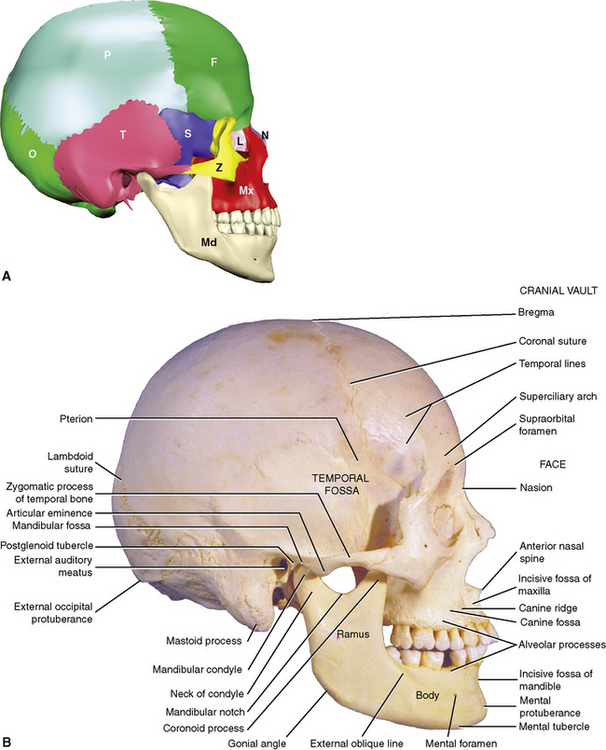
Features
The Cranial Vault Region
The Facial Region
The Infratemporal Region
The infratemporal region is obscured by the ramus of the mandible, which serves as the lateral wall of the region (Figure 6-5). With the mandible removed, the limits of the infratemporal region can be further delineated. The infratemporal region is separated from the temporal fossa above by an indistinct infratemporal crest.
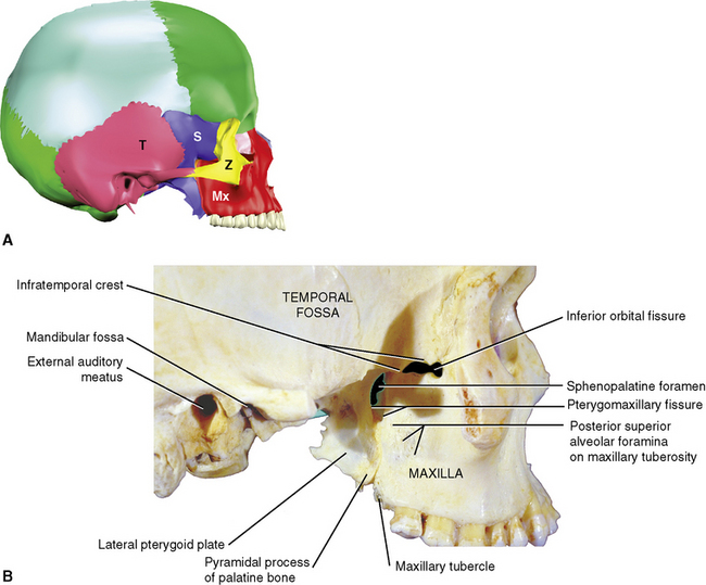
Figure 6-5 Lateral view of skull with mandible removed to expose infratemporal region. A, Bones. B, Features.
Bones and Walls
Features
SUPERIOR VIEW
Bones
The superior aspect of the skull presents a somewhat egg-shaped outline with the small end anteriorly (Figure 6-6). Only four bones are seen from this view: the frontal bone anteriorly, the right and left parietal bones laterally, and the occipital bone posteriorly.
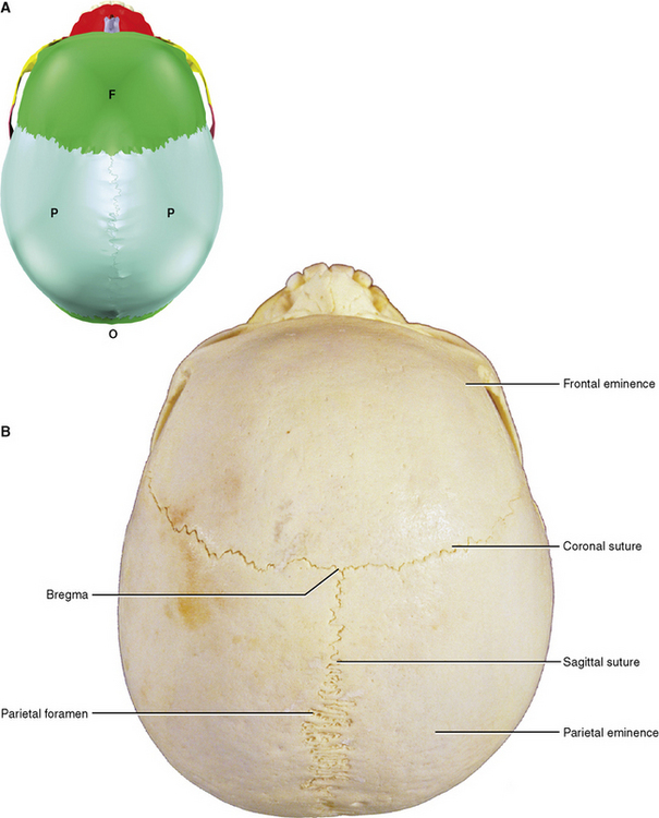
Features
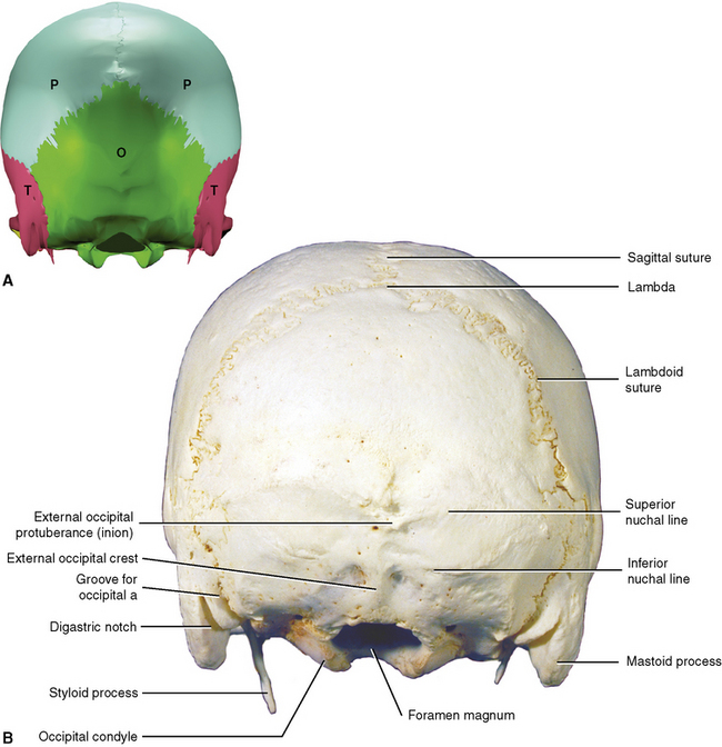
POSTERIOR VIEW
The most prominent feature of the posterior view is the rounded posterior pole of the skull, called the occiput (see Figure 6-7). Hence, the area is often referred to as the occipital area.
Features
BASAL OR INFERIOR VIEW
Bones
The bones seen from the basal aspect (Figure 6-8, A) are the right and left maxillae (palatal processes), the right and left palatine bones (palatal processes), the sphenoid bone (body, pterygoid processes, and greater wings), the vomer, the right and left temporal bones, and the occipital bone.
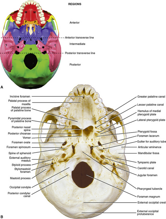
Features
Anterior Region
Intermediate Area
From lateral to medial, the features encountered in the intermediate area are as follows (see Figures 6-8 and 6-9):
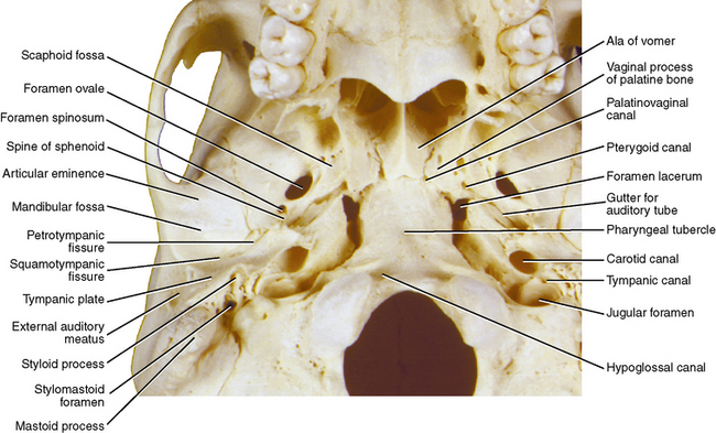
Structures Straddling the Posterior Line
From lateral to medial the features crossed by the posterior transverse line are as follows:
INTERNAL ASPECT OF THE BASE
To expose the internal features of the skull, the skullcap (calvaria) is sawn off and removed.
Bones
The bones seen in this view are the frontal bone, the ethmoid bone, the sphenoid bone (including the body, lesser wings, and greater wings), the right and left temporal bones, and the occipital bone (Figure 6-10, A).
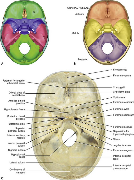
Regions
The internal configuration of the base of the skull is conveniently tiered and named as three cranial fossae (Figure 6-10, B):
Features (Figures 6-10,C and 6-11)
Middle Cranial Fossa
Features of the middle cranial fossa include the following:
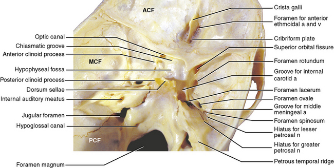
Posterior Cranial Fossa
Stay updated, free dental videos. Join our Telegram channel
VIDEdental - Online dental courses


