Anomalies of the Developing Dentition
Avariety of dental anomalies are associated with defects in tooth development precipitated by hereditary, systemic, traumatic, or local factors. Numerous systems have been used to classify dental anomalies, each with merit. The one used in this text categorizes them in terms of abnormalities in tooth number, size, shape, structure, and color.1 The advantage of this system is that the categories can be related to the stages of tooth development in which the respective anomalies are thought to originate. These stages of dental development are discussed in Chapter 12. The reader is also encouraged to review textbooks on dental histology, dental embryology, and orofacial genetics for more in-depth information.
Anomalies of Number
Alterations in tooth number result from problems during the initiation or dental lamina stage of dental development. In addition to hereditary patterns producing extra or missing teeth, physical disruption of the dental lamina, overactive dental lamina, and failure of dental lamina induction by ectomesenchyme are several examples of etiologic factors that affect tooth number.1
Hyperdontia
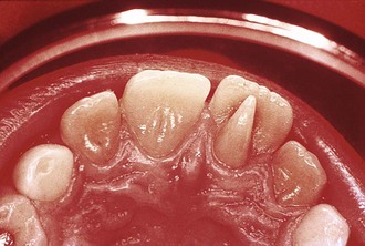
Hyperdontia and supernumerary teeth are terms describing an excess in tooth number that can occur in both the primary and permanent dentitions. Reports on the incidence of hyperdontia include values as high as 3%, with males being affected twice as frequently as females.2 Ninety percent to 98% of supernumerary teeth occur in the maxilla, with the permanent dentition being more frequently affected than the primary dentition. The most common supernumerary tooth is the mesiodens, which occurs in the palatal midline and can assume a number of shapes and positions relative to the adjacent teeth. The majority tend to be located palatal to the central incisors.3
As reported by Primosch in 1981, supernumerary teeth are morphologically classified as either supplemental or rudimentary.2 Supplemental supernumerary teeth (Figure 3-1, A) duplicate the typical anatomy of posterior and anterior teeth. Rudimentary supernumerary teeth are dysmorphic and can assume conical forms, tuberculate forms (Figure 3-1, B), or shapes that duplicate molar anatomy. From a clinical standpoint, the tuberculate, or barrel-shaped, supernumeraries generate the most severe complications with respect to difficulty of removal and adverse effects on adjacent teeth, such as impaction or ectopic eruption. Additional complications associated with supernumeraries include dentigerous cyst formation, pericoronal space ossification, and crown resorption.3 It is important in supernumerary tooth detection to rule out the presence of odontoma in light of the fact that the morphologic characteristics of a compound odontoma are similar to those of supernumerary teeth.
Cleft lip and palate (CLP) commonly demonstrates an excess or deficiency in the normal complement of teeth and provides a clear example of physical disruption of the dental lamina as an etiologic factor. Patients with CLP have been reported to have an increased incidence of hyperdontia, with reports indicating an incidence of up to 5%.4 Classic syndromes involving supernumerary teeth are summarized in Table 3-1; cleidocranial dysplasia has the highest association with this dental anomaly.
 TABLE 3-1
Syndromes Demonstrating Supernumerary Teeth
| Condition | Characteristics |
| Apert syndrome | Scaphocephaly, craniosynostosis, bilateral syndactyly, midface hypoplasia |
| Cleidocranial dysplasia | Aplastic clavicles, frontal bossing, hypoplastic midface |
| Gardner syndrome | Osteomas, epidermoid cysts, odontomas, intestinal polyps |
| Down syndrome | Brachycephaly, mental retardation, epicanthal folds |
| Crouzon disease | Craniosynostosis, exophthalmos, hypoplastic midface |
| Sturge-Weber syndrome | Angiomatosis and calcification of leptomeninges, seizures, port-wine nevi of face |
| Oral-facial-digital syndrome | Hypoplastic alar cartilage, cleft tongue, clinodactyly |
| Hallermann-Streiff syndrome | Dyscephaly, mandibular hypoplasia, hypotrichosis |
Hypodontia
Hypodontia, or congenital tooth absence, is a deficiency in tooth number. Familial heredity patterns account for the largest etiologic correlation with patterns of hypodontia. Incidence reports identify a range of 1.5% to 10% excluding third molars in U.S. populations.5 The most frequently occurring congenitally absent permanent tooth, excluding the third molar, tends to be the mandibular second bicuspid (3.4%), followed by the maxillary lateral incisor (2.2%).6
There is a high correlation between primary tooth absence and permanent tooth absence.7–9 Ectodermal dysplasia (ED) represents a group of classic syndromes characterized by oligodontia or multiple congenitally missing teeth. The most common is hypohidrotic ED, followed in descending frequency of occurrence by hidrotic ED, ectrodactyly ED plus cleft lip and palate, Rapp-Hodgkin ED, and Robinson-type ED (Figure 3-2). Other conditions involving hypodontia are summarized in Table 3-2 and those involving both hyperdontia and hypodontia in Box 3-1.
 TABLE 3-2
Syndromes Demonstrating Hypodontia
| Condition | Characteristics |
| Ectodermal dysplasia (hypohidrotic type) | Hypotrichosis, aplasia of sweat/sebaceous glands |
| Chondroectodermal dysplasia | Polydactyly, mesomelic dwarfism, hidrotic ectodermal dysplasia |
| Achondroplasia | Short-limbed dwarfism, macrocephaly, frontal bossing |
| Rieger syndrome | Iris dysplasia, midface hypoplasia, protruding umbilicus |
| Incontinentia pigmenti | Alopecia, pigmented macules, mental retardation |
| Seckel syndrome | Dwarfism, microcephaly, facial hypoplasia, low-set lobeless ears |
Anomalies of Size
Microdontia and Macrodontia
Facial hemihypertrophy can demonstrate comparatively larger teeth on the affected side (Figure 3-3). Of the many factors thought to cause this condition, vascular and neurogenic abnormalities are considered the most likely. In addition to an increase in crown and root size, affected teeth develop more rapidly and erupt earlier than on the uninvolved side. The otodental syndrome (also known as otodental dysplasia), consisting of high-frequency hearing loss and globe-shaped fused molar teeth, is another condition that involves macrodontia. Isolated teeth with macrodontia can also result from twinning abnormalities that originate during the proliferation stage of tooth development. Fusion and gemination are the most common twinning abnormalities, and both include enlarged crowns.
Fusion
Fusion has an incidence of 0.5% and is more common in the primary dentition.7 The classic definition of fusion is the dentinal union of two embryologically developing teeth. Although fused teeth can contain two separate pulp chambers, many appear as large bifid crowns with one chamber, which makes them difficult to distinguish from geminated teeth.
Anomalies of Shape
Dens Evaginatus
Dens evaginatus is an extra cusp, usually in the central groove or ridge of a posterior tooth and in the cingulum area of the central and lateral incisors (Figure 3-4). In incisors, these cusps appear talon-shaped and can approach the level of the incisal edge. This extra portion contains not only enamel but dentin and pulp tissue; therefore a pulp exposure can result from radical equilibration. It occurs with a frequency of 1% to 4% and results from the invagination of inner enamel epithelium cells, which are the precursors of ameloblasts.1
Stay updated, free dental videos. Join our Telegram channel

VIDEdental - Online dental courses


 Outline
 FIGURE 3-1
 Box 3-1
 FIGURE 3-2
 FIGURE 3-3
 FIGURE 3-4