(1)
Canberra, ACT, Australia
Summary
The decision between tooth retention using endodontic treatment and crown restoration and extraction and prosthodontic replacement is a common dilemma encountered in clinical practice. Correct treatment planning involves establishing whether the tooth in question is restorable and periodontally sound, the patient is able to tolerate the proposed treatment and the clinician has the skills required to perform the necessary treatment.
Clinical Relevance
In everyday practice the clinician along with the patient must weigh up the option of saving teeth against extraction and alternative prosthodontic replacement options that may be more predictable. A compromised tooth troubled by a combination of endodontic, periodontal and restorative problems must be carefully assessed from a risk/benefit point of view not only in terms of outcome but also costs involved.
8.1 Treatment Planning and the Decision-Making Process
The goal of endodontic treatment, re-treatment or endodontic surgery is to preserve the tooth as a functioning unit of dentition. Alternative prosthodontic replacement options following extraction, including dentures, bridges or implants, are selected for the same reasons and more often than not when the tooth is compromised (Fig. 8.1). Clinicians are often faced with the challenge of informed decisions as to which option best serves the patient. Judgements must be made not only on clinical experience but also prognostic factors that determine long-term success of the various options. Careful evaluation of all factors must be made, and occasionally a multidisciplinary approach including the endodontist, prosthodontist and/or periodontist may be required before the final decision is reached.
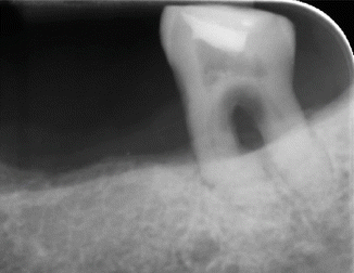
Fig. 8.1
Pre-operative parallel peri-apical radiograph of tooth 36 which was referred for endodontic management. Note moderate – severe periodontal disease (50–70 % horizontal bone loss), furcation involvement (grade III) and gross furcal caries associated with the tooth. This tooth has a poor long term prognosis and is not a candidate for “herodontics”
Having taken a case history and carried out all relevant diagnostic tests, the patient’s treatment can then be planned. The type of endodontic treatment chosen must take into account the patient’s medical condition and general dental status. Contraindications to treatment include local factors such as an unrestorable tooth, insufficient periodontal support, nonstrategic tooth, root fractures and extensive internal/external root resorption [1].
The decision-making process to decide whether the tooth is salvageable or whether extraction is preferred should be based on a logical sequence of decision-making. As such the endodontic treatment must be integrated into a comprehensive treatment plan that includes restorative and periodontal management. The difficulty of the case should be balanced with the skill and experience of the dentist in deciding whether to manage the case in general practice or to refer the patient to an endodontist. The overall treatment planning in endodontics should be in agreement with the overall dental management of the patient [2].
Overall success in root canal treatment has often been based on a strict set of criteria defining success as an asymptomatic tooth with normal periodontal architecture at the peri-apex, bony infill and the absence of infection [3]. Systematic reviews using clinical and radiographic measures of peri-apical healing revealed that the estimated weighted pooled success rates of primary root canal treatment completed at least 1 year prior to review ranged between 68 and 85 % when strict criteria (complete absence of peri-apical radiolucency) were used. The equivalent estimated weighted pooled success rates of root canal re-treatment ranged between 70 and 86 %. The reported success rates for both treatments had not improved over the last five decades. Four conditions were found to be significantly associated with better peri-apical healing following treatment. These included the absence of a peri-apical lesion, a root filling with no voids, a root filling within 2 mm of the radiographic apex and a satisfactory coronal restoration [4–6].
A further prospective study revealed that the success rate of 1° root canal treatment with complete healing occurred in 83 and 80 % after 2° root canal re-treatments. Four preoperative factors (the presence of a preoperative peri-apical lesion, the size of peri-apical lesion, the presence of a preoperative sinus and the presence of a pre-existing perforation), six intraoperative factors (patency at apical foramen, apical extent of instrumentation, additional use of chlorhexidine for irrigation, additional use of EDTA as an irrigant, the occurrence of interappointment complications (swelling or pain) and apical extent of root filling) and one postoperative factor (quality of coronal restoration) were found to be significant prognostic indicators for the success of 1° root canal treatment and 2° root canal re-treatments. Those roots with a preoperative peri-apical lesion were significantly associated with 49 % lower odds of success than roots without a lesion. The odds of success of treatment were found to decrease by 14 % for every 1 mm increase in the diameter of the preoperative lesion. The presence of a preoperative sinus or root perforation significantly reduced the odds of success by 48 and 56 %, respectively. During treatment, achieving technical patency at the canal terminus significantly increased the odds of success twofold, whereas the odds of success were reduced by 12 % for every 1 mm of the canal short of the terminus remaining ‘un-instrumented’. In contrast, a long root filling reduced the odds of success by 62 %. The use of 0.2 % chlorhexidine in addition to sodium hypochlorite solution for canal irrigation did not improve but reduced the odds of success by 53 %. Interestingly, the additional use of EDTA solution for canal irrigation had no significant effect on the success of 1°RCTx but significantly increased the odds of success of 2°RCTx by twofold. The occurrence of inter-appointment complications (swelling or pain) reduced the odds of success by 47 %. Finally, a good-quality coronal restoration significantly increased the odds of success by 11-fold [7, 8].
Surgical endodontics is primarily indicated and related to difficultly in access for conventional nonsurgical treatment or retreatment or where access to the peri-apical area is necessary to aid diagnosis, for example, for biopsy or to identify a possible root fracture/perforation. Limited evidence suggests that although surgery may offer a more favourable outcome in the short term, nonsurgical retreatment appears to offer a better long-term result. Nonsurgical retreatment may provide a better opportunity to clean the pulp space over a surgical approach and therefore remains the preferred treatment option where appropriate [9, 10].
Coupled with magnification through the use of the surgical operating microscope, refined principles of soft and hard tissue management, the use of tissue regenerative root-end filling materials and enhanced principles of wound closure and postoperative management, surgical endodontics has emerged as a highly predictable and relatively painless procedure with success rates of 94 % compared to 59 % using traditional techniques [11–14]. The outcome of repeat endodontic surgery was less favourable than that of primary endodontic surgery for posttreatment disease [15].
The long-term prognosis of an endodontically treated teeth and whether it is extracted depends on the health of the periodontium. Periodontal diseases are diagnosed on the basis of clinical signs, with radiographs assisting in treatment planning decision. The full diagnostic approach relies on periodontal probing and the response to probing [16]. Careful assessment and attempt to distinguish between true periodontal lesions, a lesion of endodontic origin, a combined periodontal-endodontic lesion or vertical root fracture is essential from both a management and prognostic point of view [17–19].
The completion of root canal treatment does not signal the end of patient management. The endodontically treated tooth needs to be restored back to form, function and aesthetics. It is clear from the literature that a well-constructed coronal cuspal coverage restoration serves to protect both the weakened tooth from catastrophic fracture and also to prevent re-infection from the oral microbiota. In one sample study of 280 patients and 400 teeth, those that were not crowned after root canal treatment were lost at a six times greater rate than those that had been crowned. A restorability assessment is therefore imperative before embarking upon root canal therapy and a clinical judgement must be made including whether additional crown lengthening procedures are indicated to attain a suitable ferrule [5, 8, 20–24].
A multitude of viable treatment options are available to the clinician and patient following the loss of a tooth. From a cost analysis point of view following extraction the option of doing nothing may be beneficial to the patient. Nevertheless, the patient must be made aware of the fact that failure to maintain the space may result in tooth positional changes such as supraeruption, tilting, rotation and lateral movement of the opposing tooth [25–27].
The decision to treat a tooth endodontically or to extract and replace with either conventional removal partial dentures or bridgework or to place a single-tooth implant should be based on criteria including prosthetic restorability of the tooth, quality of bone, aesthetic demands, cost–benefit ratio, systemic factors, potential for adverse effects and patient preferences. Endodontic treatment of teeth represents a feasible, practical and economical way to preserve function in a vast array of cases and dental implants serve as a good alternative in selected cases in which the prognosis is poor [28–31].
Whilst treatment outcomes of root canal treatment have been based upon strict criteria manifested by clinical and radiographic signs of healing, this has not been the case with implant studies. Instead survival rates have often been used not giving a true representation of the failures inherent with implants [31]. Recent advancement in implantology techniques has brought about a useful option in the management of severely compromised teeth troubled by a combination of endodontic, periodontal and restorative problems. Contraindications to implant placement include psychosis, acute infectious disease and pregnancy and cancer therapy. The risk of bisphosphonate-induced osteonecrosis has been associated with patients on systemic bisphosphonate therapy. Caution must be taken in patients with diabetes and other co-morbidities such as poor oral hygiene and smoking. Implants should be only placed when cranial growth is complete. In the anterior region single tooth implants should only be considered after the age of 25 [31–35].
Implant failure can occur as a result of the implant’s failure to integrate with the bone or bone loss subsequent to integration. Soft tissue complications such as inflammation and/or proliferation, soft tissue fenestration and/or dehiscence and fistulas have been reported. Mechanical complications such as screw loosening, screw fracture, prosthesis fracture and implant fracture also can occur. Perimucositis and periimplantitis are not uncommon following implant placement and if left untreated will result in subsequent bone loss and eventual failure [35, 36].
Patients now face a number of choices regarding the treatment of individual teeth. A tooth can be retained with root canal treatment and subsequent restoration or may be extracted and not replaced, replaced with a bridge, a removable denture or an implant. Dentists have a duty to provide a comprehensive review of the benefits and risks of each option and to ensure that the patient’s needs are addressed with the goal of improving oral and general health [37].
8.2 Peri-Apical Pathology
The presence of peri-apical pathology is sometimes the first sign that pulpal disease has occurred. The vast majority of teeth with peri-apical infection are expected to heal following routine nonsurgical root canal treatment or re-treatment. Occasionally surgical exploration/treatment may be indicated, but this is reserved for those cases that do not respond to conventional treatments (Fig. 8.2).
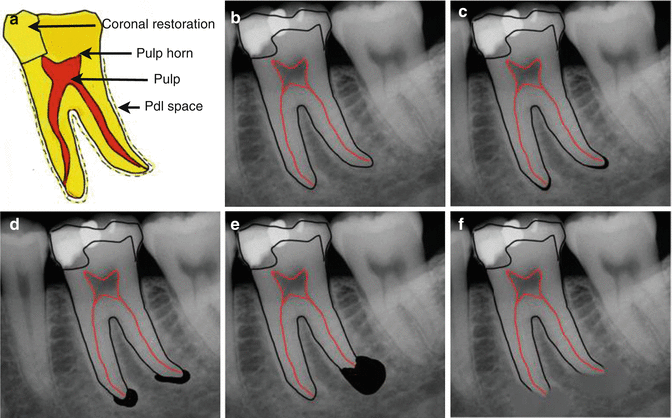
Fig. 8.2
Diagrams representing (a) extensive caries involving the pulp. (b) Normal radiographic appearance – intact periodontal ligament space. (c) Early apical changes – widening of the radiolucent periodontal ligament space. (d) Extensive destructive acute inflammation – diffuse, ill-defined area of radiolucency at the apex. (e) Long-standing chronic inflammation – well-defined area of radiolucency surrounded by dense sclerotic bone. (f) Low-grade chronic inflammation – diffuse radiopaque area at the apex (sclerosing condensing osteitis)
Evidence suggests that those teeth with peri-radicular radiolucencies present preoperatively have a poor prognosis compared to teeth with no lesions in terms of success. The prognosis for endodontic treatment of the tooth in question must be taken into account in the treatment planning phase. Classical studies have shown endodontic success to be 95 % with a vital tooth, 85 % with a tooth with peri-apical pathology, 80 % with re-treatment cases and 65 % with contemporary surgery. Clinicians should be reminded that studies reporting an overall success rate may not be applicable to the same tooth that you are treating and may be misleading to the patient. Nevertheless, if the standard of care for root canal treatment and subsequent coronal restoration is carried out methodically, then success should be achievable in the majority of cases.
8.3 Periodontal Assessment
The health of the periodontium needs to be assessed, and insufficient periodontal support may be a contraindication to carrying out root canal therapy (Fig. 8.3).
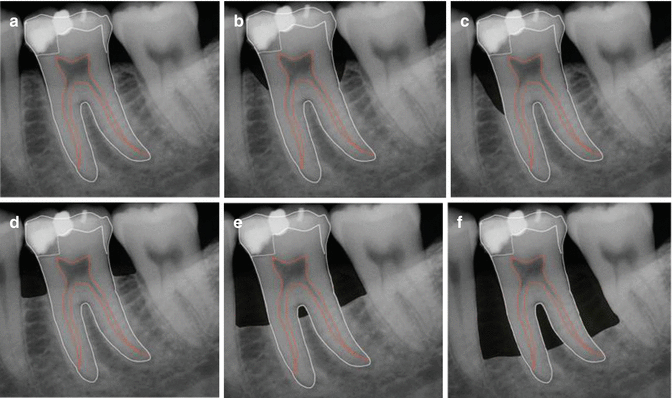
Fig. 8.3
Diagrams representing (a) healthy (b) localised bilateral infrabony vertical defects – typically seen in occlusal trauma, (c) localised vertical bone loss, (d) mild horizontal bone loss, (e) moderate horizontal bone loss with early furcation involvement, (f) severe horizontal bone loss
Current periodontal classification recognises the following categories of destructive periodontal diseases: chronic periodontitis, aggressive periodontitis, periodontitis as a manifestation of systemic disease, necrotising ulcerative gingivitis/periodontitis, abscesses of the periodontium (gingival, periodontal, pericoronal), combined periodontic–endodontic lesions and developmental or acquired deformities and conditions.
A visual inspection of the marginal periodontal tissues, plaque, calculus, overhangs, gingival bleeding and bleeding on probing, basic periodontal examination, probing depth and loss of attachment measurements, furcation involvement, mobility, suppuration, tooth drifting/migration and radiographic assessment are all necessary in order to assist in treatment planning decisions regarding root canal therapy on a prospective tooth. Those cases where a questionable prognosis is suspected may require specialist referral prior to further determine long-term prognosis. Certainly in cases where there is no doubt as to an unfavourable (poor) periodontal prognosis, then extraction will be the only option.
Stay updated, free dental videos. Join our Telegram channel
VIDEdental - Online dental courses


