Fig. 11.1
Clinical practice guidelines for correction of diastema
11.2 Orthodontic Closure of Diastema
Orthodontic closure of an anterior diastema can be accomplished either by mesiodistal and/or anteroposterior movement of the teeth. If the teeth are proclined and the overjet is increased, retraction of the incisors will automatically close the anterior spacing (Fig. 11.2). If the teeth are not protruded but laterally migrated (e.g. when the maxillary lateral incisors are congenitally missing), mesially directed forces will bring the teeth together to close a median diastema. In cases where the buccolingual position of the teeth and lips should be maintained, anterior movement of the posterior teeth would be preferred (Fig. 11.3). The decision of which orthodontic mechanics to use in order to obtain these forces and moments largely depends on the amount and localization of the diastema, the age and dentitional stage of the patient, inclinations and angulations of the teeth and the presence of an adequate overjet.
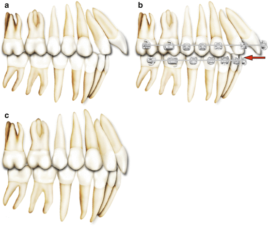
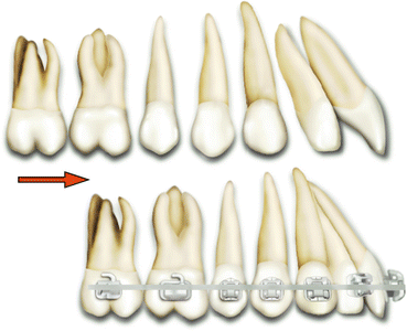
Fig. 11.2
(a–c) Diagrammatic illustration of anterior diastema closure with retraction of the flared maxillary incisors
Fig. 11.3
Diagrammatic illustration of generalised diastema closure with mesialisation of the posterior teeth when the retraction of the incisors is contraindicated
Key Note
The evaluation of overjet is important in diastema cases. If the overjet is reduced, diastema cannot be closed by palatal tipping of the maxillary incisors.
Minor diastema (less than 2 mm) caused by distal crown angulation of the teeth can easily be corrected by tipping movement with removable appliances in an adolescent patient (Fig. 11.4). The finger springs of a removable Hawley appliance create mesially directed tipping forces to bring the teeth together. As an alternative, clear plastic appliances and rubber bands can also be used to close median diastema [5]. It is important to note though whenever possible, mechanics enabling three-dimensional tooth control should be chosen especially in complex cases (e.g. large median diastema, generalised spacings, deepbite, skeletal problems, microdontia, hypodontia). Therefore, active orthodontic treatment to close a diastema is preferably achieved by bodily tooth movement generated by fixed appliances (Fig. 11.5). For this purpose, anterior segmental archwires, 2 × 4 appliances (extending from first molars to incisors) or continuous archwires can be used (Fig. 11.6). Stiff and rectangular archwires provide good control of tooth movement during space closure.
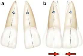
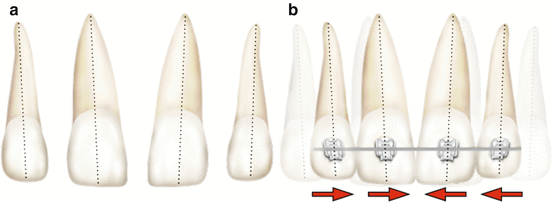
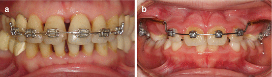
Fig. 11.4
(a) Distally tipped maxillary central incisors and minor median diastema. (b) Closure of the median diastema by mesial tipping of the incisors (note the final upright position of the teeth)
Fig. 11.5
(a) Generalised anterior diastemas. (b) Closure of the diastemas by mesially directed bodily tooth movement using fixed orthodontic appliances
Fig. 11.6
The use of anterior segmental archwire (a) and 2 × 4 mechanics (b) for orthodontic treatment of maxillary diastemas
Key Note
Never place elastics around the teeth to close a diastema without the use of orthodontic appliances. Uncontrolled subgingival dislocation of the elastics may cause severe periodontal breakdown.
11.2.1 Case 1: Maxillary Midline Diastema
The patient in Fig. 11.7 had a maxillary midline diastema in mixed dentition. She was complaining about the unpleasant appearance of the spacings and irregularities. Although the physiological process was explained, she preferred not to wait for the eruption of the canines for psychological reasons. A removable appliance with finger springs was used to close the median diastema. Once the median diastema is closed, the finger spring on the left lateral incisor will be activated. Although the left lateral incisor’s medial movement is indicated, the right lateral incisor’s mesial tipping is contraindicated because the cusps of the erupting canines are in close proximity with the roots of the lateral incisors at the early stages of dental development. Tipping the mesially inclined crowns of the lateral incisors in the ugly duckling stage will result in distal movement of the roots into the eruption path of the developing canines; thus, early orthodontic treatment may create risk of root resorption or canine impaction. Bishara [6] suggested to begin orthodontics after the cusp tips of the erupting canines have passed the apical third of the root of the lateral incisors. Therefore, orthodontic intervention should be avoided in the early mixed dentition period. The exceptions are midline diastema larger than 4 mm which may complicate eruption of the lateral incisors, presence of a midline pathology (mesiodens, odontome etc.) or bad oral habits.
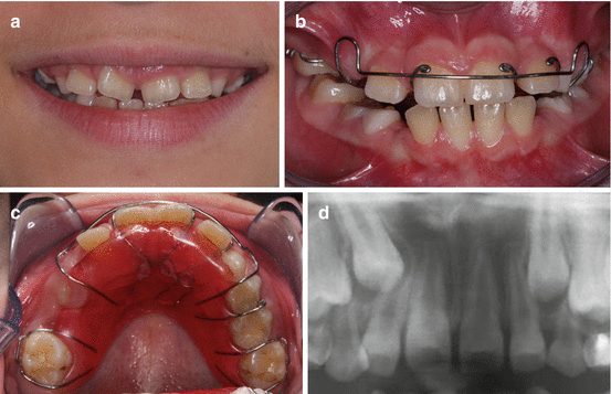
Fig. 11.7
(a) Mixed dentition patient with maxillary midline diastema. (b, c) A removable appliance with finger springs was used to close the median diastema. (d) Note the close proximity of the erupting right maxillary canine with the root of the lateral incisor. Because of the risk of root resorption, mesial tipping of the lateral incisor was avoided
11.2.2 Case 2: Generalised Diastemas
A 12-year-old boy, at the end of mixed dentition stage, presented with proclined maxillary and mandibular incisors and generalised spacings (Fig. 11.8a–d). Although the maxillary canines had not fully erupted, the tooth-arch dimension analysis (Hays Nance analysis, see Chap. 8) revealed excess spacing at permanent dentition. The maxillary and mandibular spacings were closed using fixed orthodontic appliances with elastic chains and a consolidation arch aiming to control root divergence and torque (Fig. 11.8e–g).
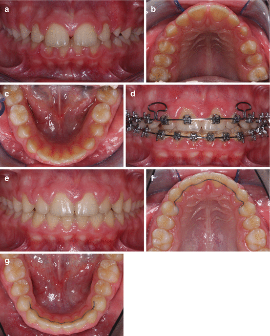
Fig. 11.8
(a–c) Frontal and occlusal intraoral views of generalised spacing before orthodontic treatment. (d) Closing the spaces with a consolidation arch in the maxillary dental arch and elastic chains in the mandibular dental arch during fixed orthodontic treatment. Headgear and intermaxillary elastics were used to reinforce the posterior anchorage. (e–g) Frontal and occlusal intraoral views after orthodontic space closure. Fixed lingual retainers were bonded to each tooth from right first premolar to left first premolar in the maxillary and mandibular dental arches following orthodontic treatment of polydiastema to prevent relapse
Key Note
In general, excessive proclination of the teeth generates spacing, whereas retraction of the teeth to their neutral position helps to close the diastemas. However, elimination of the aetiological factor is essential.
11.3 Management of Diastema Due to Abnormal Oral Habits: Breaking the Habit
The teeth are in equilibrium between forces generated by muscles, mastication and stabilisation of the periodontium [7]. If there is an alteration in the equilibrium due to an abnormal oral habit such as finger sucking, dental movement is likely to occur resulting in spacing in the dental arches in addition to other malocclusions. If the abnormal habit is stopped before the eruption of permanent teeth, normal cheek and lip pressures can establish the equilibrium and self-correction of the displaced teeth. However, if it persists in mixed dentition, orthodontic treatment may be required. In general, when the parafunctional cause is eliminated, a spontaneous correction will be observed [8]. There are several methods to stop abnormal oral habits. In orthodontics, myofunctional therapy and the use of habit breakers are very effective as a complementary to a psychological approach [8, 9]. After the patient has stopped the pernicious habit, wearing of the appliance is recommended for an additional several months.
11.3.1 Case 3: Lower Lip Sucking and Maxillary Spacing
An 11-year-old female patient presented with generalised maxillary spacing of 7 mm (Fig. 11.9a, b). Her history revealed that she was sucking her lower lip in her sleep throughout the night (Fig. 11.9c). Lower lip sucking habit caused proclination of the maxillary incisors which could not have been opposed by the forces of the upper lip. The aim was to normalise the extraoral muscle force and establish the equilibrium. A lip bumper appliance was used to break the sucking habit (Fig. 11.9d). This prefabricated appliance was placed at the level of the gingiva 2–3 mm in front of the lower incisors and 4–5 mm away from the buccal segments and was fixed to the molar tubes. It was reactivated when necessary. After 3 months of lip bumper therapy (full-time wear), the lower lip sucking habit was stopped, maxillary incisors spontaneously retroclined due to elimination of increased forces generated by sucking, and thus, maxillary diastemas were reduced (Fig. 11.9e, f). The residual diastemas were closed with fixed orthodontic therapy (Fig. 11.9g, h).
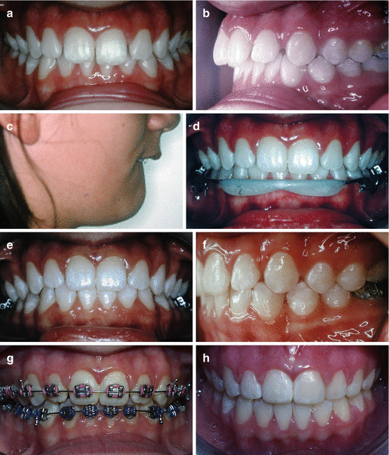
Fig. 11.9
(a, b) Generalised maxillary spacing and increased overjet. (c) Lower lip sucking exerting abnormal and unopposed force causing flaring of the upper teeth. (d) Use of lip bumper appliance to break the sucking habit. (e, f) Spontaneous reduction of maxillary spacing due to normalised muscle forces shows retroclination of the maxillary incisors after 3 months of lip bumper therapy. (g) Fixed orthodontic therapy phase. (h) Correction of the malocclusion at the end of orthodontic treatment (From Germeç and Taner [8]. Reprinted with permission from Angle Orthodontist)
11.3.2 Case 4: Finger Sucking and Maxillary Spacing
A female patient in early transitional dentition having a finger sucking habit showed increased overjet, labial inclination of the incisors and maxillary spacing (beyond physiologic diastema of mixed dentition) particularly on the right side where she inserted her right finger to suck (Fig. 11.10a–d). A myofunctional appliance was used for 3 months (only at night) to stop finger sucking habit (Fig. 11.10e). This soft appliance with vestibular screens broke the finger sucking habit and thus eliminated the abnormal pressure caused by the finger sucking and established a balance between intraoral and extraoral muscles. In addition, it generated palatally directed forces to the overproclined right maxillary incisors. The final result was decreased maxillary spacing. At the end of myofunctional therapy, the maxillary spacing was reduced but not totally eliminated (ideal for a transitional period, see Chap. 2), and the overproclined incisors and the increased overjet were corrected (Fig. 11.10f–h).
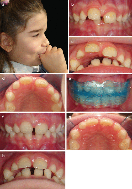
Fig. 11.10
(a–d) A 7.5-year-old patient with finger sucking habit, placing her finger on the right side of the dentition, leading to proclination of the incisor, increased diastema and overjet. (e) Soft myofunctional appliance was used to stop the habit and worn during sleep (when she used to suck her finger). (f–h) Intraoral photographs showing decrease in spacing, correction of overjet and retroclination of the incisors after 3 months of myofunctional therapy (From Tozlu and Germeç [9]. Reprint permission from 7 Tepe Klinik)
11.4 The Role of Orthodontics in the Interdisciplinary Management of Diastema Due to Tooth Size Discrepancies and Missing Teeth
Diastemas caused by tooth size discrepancies can generally be treated by restorations. This treatment approach offers a quick solution and is readily accepted by patients. However, sometimes it is not possible to correct tooth size discrepancy with restorations alone due to unfavourable tooth positions and malocclusions (Fig. 11.11). Orthodontics helps to solve these problems by tooth movement, properly positioning the teeth and enabling an infrastructure for aesthetic restorations.
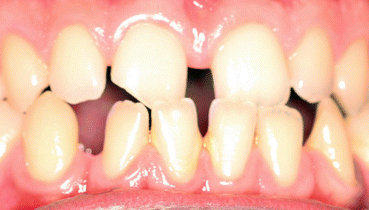
Fig. 11.11
A maxillary and mandibular generalised spacing case due to small teeth and large alveolar base. Restorative approach without orthodontics for this case presenting a Class III malocclusion with anterior crossbite, a midline diastema of 3 mm, disproportionate localisation of the maxillary and mandibular spacings between teeth may result in compromised aesthetics and impede the survival of the restorations
Interdisciplinary treatment starts with collecting accurate data, analysing it and composing a list of problems. Subsequently a treatment with alternatives is planned and discussed in the interdisciplinary team considering both treatment objectives and the patient’s needs, demands and expectations. Once the final treatment plan is agreed and upon approval of the patient, every discipline works in collaboration to achieve the stated goals. This collaborative team work has some important steps. A crucial step of interdisciplinary treatment in diastema cases is to determine the final positions of the teeth and redistribute the spaces with orthodontics, which is guided by the principles of proportion and occlusion. The tooth size should be in harmony with the adjacent teeth and dental arch. The clinician can use the tooth proportions (e.g. width/length ratio, relationship between the sizes of the adjacent teeth) and tooth size analysis (see Chap. 8) as a guide when redistributing the spaces between the teeth. A diagnostic wax set-up is also very helpful to visualise the final result (see Chap. 4
Stay updated, free dental videos. Join our Telegram channel
VIDEdental - Online dental courses


