Fig. 9.1
Single Miller type II recession, treated with connective tissue grafting before (a) with a follow-up of 15 years (b)
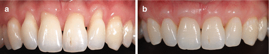
Fig. 9.2
Multiple Miller type III recessions treated with connective tissue graft before (a) and 2 years after (b). Note the changes of the interdental soft tissue. Increasing the tissue volume at the interdental space does contribute to tissue fill without restorative intervention in the presence of contact between teeth and absence of periodontal disease
Presence of a diastema results with missing interdental tissue; hence, restorative procedures to augment the diastema should also require soft tissue management. Since the interdental papilla is a small rounded protuberance in between two teeth, two implants, or a tooth and an implant or a pontic and a tooth or implant, management of any diastema consequently requires soft tissue management. Furthermore, management of the diastema may also change the mesiodistal dimensions of the clinical crown resulting in a discrepancy at the location of the zenith points (Figs. 9.3 and 9.4).
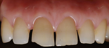
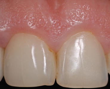
Fig. 9.3
Treatment plan of diastemas may include soft tissue management depending on the extent of proportional change in the clinical crown diameters. Cases with thin marginal gingiva with apparent zenith points require special attention
Fig. 9.4
The median diastema treated without soft tissue arrangement leaving a zenith far distal than the ideal position in tooth 11
9.1 The Papilla
The presence and shape of the interdental papilla are dependent on the existence of the contact between two lateral walls of neighboring teeth or restorative structures, healthy periodontal tissues at the base, and the shape of the lateral walls. Indeed, papillae were observed to be present at all cases when the distance between the interdental contact point and the alveolar crest was 4 mm or less. Two percent of the cases demonstrated loss of papillae with an increase of 1 mm of the distance between the contact and the alveolar crest. Generally speaking, papillae are present in almost all cases when the distance between the alveolar crest and the bone does not exceed 5 mm. However, 42 % of the cases failed to present a papilla when this distance is 6 mm, and a further 29 % lost papilla with an increase of another 1 mm, hence leaving papilla present in only 27 % of the cases observed. This demonstrates the significance of the vertical distance between the base of the papilla and the contact point between neighboring surfaces (Fig. 9.5).
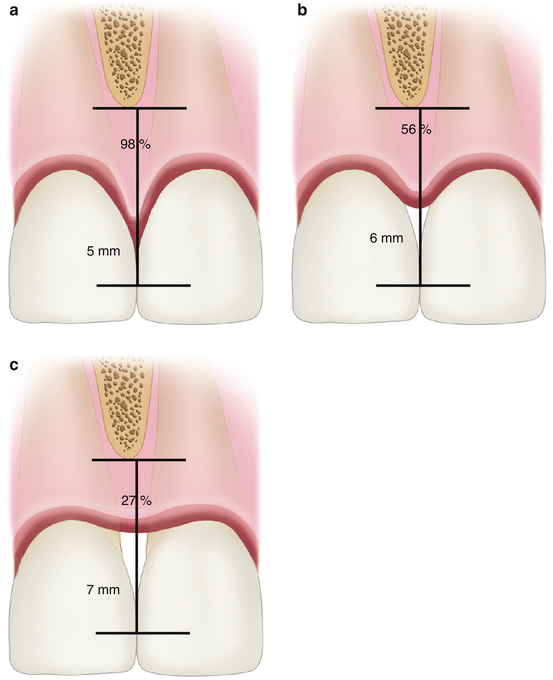
Fig. 9.5
The papilla was shown to be present 98 % of the time in the distance between the interdental contact and the tip of the alveolar bone (a), while at 6 mm, it was present 56 % of the time (b), and at 7 mm, it was only present 27 % of the time (c)
On the other hand, factors affecting the presence and shape of interdental tissue are not limited only to the distance between the bone and contact. Tooth form, distance between two teeth at bone level, angulation of the roots, and the surface geometry are among other factors as well as the health of the soft tissue and the root surfaces. Factors that may affect the position and shape of the interdental tissue may be listed as follows.
9.1.1 Position of the Contact Point or Surface
Coronal positioning of the contact between neighboring structures creates a longer distance between the base of the papilla. This consequently affects the fill of the interdental space. When the biotype of the soft tissue is thick, indicating more tissue volume, giving a better chance to fill the space even the distance increases to the contact point from the base of the papilla.
Stay updated, free dental videos. Join our Telegram channel
VIDEdental - Online dental courses


