Gummy smile cases are always esthetically demanding cases. This article presents a case treated with an interdisciplinary treatment approach and Digital Smile Approach (DSA) using Keynote (DSA), to predictably achieve an esthetic outcome for a patient with gummy smile. The importance of using questionnaires and checklists to facilitate the gathering of diagnostic data cannot be overemphasized. The acquired data must then be transferred to the design of the final restorations. The use of digital smile design has emerged as a powerful tool in cosmetic dentistry to help both practitioner and patient visualize the final outcome.
Key points
- •
Gummy smile cases are always esthetically demanding cases.
- •
This article presents a case treated with an interdisciplinary treatment approach and Digital Smile Approach (DSA) using Keynote (DSA), to predictably achieve an esthetic outcome for a patient with gummy smile.
- •
In order to formulate a treatment plan that predictably leads to a successful esthetic outcome, the final appearance of the case must be visualized and defined before the initiation of active treatment.
- •
The importance of using questionnaires and checklists to facilitate the gathering of diagnostic data cannot be overemphasized.
- •
The acquired data must then be transferred to the design of the final restorations.
- •
The use of digital smile design has emerged as a powerful tool in cosmetic dentistry to help both the practitioner and the patient visualize the final outcome.
Introduction
A smile’s attractiveness is determined by tooth shapes, position, and color, as well as the extent and healthy appearance of the gingival tissue display. The overall relationship between these elements and the face complete the esthetic determinants.
Computer design software has evolved into the major component of the communication between dentists, technicians, and patients. The power of digital technology can now be incorporated into the smile design procedure. Although there are several proprietary digital design services available in the marketplace (Smile Designer Pro, Toronto, Ontario, Canada; Digital Smile Design, Sao Paulo, Brazil). Photoshop software (Adobe Systems, San Jose, CA, USA), PowerPoint (Microsoft Corp, Redmond, WA, USA), or Keynote (Apple Inc, Cupertino, CA, USA) can also be used to facilitate patient input, conceptualization of outcomes, and laboratory communication. The design must be based upon an understanding of macroesthetic and microesthetic concepts regardless of the system used. The primary author used Keynote (as others have done) with a DSA to create a smile design incorporating correction of the patient’s chief complaint and information derived from the New York University (NYU) esthetic evaluation form.
Soft tissue surgical periodontal plastic procedures play an important role in the enhancement of smile by helping to optimize the relationships between the 3 primary components: the teeth, the lip framework, and the gingival scaffold. Kois has stated that when attempting to enhance esthetic outcomes, the influence of 2 essential biological concerns must be fully understood. The first, location of the base of the sulcus, influences the cervical termination of tooth preparation and allows for intracrevicular location of the restoration margin. The second, knowledge of location of the osseous crest, is required before altering gingival levels. The so-called gummy smile is largely a result of an unfavorable ratio between upper lip length and gingiva/tooth display. The location of the smile line is also essentially the product of this ratio. The smile line is defined as the ratio between the upper lip and visibility of the gingival tissue and teeth. Smile level is an imaginary line that follows the lower margin of the upper lip and usually has a convex appearance. Cases exhibiting excessively short teeth and a lack of tooth display are frequently encountered esthetic problems. Excessive gingival display may also be encountered in cases with short teeth. When the incisal edge position is correct, crown lengthening can be used to increase the clinical crown length as a stand-alone esthetic procedure. When the incisal edge position is inadequate and there is excessive gingival display, crown lengthening in conjunction with restorative treatment is indicated. The surgical technique involves apically positioning the gingival margin and may require the removal of supporting bone. The periodontal surgical procedure must also result in a proper biological width and adequate keratinized tissue. Gummy smile can also be the result of altered passive eruption (the alveolar crest is <2 mm from the cementoenamel junction), gingival overgrowth, inadequate length of the upper lip, muscular hyperelevation of the upper lip, and vertical maxillary excess. Cases in which there has been extrusion of the upper teeth, with an associated deep bite, present a related problem.
The initial step in establishing a correct diagnosis and a definitive plan of treatment is through a proper classification of the gingival level. Tjan and colleagues established the smile guidelines standards in the 1980s. Smiles were classified into 3 basic categories (high, average, and low) depending on the exposure of the midfacial cervical margin of the clinical crown relative to the vermillion border. This article shows a step-by-step procedure describing how to optimize the final esthetic outcome with the aid of the digital smile design approach and the proper steps to follow in the diagnosis and treatment planning of a patient with gummy smile.
Case presentation
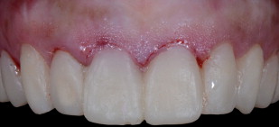
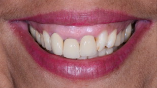
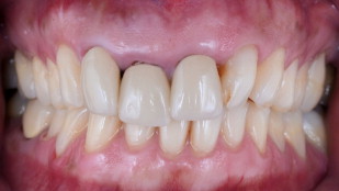
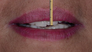
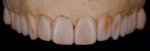
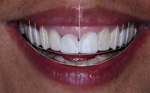
The patient, a Latin American homemaker in her mid-50s, presented with the chief complaint of wanting to improve her appearance. Specifically she sought dental treatment because she did not like her front teeth and desired to improve her smile ( Figs. 1–3 ).

A medical history was taken, and a comprehensive extraoral and intraoral examination was conducted. The patient had an extensive previous dental history, including crowns, composite restorations, and root canals. An esthetic evaluation of the patient was also performed. This evaluation included mounted models, radiographs, photographs, and an esthetic evaluation form incorporating the changes desired by the patient.
The following problem list was created from the gathered data:
- •
Localized initial periodontitis (No. 2–3 and No. 6–7)
- •
Caries No. 2, 3, 5, 13, 19, and 31
- •
Poorly filled root canals No. 2, 7, and 8
- •
Overeruption No. 8 and 9
- •
Excessive maxillary gingival display
- •
Abrasion No. 21 to 28
- •
Poor-fitting crowns No. 7, 8, and 9
- •
A narrow arch form.
Determination of the origin of the problem is extremely important in patients presenting with a gummy smile, which can be skeletal, muscular, dentogingival, or a combination of several or more factors. Knowing the origin of the problem helps to guide the treatment decisions.
Initially, a healthy oral environment was achieved by oral hygiene instruction; localized scaling and root planning on No. 2–3 and No. 6–7; endodontic re-treatment of No. 2, 7, and 8; and composite restorations on No. 2, 3, 19, and 31.
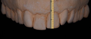
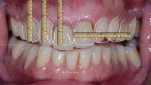
Once hard and soft tissues free of disease were obtained, the final design and position of the restorations were defined by the primary author with the aid of the DSA. The DSA was particularly useful in this case because there was insufficient room for additive mock-up material (usually composite) in the unprepared case. Precisely replicating every detail of the DSA design, strictly adhering to the data flow, enables achievement of the predicted esthetic outcome. A digital caliper was used to measure some reference points on the casts ( Fig. 4 ). With the aid of a calibrated virtual digital ruler, the reference points are subsequently transferred to the computer photographs of the patient ( Figs. 5–7 ). The newly established incisal edge position, as always, dictated the design of the restorations.
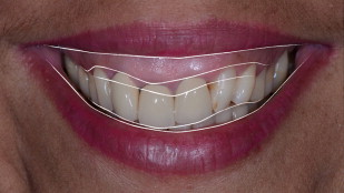
The digitally designed images allowed the patient to visualize the final result and comprehend the issues presented by her current oral condition ( Figs. 8 and 9 ). The number of teeth requiring restoration and the need for periodontal surgeries became apparent. The patient’s approval to proceed with the treatment was based upon viewing the potential outcome via the DSA software.
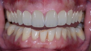

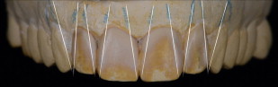
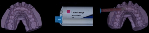
The first wax-up was created based on the DSA measurements ( Fig. 10 ). The restorations proposed in the wax-up were transferred to the patient’s mouth (the mock-up) through the use of a silicone putty matrix (Coltene Lab-Putty, Coltène/Whaledent Inc Cuyahoga Falls, OH, USA) and bisacryl (Luxatemp Ultra, DMG America, Englewood, NJ, USA). The incisal edge position and parallelism to the horizontal reference line were verified. A few minor intraoral modifications were made and followed by an impression of the mock-up. Models were poured on which the final wax-up was created. Indexes fabricated from this wax-up were used as the surgical and preparation guides ( Figs. 11–13 ).
The esthetic crown lengthening surgery was accomplished, with the aid of the guides, correcting the gingival margin levels. To increase predictability, the procedure was divided into 2 distinct phases. Initially, the old crowns were removed, a mock-up was performed ( Fig. 14 ), a gingivectomy was accomplished ( Fig. 15 ), and the provisionals were inserted ( Fig. 16 ). The biological width was deliberately violated. Dividing the overall periodontal procedure into 2 phases meant shorter appointments and enabled tooth preparation margin position correction. Osseous recontouring to establish an acceptable biological width was accomplished 3 weeks later. A full-thickness flap was raised to allow visualization during the osteoplasty and permit accurate positioning of the gingival margin ( Figs. 17 and 18 ).