Introduction
The immutability of intercanine width has long been the subject of discussion. The aims of this study were to describe the longitudinal intercanine width changes of children from 6 to 14 years of age and to interpret them with a 3-dimensional method.
Methods
Complete dental stone casts were annually prepared for 66 subjects (50 girls, 16 boys) from 6 to 14 years of age. By using 3-dimensional laser scanning and reconstruction software, virtual casts were constructed. Intercanine width was measured as well as the related 3-dimensional measurements, such as the area of the intercanine triangle, the intercanine angle, the radius of the inscribed circle, and the angles formed by the virtual axes of the canines and the occlusal plane. The measurement changes over time were analyzed by using mixed-effects analysis for longitudinal data.
Results
There were slight decreases in intercanine widths for both sexes and both arches. However, the amounts of change were relatively small when compared with the initial values and individual random variability. The values of area, the angles formed by the virtual axes of the canines and the occlusal plane, and the radius showed decreasing trends, whereas the intercanine angle exhibited increasing trends during the observation period. Although the intercanine width changed over time, it was not clinically significant, showing relative stability.
Conclusions
The intercanine width of an untreated subject after stabilization in the mouth is considered to be quite stable, even though individual variation is great.
It is well known that the development of the human dentition is a continuous and complex biologic process. The dentoalveolar processes undergo visible alterations during growth, become part of the craniofacial complex, and are influenced by changes in various parts of the skull. Orthodontists can benefit from understanding these changes during every stage of human development and have a special interest in the relative dimensional changes of the dental arches.
Understanding the changes in the dental arch resulting not only from growth, but also from orthodontic treatment is useful in treatment and retention planning. Long-term changes in tooth positions and dental arch dimensions have been reported after orthodontic treatment and in samples of untreated subjects. However, the inability to determine whether these changes occurred primarily as a result of orthodontic therapy or were part of the normal developmental process, as stated by Sinclair and Little, is an intriguing question.
Moorrees et al pointed out that considerable individual variation in the arch form occurs with normal growth, and it has been reported that the growth and development period is affected by various environmental factors. Therefore, it is difficult to predict the growth potential in each patient. Some informative reports are available on the average changes in arch dimensions in untreated samples, although the results of those studies are inconclusive. The reason for that might be attributed to the differences in the subjects among the studies such as ethnicity, age, and sample selection. In addition, the designs of these studies were varied, and it is difficult to determine whether all had adequate sample sizes.
On the other hand, for various reasons, including difficulties in measuring and describing the curvature, the longitudinal changes of dental arches have generally been described as dimensional changes in intercanine and intermolar widths and arch lengths, although these changes might not always lead to a change in the arch form. In addition, the immutability of intercanine width has long been the subject of heated discussions in the orthodontic literature. Those measurements are usually made by using traditional methods, such as with calipers, and further description of arch changes other than distance is difficult.
Recently, 3-dimensional scanners have been introduced in dentistry for various applications with reconstructed virtual models. Studies of 3-dimensional reconstructions have reported accurate and reliable techniques for restorative procedures and for orthodontic dental and facial analyses to aid clinicians in planning more effective treatments. The 3-dimensional virtual model and its specialized software make possible measurements that were once impossible to analyze with traditional tools and 2-dimensional settings, such as the area and the angle between the line and surface. In addition, this technique is adequate for the management and storage of numerous dental casts for growth studies.
Our investigation was designed to describe the longitudinal dental arch changes of children from 6 to 14 years of age in terms of intercanine width with 3-dimensional reconstructed virtual models and specialized software programs and to interpret the changes with a previously unused 3-dimensional method. The aims of this study were to determine whether the intercanine width changes from the mixed dentition to the early permanent dentition, and to evaluate the changes of the anterior dentition 3 dimensionally.
Material and methods
The material in this study consisted of complete dental stone (Neoplumstone; Mutsumi Chemical Industry, Yokkaichi, Japan) cast sets that were prepared for 232 children annually from 1995 to 2003 as a part of the Korean Dental Growth Study. The casts of 66 children were selected for this study, including 16 boys and 50 girls of Korean origin followed from 6 to 14 years of age. They had no orthodontic treatment before and during the observation period. These subjects were judged to have clinically good occlusion rather than orthodontic ideals. All selected children had Class I or near Class I buccal relationships of molars. Children missing teeth other than the third molars and with more than 3 mm of crowding or spacing and those who did not have full sets of casts of 9 years were excluded from the original pool. All subjects and parents or guardians provided written informed consent. The Institutional Review Board for the Protection of Human Subjects reviewed and approved the research protocol (S-D2010013).
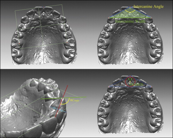
All casts selected were scanned by a 3-dimensional scanner (optoTOP-HE; Breckmann, Meersburg, Germany) with a point accuracy of ±0.001 mm, and a resolution of 0.040 mm in the x and y directions and 0.002 mm in the z direction. Each cast was scanned from 10 or more views that were then combined and rendered into 3 dimensions by using specialized software (Rapidform XO, version 2.0.1.0; INUS Technology, Seoul, South Korea) ( Fig 1 ). The virtual 3-dimensional models were measured and analyzed with specialized software (Rapidform 2004, version PP2; INUS Technology). The measurements of each cast were made at 3 separate times by 1 observer (Y.S.P.) over a 3-week period.
Before the measurements, it was found that there was no 7-year-old or younger subject with a measurable bilateral permanent canine at the initial examination in our data set. Because we focused on the changes of the permanent dentition primarily, we did not measure the samples for this age. However, samples of 6-year-olds were analyzed as a representation of intercanine width in the deciduous dentition for comparision.
The intercanine width was measured on the virtual casts to determine the linear changes of the dental arch as the distance between the crown tips of the permanent or deciduous canines. In the case of intercanine width for the permanent canine, a clinical crown length of at least 7 mm from the labial side was the inclusion criterion. Casts that did not meet this criterion were considered to have missing values.
To 3-dimensionally depict the dental arch changes, 2 novel attempts were made. First, a new reference point was created at the midpoint between the mesial incisal tips of both central incisors. A triangle was then created with both canine tips as the new reference point. The area of the triangle (area), the angle formed by the midpoint of the mesial incisal tips of both central incisors and both canine tips (intercanine angle), and the radius of the inscribed circle (radius) were calculated with the software ( Fig 1 ).
Second, a new reference plane was established for each virtual cast from 3 points: the midpoint between the proximal tip of the central incisors, and the most occlusal point of each mesiolingual cusp of the first molars. New reference points were then created at the center between the most cervical points of the canines from the buccal and lingual aspects. After that, a midpoint created from these 2 reference points and the cusp tip of the canine were connected as a new vector and defined as the long axis of the canines. This definition is valid only in this study. The angles formed between the reference plane and the apical direction of the long axis of the canines from the canine mesiodistal perspective were measured (RC angle, the right canine axis angle; LC angle, the left canine axis angle). Detailed descriptions of the measurements are presented in Figure 1 . To test the reliability, 3-dimensional scans (n = 10) were randomly selected and measured again on separate days 2 months after the initial measurement.
Statistical analysis
Descriptive statistics of the changes in the measurements were calculated annually. Each subject provided multiple repeated observations for linear, angular, and area measurements. Because the serial measurements were correlated according to each subject, a mixed-effects analysis was applied. The analysis equation for the data was written as y ij = μ + β 1 sex i + β 2 age ij + β 3 sex i *age ij + b i1 + b i2 *age ij + e ij , where the terms in the model are μ , total mean; β 1 , sex effect; β 2 , age effect; β 3 , interaction effect between sex and age; and b i (i = 1, 2, . . . . , 66), random effect of a subject. Statistical analyses were performed by using the language R, and a P value less than 0.05 was predetermined to be statistically significant.
Results
The intraexaminer reliability coefficients ranged from 0.998 to 1.000. In terms of root mean squares, the random errors of estimation were lower than 0.05 mm for linear measurements and 0.45° for angular measurements. No variables showed a statistically significant difference between the test and retest measurements. Means and standard deviations of all measurements from 6 to 14 years are summarized according to sex in Tables I and II . The measurements were separately recorded for bilateral deciduous canines. There was a time period during which some subjects did not have adequately erupted bilateral deciduous or permanent canines because of exfoliation and subsequent eruption. In this case, they were considered to have missing values.
| Age | Girls (total n = 50) | Boys (total n = 16) | ||||||||||||
|---|---|---|---|---|---|---|---|---|---|---|---|---|---|---|
| n | Intercanine width (mm) | Area (mm 2 ) | RC angle (degrees) | LC angle (degrees) | Intercanine angle (degrees) | Radius (mm) | n | Intercanine width (mm) | Area (mm 2 ) | RC angle (degrees) | LC angle (degrees) | Intercanine angle (degrees) | Radius (mm) | |
| Deciduous dentition | ||||||||||||||
| 6 | 39 | 32.11 (1.95) | 125.50 (30.04) | 116.67 (8.95) | 113.91 (8.29) | 128.64 (7.37) | 3.66 (0.67) | 14 | 32.33 (2.71) | 126.90 (31.96) | 111.96 (7.13) | 113.89 (8.78) | 128.52 (5.82) | 3.68 (0.61) |
| 8 | 34 | 33.97 (1.88) | 170.62 (25.03) | 121.71 (11.74) | 118.67 (11.99) | 118.92 (6.51) | 4.63 (0.52) | 14 | 34.33 (2.36) | 168.38 (32.52) | 120.17 (8.76) | 122.74 (15.67) | 120.95 (5.69) | 4.53 (0.60) |
| 9 | 12 | 33.69 (1.80) | 175.12 (20.21) | 120.73 (9.88) | 117.10 (9.49) | 116.57 (5.83) | 4.77 (0.41) | 7 | 32.92 (2.58) | 161.23 (22.53) | 131.86 (7.72) | 131.17 (11.25) | 125.72 (7.12) | 4.62 (0.84) |
| 10 | 3 | 35.14 (0.92) | 182.59 (8.97) | 120.25 (7.74) | 114.95 (9.10) | 118.72 (4.65) | 4.81 (0.27) | 5 | 33.77 (1.56) | 173.46 (35.05) | 121.10 (7.44) | 119.15 (10.21) | 117.80 (7.19) | 4.70 (0.70) |
| 11 | 0 | 1 | 33.53 | 169.44 | 120.61 | 131.67 | 117.82 | 4.66 | ||||||
| Permanent dentition | ||||||||||||||
| 8 | 5 | 34.30 (1.76) | 172.35 (31.86) | 113.10 (6.41) | 111.39 (7.45) | 119.65 (6.12) | 4.63 (0.61) | 0 | ||||||
| 9 | 12 | 35.14 (2.85) | 169.57 (30.75) | 121.56 (14.53) | 117.96 (8.15) | 122.52 (7.28) | 4.49 (0.59) | 2 | 39.58 (0.65) | 198.30 (55.23) | 124.49 (8.13) | 130.33 (17.10) | 126.25 (14.17) | 4.671 (1.19) |
| 10 | 39 | 35.78 ∗ (2.37) | 172.47 (23.56) | 116.97 (9.19) | 116.97 (9.05) | 123.32 (6.43) | 4.50 (0.47) | 7 | 38.36 ∗ (2.79) | 188.65 (29.84) | 126.88 (13.29) | 123.88 (14.06) | 125.75 (6.38) | 4.62 (0.54) |
| 11 | 49 | 35.85 (2.31) | 169.67 (28.14) | 116.12 (8.94) | 116.27 (7.99) | 124.43 (7.28) | 4.42 (0.58) | 12 | 37.45 (3.17) | 181.17 (36.39) | 120.29 (10.50) | 119.67 (12.73) | 125.65 (5.85) | 4.52 (0.60) |
| 12 | 50 | 35.75 ∗ (2.42) | 165.86 (25.62) | 115.31 (8.43) | 114.92 (7.63) | 125.16 (7.21) | 4.35 (0.54) | 15 | 37.30 ∗ (2.83) | 182.52 (36.41) | 118.30 (16.62) | 115.64 (10.86) | 124.95 (6.39) | 4.57 (0.63) |
| 13 | 50 | 35.70 ∗ (2.52) | 163.01 (27.64) | 114.87 (8.99) | 115.22 (7.63) | 125.91 (7.39) | 4.28 (0.57) | 16 | 37.24 ∗ (2.71) | 177.11 (36.61) | 117.51 (12.51) | 115.63 (10.93) | 126.32 (5.67) | 4.45 (0.63) |
| 14 | 50 | 35.58 ∗ (2.60) | 161.79 (27.76) | 114.66 (8.54) | 114.20 (7.14) | 125.91 (7.67) | 4.26 (0.58) | 16 | 37.15 ∗ (2.63) | 178.13 (35.63) | 116.37 (12.27) | 113.95 (10.98) | 125.83 (5.70) | 4.48 (0.62) |
∗ There were statistically significant differences between the sexes ( P <0.05).
| Age | Girls (total n = 50) | Boys (total n = 16) | ||||||||||||
|---|---|---|---|---|---|---|---|---|---|---|---|---|---|---|
| n | Intercanine width (mm) | Area (mm 2 ) | RC angle (degrees) | LC angle (degrees) | Intercanine angle (degrees) | Radius (mm) | n | Intercanine width (mm) | Area (mm 2 ) | RC angle (degrees) | LC angle (degrees) | Intercanine angle (degrees) | Radius (mm) | |
| Deciduous dentition | ||||||||||||||
| 6 | 49 | 25.83 (1.97) | 59.89 (14.85) | 111.41 (12.90) | 110.42 (11.35) | 140.68 (8.10) | 2.23 (0.48) | 15 | 27.20 (2.38) | 69.54 (19.51) | 114.95 (12.91) | 115.68 (11.94) | 139.36 (7.23) | 2.44 (0.53) |
| 8 | 25 | 27.45 (2.07) | 72.46 (18.19) | 117.14 (15.65) | 118.88 (16.29) | 138.56 (6.65) | 2.52 (0.49) | 9 | 28.46 (3.28) | 79.75 (28.63) | 114.53 (6.89) | 101.74 (34.50) | 138.34 (6.82) | 2.64 (0.66) |
| 9 | 4 | 26.97 (1.53) | 67.89 (18.66) | 132.19 (14.86) | 133.40 (16.76) | 139.66 (6.88) | 2.41 (0.53) | 3 | 27.27 (2.52) | 73.53 (17.32) | 118.48 (16.32) | 112.55 (19.69) | 132.60 (6.83) | 3.17 (0.82) |
| 10 | 0 | 2 | 29.40 (3.72) | 108.26 (33.92) | 116.47 (3.41) | 115.02 (10.26) | 127.40 (2.78) | 3.44 (0.63) | ||||||
| Permanent dentition | ||||||||||||||
| 8 | 14 | 26.60 (1.69) | 79.90 (12.95) | 104.19 (7.81) | 106.45 (9.65) | 131.33 (7.13) | 2.86 (0.39) | 1 | 31.131 | 100.67 | 138.97 | 129.41 | 125.06 | 3.40 |
| 9 | 35 | 27.47 (1.92) | 83.58 (15.96) | 109.64 (15.28) | 111.43 (14.27) | 132.10 (8.99) | 2.90 (0.50) | 4 | 29.05 (2.68) | 96.83 (7.75) | 112.99 (10.92) | 111.47 (12.15) | 128.75 (7.73) | 3.27 (0.19) |
| 10 | 49 | 27.32 ∗ (1.72) | 83.68 (16.20) | 105.77 † (6.80) | 105.72 (7.69) | 132.03 (6.78) | 2.90 (0.44) | 12 | 29.28 ∗ (2.28) | 93.26 (18.46) | 112.65 † (8.78) | 112.38 (11.18) | 132.93 (8.17) | 3.04 (0.51) |
| 11 | 50 | 27.47 † (1.70) | 81.53 (16.19) | 105.27 (7.96) | 104.43 (6.67) | 133.44 (7.62) | 2.82 (0.47) | 14 | 28.92 † (2.37) | 83.05 (17.97) | 108.01 (10.47) | 105.36 (8.81) | 136.71 (8.07) | 2.75 (0.50) |
| 12 | 50 | 27.33 † (1.76) | 79.84 (16.79) | 104.67 (6.36) | 103.89 (5.89) | 133.85 (7.80) | 2.78 (0.49) | 15 | 28.78 † (2.40) | 85.35 (20.07) | 105.25 (9.98) | 105.37 (9.68) | 135.45 (8.23) | 2.83 (0.54) |
| 13 | 50 | 27.38 † (1.72) | 76.70 (16.47) | 106.10 (7.01) | 106.01 (6.89) | 135.54 (7.90) | 2.68 (0.49) | 16 | 28.92 † (2.38) | 84.44 (23.88) | 105.30 (10.37) | 104.30 (9.85) | 136.45 (9.21) | 2.78 (0.64) |
| 14 | 50 | 27.14 ∗ (1.74) | 76.30 (17.31) | 105.12 (6.84) | 104.17 (6.74) | 135.02 (8.43) | 2.68 (0.52) | 16 | 29.17 ∗ (2.40) | 85.31 (23.41) | 105.11 (8.86) | 104.34 (8.35) | 136.86 (9.04) | 2.78 (0.64) |
∗ There were statistically significant differences between sexes ( P <0.01).
† There were statistically significant differences between the sexes ( P <0.05).
Stay updated, free dental videos. Join our Telegram channel
VIDEdental - Online dental courses


