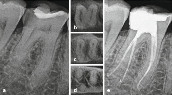
Fig. 4.1
Periapical radiograph of a right mandibular second molar with associated apical pathosis (a) and the post-endodontic periapical radiograph with obturation material demonstrating the complex nature of the root canal system (b)
4.2 Methods for Studying Tooth Anatomy
Methods used to evaluate tooth and root anatomy can be categorized as in vitro or in vivo. In vitro methods allow for detailed microscopic and histologic examination of teeth in all dimensions and include demineralization, clearing, sectioning, modeling, scanning electron microscopy, and micro-computed tomography [3]. In vivo methods have more clinical implications and consist of traditional radiography, spiral computed tomography, optical coherence tomography, tuned aperture computed tomography, cone beam computed tomography, ultrasound, and magnetic resonance imaging. In vitro histologic evaluation via canal staining and tooth clearing is considered to be the gold standard when evaluating root canal anatomy and morphology [4, 5]. However, these methods are destructive and limited to ex vivo (“bench top”) examination. The accuracy of CBCT has been reported to be equivalent to that of histologic sectioning and staining for practical clinical purposes (i.e., identifying canals and unusual anatomy). A study directly comparing tomographic scans to that of the histologic sectioning of 152 maxillary molar teeth found identical canal configurations using the two methods [6]. A 2010 study of 233 tooth sections found a “strong to very strong correlation” between CBCT imaging and histologic sections [7]. Neelakantan et al. compared the efficacy of six different imaging methods to that of canal staining and clearing. CBCT scanning was reported to be as accurate in the identification of root canal systems as the modified staining and clearing techniques and significantly more accurate than conventional radiographic methods [8]. In another study comparing traditional radiographic techniques to that of CBCT scanning and clinical sectioning, Blattner et al. reported that CBCT scanning was a reliable method for canal detection and represents a highly accurate, in vivo method for the evaluation of tooth anatomy equivalent to that of the histologic gold standard [9] (Fig. 4.2).
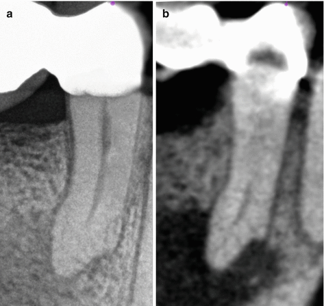

Fig. 4.2
Periapical radiograph of a right mandibular first premolar (a) with a corresponding sagittal CBCT slice demonstrating visible complex internal anatomy in the apical third of the root canal system (b)
4.3 Clinical Methods for the Evaluation of Tooth Anatomy
4.3.1 Common Tooth Forms and Anatomical Landmarks
The collective information gained from numerous anatomic and morphologic studies can be applied clinically by establishing a thorough knowledge of commonly known root variations. Average statistics for permanent human teeth with respect to type and location as well as common canal configurations can be found in many published endodontic texts. In addition, several studies have classified various canal configurations identified within a single root [3, 10]. Studies have also attempted to identify common morphologic and anatomic features of roots and root canal systems as they relate to gender, ethnicity, and geographic location [11, 12].
A study examining external landmarks of teeth and their association with internal anatomic features found specific observable patterns relating the pulp chamber to the clinical crown as well as the canal orifices to the pulp chamber floor. Seven distinct features were observed on the pulpal floor that provide clues as to the location of the canal orifices. The authors of this study proposed nine “laws” to assist in the identification root canal anatomy during endodontic treatment [13].
Studies that attempt to categorize tooth anatomy and morphology can only provide clinical treatment recommendations and guidelines as they relate to general circumstances. These guidelines are based on weighted averages and common observations. Averages are useful when studying characteristics of populations, but of limited predictive value for treatment of the individual patient. CBCT can capture information, which previously required multiple radiographs, in a more precise way than is possible with two-dimensional radiographs.
CBCT imaging overcomes several limitations found with conventional radiographic methods. Dimensionally accurate image acquisition and viewing in all three spatial planes eliminates superimposition of multiple roots and/or surrounding anatomic structures and provides more sensitivity with regard to the detection of periradicular pathosis, root resorption, and root fractures [14]. Visualization of multidimensional root curvature allows for precision treatment planning and predictable canal management [15] (Fig. 4.3). Geometrically accurate measurement capabilities assist in the determination of root length as well as establish precise intra- and inter-canal distance relationships [16]. Additionally, information obtained from a cone beam scan prior to endodontic treatment has been reported to improve diagnostic capabilities and prompt endodontic treatment plan changes [17, 18].
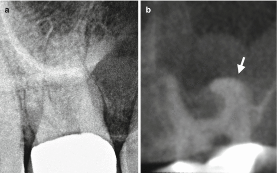

Fig. 4.3
Periapical radiograph of a maxillary right first molar (a) with a corresponding coronal CBCT slice demonstrating a significant curvature of the mesiobuccal root in the buccopalatal plane (white arrow) (b)
A preoperative CBCT scan provides an undistorted reconstruction of a tooth’s internal and external morphologic features. The ability to detect more roots and root canals with greater accuracy and higher frequency when a CBCT scan is available has been reported [19] (Fig. 4.4). Figure 4.5 represents the periapical radiographs of two different maxillary second molars and their corresponding axial CBCT slices (below). Although both teeth appear radiographically similar, the corresponding axial CBCT slices reveal two very different morphologic root features and canal configurations.
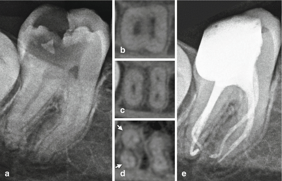
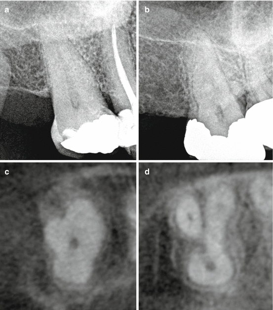

Fig. 4.4
Preoperative periapical radiograph of a right mandibular second molar (a) with corresponding axial CBCT slices captured from the coronal third (b), mid-root (c), and apical third (d). The axial CBCT slices demonstrate two distinct mesial canals as well as a division of the distal root in the apical third (white arrows). The post-endodontic radiograph demonstrates all identified root canals treated (e)

Fig. 4.5
Preoperative periapical radiographs of two different right maxillary second molars with conically shaped roots (a, b) and their corresponding axial CBCT slices (below) demonstrating different external root forms and different internal canal configurations (c, d)
4.3.2 Endodontic Access Design
Gaining access to the pulp chamber to facilitate the identification of all canal orifices represents the first technical step in root canal treatment that irreversibly alters tooth structure. Traditional access designs tend to be somewhat standardized per tooth type and are typically depicted in most texts with easily identifiable canal orifices at the base of a large pulpal floor. Many teeth requiring endodontic treatment present with pulp chambers that have significant alterations in volumetric space and shape due to excess deposits of tertiary dentin in response caries, bacterial ingress, dental procedures, and/or surface tooth loss over time. The integration of modern endodontic techniques and technology can assist in shaping the endodontic access based on the specific pulp chamber morphology of the tooth being treated.
Traditional access designs are treatment planned from parallel periapical and bitewing radiographs as well as clinical anatomic landmarks. This limits radiographic access design to a single plane and creates reliance upon the suggested access design standardizations. A more conservative and customized endodontic access can be designed from the anatomical information available on the CBCT volume. Precision vertical depth measurements can be determined from the occlusal plane to multiple levels within the pulp chamber in both the coronal and sagittal planes. Additionally, the lateral extension of the access as it relates to the occlusal surface can be determined from measurements available in the axial plane (Fig. 4.6). Maintaining a conservative endodontic access minimizes the unnecessary removal of tooth structure and may improve overall fracture resistance [20].
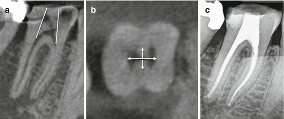

Fig. 4.6
Treatment plan for a conservative access design of a mandibular right first molar based on the exact pulp chamber morphology as determined via the CBCT scan. The sagittal CBCT slice demonstrates the planned minimum mesial/distal outline form (white lines) (a), and the axial CBCT slice captured at the level of the canal orifices demonstrates the planned mesial, distal, buccal, and lingual access extensions (b). The post-endodontic radiograph demonstrates a minimal access preparation and appropriate root canal system management (c)
4.3.3 Evaluation of Complex Roots and Root Canal Variations
Many anatomic and morphologic variations in tooth anatomy exist within the global population. A detailed description of common statistical anatomic averages per tooth type can be found in previously published endodontic texts. In vivo identification of more complex anatomical root forms and root canal configurations can be best observed using CBCT imaging. Some variations have been reported to occur at higher frequencies and have been associated with specific geographic populations and/or ethnic heritage [11]. Multiple published case reports demonstrate the significant advantages of CBCT over traditional radiographic imaging for the detection and management of a wide variety of anatomic and morphologic tooth and root variations [21].
4.4 Maxillary Molar Teeth
4.4.1 The Mesiobuccal Root Complex
The mesiobuccal root complex of the maxillary first permanent molar is one of the most well-documented and studied root canal systems in the endodontic literature. It is generally broad buccolingually with prominent depressions on the mesial and distal surfaces and usually contains two canals. One ex vivo study reported the presence of two mesiobuccal canals in the coronal half of maxillary first molars to be as high as 95.2 % with 71 % having separate apical portals of exit [22]. Similar variations occur in the maxillary second molars, although with less reported frequency [23]. The clinical detection and treatment of second mesiobuccal canals (MB2) can be enhanced with the acquisition of a CBCT scan [19]. The presence of an additional root and/or root canal can often be detected by studying the buccopalatal extent of the mesiobuccal root complex in the axial and coronal planes (Fig. 4.7).
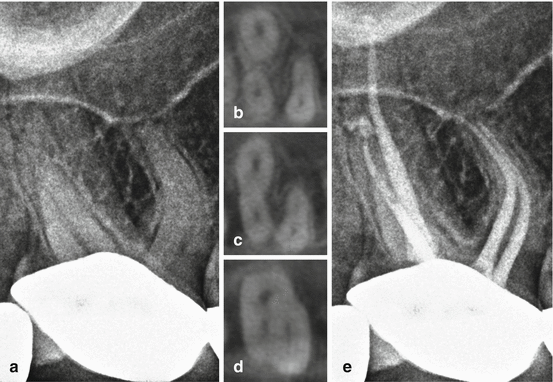

Fig. 4.7
Preoperative periapical radiograph of a maxillary right first molar (a) with corresponding axial CBCT slices captured from the apical third (b), mid-root (c), and coronal third (d) demonstrating a mesiobuccal root with two identifiable root canal systems. The post-endodontic periapical radiograph demonstrates all identified root canals treated (e)
4.4.2 Multiple Palatal Roots and Canals
A maxillary permanent molar with four roots and/or four root canals is not unusual when the additional root or root canal is located within the mesiobuccal root complex, but occasionally an additional root or root canal can be associated with the palatal root (Figs. 4.8 and 4.9). This morphologic variation has been suggested to occur more frequently in maxillary second molars with a reported frequency of 0.4–1.4 % [28, 29]. A CBCT scan can aid in the recognition of this rare anatomical variation as well as determine the exact location of the additional canal orifice for maximum conservation of tooth structure during endodontic access preparation.
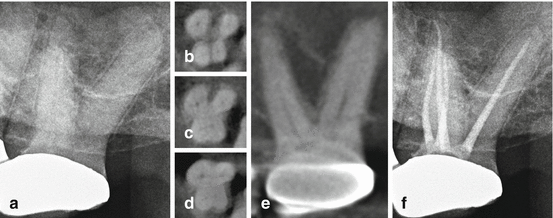
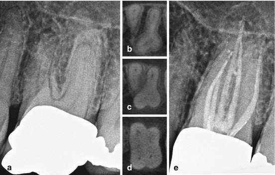

Fig. 4.8
Preoperative periapical radiograph of a maxillary left second molar (a), with corresponding axial CBCT slices captured from the apical third (b), mid-root (c), and coronal third (d) demonstrating four separate canals and a morphologic root division into four separate roots. The sagittal CBCT slice demonstrates two separate palatal roots (e). The post-endodontic periapical radiograph demonstrates all identified root canal systems treated (f)

Fig. 4.9
Preoperative periapical radiograph of a maxillary left first molar (a) with corresponding axial CBCT slices captured from the apical third (b), mid-root (c), and coronal third (d) demonstrating five separate root canal systems (two canals in the mesiobuccal root and two canals in the palatal root). The post-endodontic periapical radiograph demonstrates all identified root canal systems treated (e)
4.5 Mandibular Molar Teeth
4.5.1 Radix Entomolaris and Radix Paramolaris
Mandibular permanent molars will occasionally present with an additional separate root located either to the lingual (radix entomolaris) or to the buccal (radix paramolaris). The literature suggests that the highest incidence of these morphologic variations is present in populations with Mongoloid ancestry (such as Chinese, Eskimo, and Native Americans) and occurs at a frequency of 5–40 % [24]. Although traditional two-dimensional imaging at various angles can reveal clues as to the presence of these additional roots, exposing the anatomic location of the additional canal orifice during endodontic access can pose a clinical challenge. A CBCT scan will not only confirm the presence of these additional roots but also assist in determining the exact location of the canal orifice so that a proper access design can be planned (Fig. 4.10).

