Fig. 2.1
(a–d) Various methods used to demonstrate facial changes using lateral cephalometrics. (a) Facial angles. These are just a few of the angles which describe changes in the skeletal profile. There are many more angles and linear measurements which can be used to relate the maxilla to the mandible and both jaws to the cranial base. (b) Facial polygon. This is a graphic method used to describe the boundaries of the skeletal face. (Pogonion constructed, Po’, is the same point as gnathion.) Facial growth changes can be shown by superimposing each succeeding polygon on the anterior cranial base (SN) and registering on sella turcica (S). (c) Projecting facial landmarks to a constructed Frankfort horizontal line which is arbitrarily drawn 6° off the SN line. This angle can vary with steepness of the anterior cranial base. This graphic method will show the relative contribution of various structures within the maxilla and the mandible to the profile. (d) Basion horizontal created facial polygon (Coben 1986). This method of superimposing tracings graphically reflects his overall concept of fixed growth. A plane at the level of the anterior border of foramen magnum (basion) parallel to Frankfort horizontal where Basion is the point of reference for the analyses of craniofacial growth
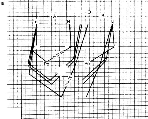
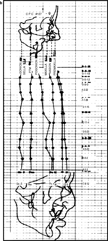
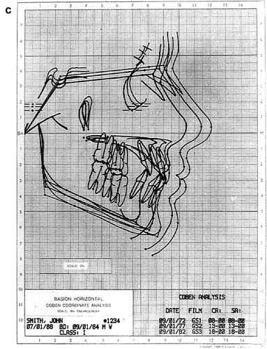
Fig. 2.2
(a) Case CP #127 (CPCLP). Superimposed facial polygons from 1 month, 18 days of age to 7 years, 8 months, and 12 days of age. A result of the mandible’s downward growth increments exceeding its horizontal growth increments, this is an example of “poor” facial growth in that the profile fails to flatten as the mandible remains retrognathic. Note that in this and the following illustration, the forward projection of the premaxilla does not increase after 1 year, 2 months, and 22 days (b) Case CP #127. Projecting facial landmarks to a constructed Frankfort horizontal 6° off the SN line. Although each of the skeletal structures except for the mandible has increased in size, the relative position of midfacial structures to the anterior cranial base has remained relatively stable (c) Case CP #127. Basion horizontal coordinate computer craniofacial serial tracings at ages 8, 13, and 18 years. Tracings are registered at Basion and oriented in Frankfort horizontal. Serial tracings maintain a constant S-N/FH relationship. S-N and FH planes are parallel. Tracings depict Coben’s growth philosophy, which states that craniofacial growth is reflected away from the foramen magnum (basion) and the vertebral column (Reprinted from Coben (1986))
The use of “landmark,” or baseline, images associated with the basicranium to show the composite results of facial growth can provide meaningful information because it is the enlargement of the face relative to the cranial base that is being evaluated. In the child, further growth changes in the anterior part of the cranial base slow considerably at about 5–6 years of age, whereas facial growth continues actively through adolescence or beyond. Comparing the relative growth between these two regions, rather than simply focusing on a single fixed point, provides clinically useful information when cephalometrically evaluated.
2.2 The Beginning of Longitudinal Cleft: Palate Research Studies
Two major research problems were common in cleft palate surgical studies prior to the 1950s. Pruzansky (1953, 1955) commented on the surgeon’s tendency to group all types of clefts together in research and clinical treatment. He also stated that surgeons were limited in their study of pathologic anatomy of clefts due to the unavailability of serial dental casts, cephaloradiographs, and photographic records.
The need for clinical records was apparent to many researchers, and within a decade, many retrospective clinical data sets were developed. These data sets spawned many investigators to determine the long-term influences of surgical and neonatal maxillary orthopedic procedures on palatal and facial growth and development. As a result of these early studies, useful diagnostic and prognostic information was obtained that provided a rationale for the management of individual cleft cases. These clinical records offered an accurate means for measuring and recording individual variation and for plotting the progress of each case in terms of growth and response to various treatments. As a result of these findings, the quality of care improved, resulting in more aesthetic and functional outcomes. Proper documentation, using objective records and individual treatment outcomes, has extended to many more modifications where it is possible to perform multicenter retrospective studies.
2.3 Research Methods (Atkins 1966; Byse et al. 1983)
2.3.1 Retrospective Studies
In a retrospective study, the nature of the study group must be delineated precisely. Definite criteria should be established so that there is no ambiguity about types of cases and stages of growth development to be included in, or excluded from, the study. The choice of the case and control groups should be guided by concerns of validity. The advantages of retrospective studies are that they can be conducted relatively rapidly because the records of patients whose treatment is already complete can be used. The investigator is protected against the circumstance of “subject dropout” during the course of treatment, and they are relatively economical.
2.3.2 Prospective Studies
The advantages and disadvantages of prospective studies are in essence the inverse of those of retrospective studies. Provided that ethically and logistically satisfactory plans for random assignment to treatment can be developed, prospective trials afford an opportunity to control selection bias and to define and control the records acquisition process.
The main disadvantages of prospective trials are that they are expensive and a great deal of time must inevitably elapse between project initiation and the point at which data on most of the main outcome variables become available for analysis.
Stay updated, free dental videos. Join our Telegram channel
VIDEdental - Online dental courses


