The uvulopalatal flap

Chapter 34 The uvulopalatal flap

1 INTRODUCTION

This book includes a number of chapters which describe a variety of palatal surgical procedures for sleep disordered breathing, including traditional uvulopalatopharyngoplasty (UPPP) techniques, Z-palatoplasty (ZPPP), transpalatal advancement pharyngoplasty, laser-assisted uvulopalatoplasy (LAUP), pillar implantation, etc. Most of these procedures can be divided into two mutually exclusive groups based upon several different criteria: by their action on the palate, the setting in which they are performed, and how the surgeon is reimbursed. First, these various procedures usually work by one of two different ways: by either shortening the soft palate or by stiffening it. They also differ in where they are performed: those that are more invasive and which are used for more significant obstructive sleep apnea (OSA) are generally done in the operating room under general anesthesia, and those that are less invasive and which are advocated for less severe forms of sleep disordered breathing (such as primary snoring) are done in the office under local anesthesia. Furthermore, the insurance industry makes a distinction between these two groups of procedures in regards to reimbursement: those done in the OR for OSA are generally reimbursed by insurance, while those done in the office for primary snoring and mild OSA are relegated to self-pay status.

This chapter describes a palatal surgical technique which in many regards bridges these divides, with applicability in both the operating room as an OSA procedure and in the office setting for primary snoring or mild OSA. It is a variation of UPPP which is easily learned, gives reproducible results, and which can be done under local anesthesia in the office in select cases. The procedure can be performed as a limited palatal procedure for snoring or mild OSA, or can be extended to more effectively treat the palate and tonsillar fossas for more significant OSA. In addition, it forms the basis for another effective palatal procedure, the Z-palatoplasty, as described elsewhere in this atlas.

2 THE PROCEDURE

The uvulopalatal flap (UPF) procedure involves the shortening of the soft palate by folding the distal soft palate with uvula forward upon itself. The intervening mucosal surfaces of the folded palate are removed, and the palate is sutured in its new position in two layers with interrupted sutures. The resultant palatal shortening creates a surgical result virtually indistinguishable from a traditional UPPP, but with several important potential differences over UPPP and some of the other procedures, as outlined below.

The UPF procedure was first described by Powell et al.2 in 1996 as a UPPP variation for the operating room. A similar technique was described in 1993 by Bresalier and Brandes,3 referred to as the imbrication technique of UPPP in 1999. In 2000, I described the use of this technique in the office under local anesthetic,4 which was presented in more detail in 2003 by Neruntarat.5 The procedure can also be expanded under general anesthesia to include greater effects in the laterally, by including concurrent tonsillectomy with tonsillar pillar closure4,6 or by extending incisions superolaterally from the apices of the tonsillar fossas toward the third molar region;4 Li et al.7 call this modification an extended uvulopalatal flap. The principles of the UPF have been further extended by Friedman with the Z-palatoplasty,8 which is the subject of Chapter 33. The Z-palatoplasty is essentially a UPF procedure in which the uvula and distal soft palate are split in the midline. This creates two flaps which are advanced not only anteriorly but laterally to further augment the retropalatal airway in its lateral dimensions. Because of the versatility of the procedure, therefore, the UPF is a useful tool which should be part of the armamentarium of all surgeons who treat sleep disordered breathing.

3 TECHNIQUE

3.1 AWAKE, LOCAL ANESTHESIA, OFFICE PROCEDURE

The procedure can be performed with the patient sitting in an examination chair in the upright or recumbent position. Topical local anesthetic is applied to the entire soft palate and uvula, and additional anesthesia to the nasal surface of the palate can be obtained by spraying the nasal cavities with a 1:1 mixture of tetracaine hydrochloride and phenylephrine hydrochloride. After allowing sufficient time for the topical anesthetic to take effect, the surgical site is infiltrated with 2–4 ml of injectable anesthetic with adrenaline. It is important not to distort the tissue or create blebs of submucosal anesthetic by injecting too much solution or by injecting too superficially. In addition to making the patient more comfortable during the procedure, meticulous injection of anesthetic results in enough vasoconstriction that the surgical field is surprisingly bloodless; any oozing can easily be controlled with a battery-operated ophthalmic cautery unit. Electrosurgical cautery should not be necessary except when the procedure is performed under general anesthesia with concurrent tonsillectomy.

The extent of reflection of the uvula and distal palate and the extent of resection of the uvular tip are then determined by grasping the uvula with medium-length forceps and reflecting it cephalad toward the junction of the hard and soft palate while simultaneously examining the retropalatal airway diameter with a number 5 laryngeal mirror. The uvula is retracted sufficiently to create a crease between the intervening mucosal edges. Standard UPPP principles are used to determine the extent of shortening desired. Because the patient is awake and able to phonate, the palatal dimple point is easily identified; VPI is more likely if the palate is shortened much beyond this point. Though varying significantly between patients, the final position of the repositioned uvular tip is generally 5–10 mm from the hard–soft palate junction.

If necessary, relaxing incisions can be made extending cephalad from the apices of the tonsillar fossas. This might be necessary if the palate is very low hanging or is tethered to the lateral pharynx by post-tonsillectomy scarring. Note that these incisions, which can measure 5–10 mm, are made further laterally than are the vertical trenches that are part of the classic LAUP. Additional advancement of the lateral soft palate can be achieved by increasing the amount of mucosal resection at the lateral aspect of the incision. Furthermore, the uvula and distal soft palate can be divided in the midline, as described by Friedman,8 to create two separate uvulopalatal flaps, which when rotated superolaterally open the oropharyngeal inlet greater in side to side dimensions. Note that though I have done many Z-palatoplasties in the operating room setting, I have done just one in the office setting under local anesthesia.

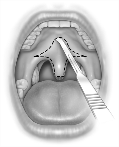
While still grasping the uvula in its new position, the planned incision is outlined with a marker or with a number 12 blade, as shown in Figure 34.1. This gothic arch-shaped incision generally has its apex within 5–10 mm of the hard–soft palate junction and flares laterally to allow for advancement of the lateral palate. The farther laterally these incisions extend, the greater the elevation of the lateral aspects of the palate. The incision will be carried caudally onto the uvula in a mirror image of the palatal incision. Unless vertical relaxing incisions are necessary, as described previously, it is recommended that the incisions be kept away from the free edge of the palate, to lessen the chance of scar contracture.

< div class='tao-gold-member'>

Only gold members can continue reading. Log In or Register to continue

Stay updated, free dental videos. Join our Telegram channel

Jul 24, 2016 | Posted by in General Dentistry | Comments Off on The uvulopalatal flap

VIDEdental - Online dental courses

Get VIDEdental app for watching clinical videos