Successful endodontic treatment depends on the restorative treatment that follows. The connection between endodontic treatment and restorative dentistry is well accepted, but the best restorative approaches for endodontically treated teeth have always been somewhat controversial. A plethora of information from various sources contributes to the controversy and much of it is contradictory. With the emergence of implants in mainstream dentistry, there has been more emphasis on long-term outcomes and on evaluating the “restorability” of teeth prior to endodontic treatment. The long-term viability of endodontically treated teeth is no longer a “given” in the implant era. In consequence, some teeth that might have received endodontic treatment in the past are now extracted and replaced with implant-supported prostheses if they are marginally restorable or it makes more sense in the overall treatment plan. As it is not possible to review here all the literature on the restoration of endodontically treated teeth, this article focuses primarily on current concepts based on the literature from the past 10 years or so, and provides treatment guidelines based on that research.
The primary goals of endodontic treatment are straightforward: to debride and disinfect the root canal space to the greatest possible extent, and then seal the canals as effectively as possible. The materials and techniques change somewhat over time, but not the ultimate goals. The primary goals of restorative treatment are to restore teeth to function and comfort and in some cases, aesthetics. Once again, the materials and techniques change, but not the ultimate goals of treatment. Successful endodontic treatment depends on the restorative treatment that follows. The connection between endodontic treatment and restorative dentistry is well accepted, but the best restorative approaches for endodontically treated teeth have always been somewhat controversial. The topic is no less controversial today, despite the massive (and ever growing) amount of information available from research, journal articles, courses, “expert” opinions, and various sources from the Internet. In fact, information overload contributes to the controversy because so much of it is contradictory.
With the emergence of implants into the mainstream of dentistry, there has been more emphasis on long-term outcomes and on evaluating the “restorability” of teeth prior to endodontic treatment. Patients are not well served if the endodontic treatment is successful but the tooth fails. The long-term viability of endodontically treated teeth is no longer a “given” in the implant era. In consequence, some teeth that might have received endodontic treatment in the past are now extracted and replaced with implant-supported prostheses if they are marginally restorable or it makes more sense in the overall treatment plan. It is not possible to review in one article all the literature on the restoration of endodontically treated teeth. This article therefore focuses primarily on current concepts based on the literature from the past 10 years or so, and provides treatment guidelines based on that research.
The relationship between endodontics and restorative dentistry
Long-term success of endodontic treatment is highly dependent on the restorative treatment that follows. Once restored, the tooth must be structurally sound and the disinfected status of the root canal system must be maintained. Because microorganisms are known to be the primary etiologic factor for apical periodontitis and endodontic failure, contamination of the root canal system during or after restorative treatment is considered an important factor in the ultimate success or failure. Exposure of gutta-percha to saliva in the pulp chamber results in migration of bacteria to the apex in a matter of days. Endotoxin reaches the apex even faster. The importance of the coronal restoration in successful endodontic treatment has been shown in several studies. Delayed restoration has been show to result in lower success rates.
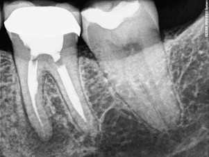
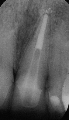
Successful restorative treatment is also greatly influenced by the execution of the endodontic procedures. Radicular and coronal tooth structure should be preserved to the greatest possible extent during endodontic procedures. Root canal preparations should attempt to preserve dentin in the coronal one-third of the root. There is no reason to prepare a “coke bottle” type of canal preparation ( Fig. 1 ) that weakens the tooth unnecessarily. Access preparations similarly should be made in such a way that cervical dentin is preserved. The roof of the pulp chamber should be removed carefully, preserving the walls of the chamber as much as possible. The chamber walls should be prepared only to the extent that is necessary for adequate access for endodontic treatment.
Many, if not most endodontically treated teeth today are restored with adhesive materials. Adhesive materials provide an immediate seal and some immediate strengthening of the tooth. These materials are generally not dependent on gross mechanical retention, so tooth structure can be preserved. The sections that follow discuss basic principles of adhesive dentistry and some of the limitations, pitfalls, and special problems presented by endodontically treated teeth.
Bonding to enamel
Enamel is a highly mineralized tissue that is often present along the margins of access preparations of anterior teeth and sometimes in posterior teeth. Effective bonding procedures for enamel were first reported in 1955. An acid, such as 30% to 40% phosphoric acid, when applied to enamel will cause selective dissolution of the enamel prisms. Microporosities are created within and around the enamel prisms, which can be infiltrated with a low-viscosity resin and polymerized, creating resin “tags” that provide micromechanical retention and a strong, durable bond. It is important to prevent contamination of etched enamel with blood, saliva, or moisture that will interfere with bonding. Poorly etched enamel leads to staining at the margins of the restoration. A good enamel bond protects the less durable underlying dentin bond.
Bonding to metal-ceramic and all-ceramic restorations
Access cavities are often made through metal-ceramic or all-ceramic materials, so attaining an effective, durable bond is important. Like enamel, the porcelain margins can be etched (usually with a 1-minute etch of 10% hydrofluoric acid) to create microporosities, which may be infiltrated with resin and polymerized. Application of silane to the etched porcelain surface enhances the bond. Etched ceramic materials form a strong, durable bond with resin.
Bonding to dentin: resin-based materials
A smear layer is formed when the dentin surface is cut or abraded with hand or rotary instruments. The smear layer adheres to the dentin surface and plugs the dentinal tubules; it consists of ground-up collagen and hydroxyapatite and other substances that might be present such as bacteria, salivary components, or pulpal remnants. The smear layer cannot be rinsed or rubbed off, but can be removed with an acid or chelating agent. Some dentin adhesives remove the smear layer, whereas others penetrate through the layer and incorporate it into the bond. Both approaches may be used successfully.
Bonding to dentin is more complex than bonding to enamel or ceramic. Dentin is a wet substrate and restorative resins are hydrophobic (“water hating”). Dentin consists of approximately 50% inorganic mineral (hydroxyapatite) by volume, 30% organic components (primarily type 1 collagen), and 20% fluid. The wet environment and relative lack of a mineralized surface made the development of effective dentin adhesives a challenge.
The first successful strategy for dentin adhesion was reported by Nakabayashi and colleagues in 1982. Their ideas were not widely accepted until later in the decade. Nakabayashi showed that resin could be bonded to dentin by demineralizing the dentin surface and applying an intermediate layer that would bond to dentin and restorative materials. Although not as durable and reliable as enamel bonding, dentin bonding forms the foundation for many of today’s restorative procedures. Nakabayashi’s technique was later simplified by combining some of the steps.
The limitations of dentin bonding
From the restorative literature it is known that dentin bonding materials have limitations, many of which are related to polymerization shrinkage. When resin-based materials polymerize, individual monomer molecules join to form chains that contract as the chains grow and intertwine, and the mass undergoes volumetric shrinkage. Resin-based restorative materials shrink from 2% to 7%, depending on the volume occupied by filler particles and the test method. The force of polymerization contraction often exceeds the bond strength of dentin adhesives to dentin, resulting in gap formation along the surfaces with the weakest bonds. Resins, even in thin layers, generate very high forces from polymerization contraction.
Another limitation of dentin bonding is deterioration of the resin bond over time. This process is well documented in vitro and in vivo. Loss of bond strength is first detectable in the laboratory at 3 months. Interfacial leakage increases as the bond degrades. Functional forces have been shown to contribute to the degradation of the resin bond in restorative applications.
The limitations of bonding in the root canal system
The root canal system has an unfavorable geometry for resin bonding. Configuration factor or C-Factor, the ratio of bonded to unbonded resin surfaces, is often used as a quantitative measure of the geometry of the cavity preparation for bonding. The greater the percentage of unbonded surfaces, the less stress is placed on the bonded surfaces from polymerization contraction. The unbonded surfaces allow plastic deformation or flow within the resin mass during polymerization. A Class 4 cavity preparation, for example, has a favorable geometry with a ratio of less than 1:1. There are few if any walls that directly oppose each other, and more than half of the resin surfaces are not bonded. In the root canal system the ratio might be 100:1, because virtually every dentin wall has an opposing wall and there are minimal unbonded surfaces. Any ratio greater than 3:1 is considered unfavorable for bonding. Because of this unfavorable geometry, it is not possible to achieve the gap-free interface with current materials. Interfacial gaps are virtually always present in bonded restorations in restorative dentistry, obturating materials, and bonded posts, and gap formation increases with time.
The potential problems of using adhesive materials deep in the root canal system
Performing the bonding steps is problematic deep in the root canal system. Uniform application of a primer or adhesive can be difficult. Once the primer is applied, the volatile carrier must be evaporated. This process can also be problematic deep in the canal. If the acetone or alcohol carrier is not completely removed, the bond is adversely affected. An in vitro post study by Bouillaguet and colleagues reported lower bond strengths were achieved bonding in the root canal system than bonding to flat prepared samples of radicular dentin.
Compatibility problems with dual-cure and self-cure resins
Because penetration with a curing light is limited in the root canal system, dual-cure or self-cure resin adhesives must be used. Dual-cure resins contain components that provide rapid light polymerization in those areas where the curing light penetrates effectively and a slower chemical polymerization in those areas where the light is not effective. Adhesives and sealers that contain a self-cure component have compatibility problems with self-etching dentin adhesive systems (ie, sixth and seventh generation), so they should be used with “fourth generation” etch-and-rinse adhesives.
Irrigating solutions and medicaments
Sodium hypochlorite is commonly used as an endodontic irrigant because of its antimicrobial and tissue dissolving properties. The antimicrobial properties of sodium hypochlorite are largely due to it being a strong oxidizing agent, but as a result it leaves behind an oxygen-rich layer on the dentin surface. The same applies to chelating agents that contain hydrogen peroxide. Oxygen is one of the many substances that inhibit the polymerization of resins. When dentin bonding agents are applied to an oxygen-rich surface, low bond strengths are achieved and microleakage is increased. A reducing agent, such as ascorbic acid and sodium ascorbate, applied to the dentin surface will reverse the negative affects of sodium hypochlorite. A final soak with ethylenediamine tetra-acetic acid (EDTA) has also been reported to be effective.
Basic principles for restoring endodontically treated teeth
Although many aspects of the restoration of endodontically treated remain controversial, there are several areas of general agreement. One of the best documented principles is cuspal coverage. Several studies evaluated factors that affected the survival of endodontically treated teeth. Cuspal coverage was the most consistent finding. In one study, teeth with cuspal coverage had a 6 times greater survival rate than teeth without cuspal coverage. Another study showed teeth without cuspal coverage had only a 36% survival rate after 5 years.
Another important principle is preservation of tooth structure. Coronal tooth structure should be preserved to support the core buildup. Several studies identify remaining coronal tooth structure as the most important factor in tooth survival in teeth with posts.
As stated previously, radicular tooth structure should also be preserved. For most teeth that are to receive posts, no additional dentin should be removed beyond what is necessary to complete the endodontic treatment. If a tooth is prepared for a 0.06 tapered preparation, a 0.06 tapered post should “drop right in” without removing additional radicular dentin.
There is wide general agreement that the “ferrule effect” is important. In dentistry, the ferrule refers to the cervical tooth structure that provides retention and resistance form to the restoration and protects it from fracture. In one study, teeth with a ferrule of 1 mm of vertical tooth structure doubled the resistance to fracture compared with teeth restored without a ferrule. Other studies have shown maximum beneficial effects from a ferrule of 1.5 to 2 mm. The “ferrule effect” is important to long-term success when a post is used. In anterior teeth, the lingual aspect of the ferrule is the most important part. If the height of the remaining dentin is not sufficient to create an adequate ferrule, crown lengthening, orthodontic extrusion, or extraction may be indicated.
Teeth restored with posts
Endodontically treated teeth often have substantial loss of tooth structure and require a core buildup. If retention and resistance of the core are compromised, a post may also be necessary. Custom cast posts and cores or prefabricated metal posts were the standard for many years. In the past 10 years or so, fiber-reinforced composite posts have gained popularity.
Indications for a post
The primary function of a post is to retain a core in a tooth with extensive loss of coronal tooth structure. Posts should not be placed arbitrarily, however, because preparation of a post channel adds a degree of risk to a restorative procedure:
- •
Disturbing the seal of the root canal filling, which may lead to microleakage
- •
Removal of sound tooth structure, which weakens the root and may result in premature loss due to root fracture
- •
Increased risk of perforation.
Some studies report higher failure rates in endodontically treated teeth with posts than without. The finding was not universal, however.
Traditional thought has been that posts do not “reinforce” the root; this was apparently true for metal posts, but there is a growing body of evidence that fiber posts may strengthen the root and make it more resistant to fracture. To date, 9 studies have shown a strengthening effect while 3 have shown no effect.
Metal posts have a high modulus of elasticity, which means that they are stiff and able to withstand forces without distortion. When a force is placed on a tooth containing a stiff post, it is transmitted to the less rigid root dentin, and concentrates at the apex of the post. Stress concentration in the post/root complex increases the chances of fracture.
To overcome the concerns about unfavorable stress distribution generated by metal posts, fiber-reinforced composite resin posts were introduced in 1990, with the aim of providing more elastic support to the core. The reduced stress transfer to tooth structure was claimed to reduce the likelihood of root fracture. Posts made of materials with a modulus of elasticity similar to dentin are more resilient, absorb more impact force, and distribute the forces better than stiffer posts.
Types of posts
Posts can be categorized by modulus of elasticity, composition, fabrication process, shape, and surface texture.
Rigid Post Systems
- •
Metal
- –
custom cast
- –
prefabricated
- –
- •
Zirconium and ceramic.
Posts traditionally were made of metal, and were either custom cast or prefabricated. Custom cast posts and cores are made of precious or nonprecious casting alloys; prefabricated posts are typically made of stainless steel, nickel chromium alloy, or titanium alloy. With the exception of the titanium alloys, they are very strong.
Parallel metal posts are more retentive than tapered posts and induce less stress into the root, because they have less wedging effect and are reported to be less likely to cause root fractures than tapered posts. In a retrospective study, Sorensen and Martinoff reported a higher success rate with parallel metal posts than tapered posts. Tapered posts, on the other hand, require less dentin removal because most roots are tapered.
Prefabricated posts can be further divided in active or passive posts. Most active posts are threaded and intended to engage the walls of the canal, whereas passive posts are retained primarily by the frictional retention of the luting agent. Active posts are more retentive than passive posts, but introduce more stress into the root than passive posts. Active posts have very limited indications, and are only recommended when the need for retention is the overriding factor.
One factor that has reduced the use of metal posts is aesthetics. Metal posts may be visible through translucent all-ceramic restorations, and even with less translucent restorations may cause the marginal gingiva to appear dark. These concerns have led to the development of posts that are white or translucent. Among the materials used for “aesthetic” posts are zirconium and other ceramic materials. These posts will work clinically, but have several disadvantages.
Among rigid posts, zirconium is stiffer and more brittle than metal. Zirconium posts were shown to cause significantly more root fractures than fiber posts in vitro. When compared with custom cast and fiber posts, ceramic posts had a lower failure load in vivo and in vitro. As a group, they tend to be weaker than metal posts, so a thicker post is necessary, which may require removal of additional radicular tooth structure. Zirconium posts cannot be etched, therefore it is not possible to bond a composite core material to the post, making core retention a problem. Retrieval of zirconium and ceramic posts is very difficult if endodontic retreatment is necessary or if the post fractures. Some ceramic materials can be removed by grinding away the remaining post material with a bur, but this is a tedious and risky procedure. It is impossible to grind away a zirconium post. In many cases, excessive removal of dentin is necessary to remove a zirconium post. For these reasons, ceramic and zirconium posts should be avoided.
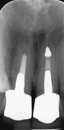
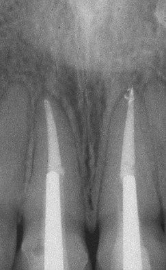
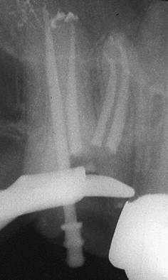
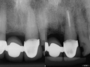
Metal and zirconium posts are all radiopaque and clearly visible on a radiograph ( Figs. 2 and 3 ). The radiopacity of titanium is similar to that of gutta-percha, and therefore sometimes the presence of a titanium post is difficult to detect on radiographs ( Fig. 4 ).
Nonrigid Post Systems: Fiber Posts
- •
Carbon fiber
- •
Glass fiber
- •
Quartz fiber
- •
Silicon fiber.
The first composite reinforced fiber posts were made with carbon fibers, which were arranged longitudinally and embedded in an epoxy resin matrix. The black carbon fibers were rapidly replaced by more esthetic white and translucent glass and quartz fibers, which are now the standard components in fiber posts. These posts are commonly used in aesthetically demanding areas.
The main advantage of fiber posts is the uniform distribution of forces in the root, which results in fewer catastrophic failures than occur with metal posts if an adequate ferrule is present. Several in vitro studies report that teeth restored with nonrigid posts have fewer catastrophic, irreparable root fractures when tested to failure. Clinical studies of fiber post systems also report successful multiyear service with few or no root fractures. A retrospective clinical study of carbon fiber posts and custom cast posts reported root fractures in 9% of teeth restored with cast posts, and no root fractures in teeth restored with fiber posts after 4 years. In a long-term retrospective study of the clinical performance of fiber posts by Ferrari and colleagues, a 7% to 11% failure rate was reported for 3 different types of fiber posts after a service period of 7 to 11 years. Half of the failures were classified as endodontic failures, the other half were mechanical failures. Out of 985 posts evaluated, the nonendodontic failures consisted of one root fracture, one fiber post fracture, 17 crown dislodgements, and 21 failures due to post debonding. The mechanical failures were always related to the lack of coronal tooth structure. In a review by Dietschi and colleagues it was concluded that nonvital teeth restored with composite resin or composite resin combined with fiber posts currently represent the best treatment option.
Although fiber posts offer several advantages, they do have limitations. Posts and core foundations are subjected to repeated lateral forces in clinical function. Because nonrigid posts have a modulus of elasticity and flexural strength close to that of dentin, they flex under occlusal load. When there is an adequate ferrule, the cervical tooth structure itself resists lateral flexion. However, in structurally compromised teeth that lack cervical stiffness from dentin walls and an adequate ferrule, a flexible post may result in micro-movement of the core and coronal leakage, which in turn may lead to caries or loss of the core and crown.
Fiber posts were shown to lose flexural strength if they are submitted to cyclic loading or to thermocycling due to degradation of the matrix in which the fibers are embedded. The strength of fiber posts varied between brands, but was directly related to post diameter and was reduced by thermocycling.
Parallel fiber posts are more retentive than tapered posts. However, in a clinical study by Signore and colleagues no difference was found in the long-term survival rate of maxillary anterior teeth restored with tapered or parallel-sided glass-fiber posts and full-ceramic crown coverage. The overall survival rate was reported to be 98.5%. Most fiber posts are relatively radiolucent and have a different radiographic appearance than traditional metal posts ( Fig. 5 ).
It has been shown that the retention of fiber posts relies mainly on mechanical (frictional) retention rather than bonding, similar to metal posts. Several in vitro studies have confirmed the presence of gaps in the interface between the luting composite resin of the fiber post and the root canal wall, and that the bond strengths between fiber posts and dentin are low, about 5 to 6 MPa. This situation is due primarily to the unfavorable bonding environment of the root canal system, as discussed earlier.
Post length and remaining root canal filling
The length of a post is dictated by several factors, some of which are conflicting. Most of the studies on optimum post length were done with metal posts, but there is no compelling evidence that the principles of post length are different for fiber posts.
Braga and colleagues evaluated the force required to remove glass fiber and metallic cast posts with different lengths. Irrespective the post type, posts with 10-mm length had higher retention values than posts with 6-mm length. In a study by Büttel and colleagues, teeth restored with glass-fiber posts with insertion depths of 6 mm resulted in significantly higher mean failure than teeth with post space preparation of 3 mm. The retention of fiber posts was shown to be directly proportional to the insertion length in resin cubes.
Several “rules” have been suggested for passively fitting posts:
- •
The post length below the alveolar crest should be at least equal to the length above the alveolar crest. Sorensen and Martinoff reported 97% success if post length at least equaled the crown height.
- •
The post should end halfway between the crestal bone and the root apex.
- •
A post should extend at least apical to the crest of the alveolar bone.
Another factor that influences post length is the length of the remaining apical root canal filling. Several studies have investigated apical seal following post space preparation and have reported that 3 to 5 mm of gutta-percha is the minimum recommended, and longer is better ; this is sometimes dictated by the length of the canal. Post placement in a long root, for example, a canine of 28 mm, allows more apical root canal filling, as placing a 23-mm post is unnecessary. When using the criterion that the post should extend beyond the apical crest, teeth with bone loss need longer posts than teeth with normal bone height.
Light-transmitting fiber posts
Although it seems logical that translucent posts would transmit light for enhancement of cure deeper in the canal, there seems to be no consensus in the literature on this issue. The use of a light-transmitting translucent fiber post was reported to increase the depth of resin cure in several in vitro studies, but other studies reported minimal or no benefits from translucent posts. One study evaluated the influence of fiber-post translucency on the degree of conversion of a dual-cure composite. Low degrees of conversion were found for the medium and deep depths. Another in vitro study measured light transmission through 4 different posts of a standard length of 10 mm. All posts evaluated showed some light transmission capacity, but with values lower than 40% of incident light. One post demonstrated less than 1% light transmission. Goracci and colleagues evaluated the light transmission of several fiber posts. These investigators reported no light transmission through 2 posts, and for all other posts light intensity decreased from coronal to apical, and rose again at the apical tip. Light transmission was significantly higher at the coronal level. Another study showed that even without a post, the luminous intensity inside the canal decreased to levels that are insufficient for polymerization, especially in the apical third. Based on these findings, the use of light-cured resin cements for post placement cannot be recommended. The benefits of light-transmitting posts are unclear.
Is there benefit to placing a post after endodontic treatment of a tooth with a crown?
In most cases, when preparing endodontic access through a crown there is no way of knowing the amount or strength of the underlying tooth structure, which is a particular concern in small teeth and bridge abutments.
When an access preparation is made through a crown, retention is lost. When the access opening is restored with amalgam or composite resin, the retention values are restored. When the access opening is restored with a post, the retention is greater than before the access was prepared.
There is growing evidence that the insertion of a fiber post can also increase fracture resistance of teeth with crowns. An in vitro study has shown that placement of fiber posts can improve fracture resistance in maxillary premolars under full-coverage crowns. The use of fiber posts in endodontically treated maxillary incisors with different types of full-coverage crowns increased their resistance to fracture and improved the prognosis in case of fracture. The type of crown was not a significant factor affecting fracture resistance, whereas the presence of a post was. D’Arcangelo and colleagues showed that fiber posts significantly increased mean load values for maxillary central incisors prepared for veneers.
Based on these findings, it seems retention will be enhanced by a post, and fracture resistance will probably be improved as long as no additional tooth structure is removed. The authors routinely place fiber posts in bridge abutments and small teeth with crowns ( Fig. 6 ).
Post placement
Advantages of Immediate Post Placement
The literature on the timing of the post space preparation is inconclusive. Some studies showed less leakage after immediate post space preparation, whereas other articles showed no difference Some in vitro studies showed that delayed cementation of a fiber post resulted in higher retentive strengths. Scanning electron microscopy examination revealed a more conspicuous presence of sealer remnants on the walls of immediately prepared post spaces. Remnants of sealer and gutta-percha may impair adhesive bonding and resin cementation of fiber posts. Therefore, it is important to clean the root canal walls before conditioning the dentin for post placement. Acid-etching of the prepared post space and EDTA irrigation combined with ultrasonics are reported to be an effective method. The use of magnification can facilitate inspection of the post space for cleanliness.
Immediate preparation for post placement following obturation has several advantages. The operator has a great familiarity with the root canal morphology, working lengths, and reference points of the root canal system. In addition, placement of a temporary post and restoration can be avoided, as maintaining the temporary seal can be difficult. In vitro studies by Fox and Gutteridge and by Demarchi and Sato showed that teeth restored with temporary posts leaked extensively.
Luting fiber posts
Fiber posts are usually luted with lightly filled composite resins. Light penetration is limited, so dual-cure of self-cure luting resins must be used. Some luting resins are used with a separate etchant and primer (total-etch method), whereas others contain an acidic primer in the luting cement (self-etching method). More recently a third category has been added (self-adhesive method), in which there is no etching and no primer. Several studies have evaluated these luting cements.
Goracci and colleagues reported that the values achieved by total-etch method were significantly higher than self-etch resin cements. Transmission electron microscopy analysis revealed that the acidic resin monomers responsible for substrate conditioning in self-etch and self-adhesive resin cements did not effectively remove the thick smear layer created on root dentin during post space preparation. Valandro and colleagues similarly concluded that more reliable bond strengths in the dowel space might be achieved when using total-etch adhesive systems instead of self-etching adhesives. A study by Radovic and colleagues concluded that the use of self-etching resin luting systems offer less favorable adhesion to root canal dentin in comparison with the total-etch and self-adhesive approaches.
Self-adhesive cements were introduced in 2002 as a new subgroup of resin cements. Self-adhesive cements do not require any pretreatment of the tooth substrate. The cement is mixed and applied in a single clinical step. The application of self-adhesive cements to radicular dentin does not result in the formation of hybrid layer or resin tags. Lührs and colleagues found the shear bond strength of self-adhesive resin cements to be inferior compared with conventional composite resin cements. The sealing ability of 2 self-adhesive resin cements was shown to be significantly lower than a self-etching and 2 conventional dual-cure resin cements. The investigators concluded that although the bonding effectiveness of self-adhesive cements seems promising, their interaction with root dentin might be too weak to minimize microleakage at the post-cement-dentin interface. In another study by Vrochari and coworkers, the degree of cure of 4 self-etching or self-adhesive resin cements in their self-curing mode was very low. The values obtained in the dual-curing mode were also low.
Self-adhesive cements offer a new, simpler approach, but the efficacy of many recently marketed products is not known, and there are few data in the literature regarding their in vitro or clinical performance. At this point in their development, the literature generally shows them to be inferior to the total-etch method.
Stay updated, free dental videos. Join our Telegram channel
VIDEdental - Online dental courses


