Fig. 8.1
(a) Enamel surface near the enamel-cementum junction. Prints or depressions of the proximal zone of ameloblasts form alignments (asterisk). (b) In the cervical zone, prisms (P) are aligned and parallel. The dentinoenamel junction (black arrows) separates the cervical enamel from dentin (D). In (c), the prismatic enamel (P) is covered by a thin layer of aprismatic enamel (Apr), ending at the enamel surface (S). In (d), in older enamel, the same distribution is visible
Non-carious cervical lesions refer to the loss of tooth structure at the cemento-enamel junction, and they are unrelated to bacterial action (see Chapter 2 of this book). Horizontal scratch marks are characteristics of the abrasion processes. Shallow, grooved, and wedge-shaped cervical lesions were identified. Corrosion induces a smoother appearance. Enamel displays a honeycomb structure, whereas dentin exhibits an undulating or rippled surface. In vitro study notes fracture and chipping of enamel in the cervical region (Nguyen et al. 2008). The AFM nanoindentation was used to probe the mechanical and tribological properties of enamel rods. Microhardness and elastic properties are significantly higher in the head region of the rod rather than in the tail region (Jeng et al. 2011).
Enamel, dentin, and cementum are coupled at the DEJ. The DEJ is a complex structure associating two calcified tissues, preventing the propagation of cracks from enamel to dentin. It is a scalloped structure with convexities toward dentin and concavities toward enamel (Shimizu & Macho 2007, Brauer et al. 2010, Beniash et al 2006) (Figs. 8.2 and 8.3).


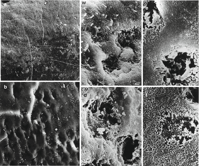
Fig. 8.2
(a) Water-treated enamel surface. (b) Enamel surface after water treatment. (c) Water treatment (d) enamel surface treated with chloroform-methanol. (e) Water treatment revealed the end of rods. (f) Chloroform-methanol treatment of enamel surface. The distal ending of rods displays large porosities, whereas in the interrod enamel, inter-crystallite porosities appear after the removal of extracellular lipids

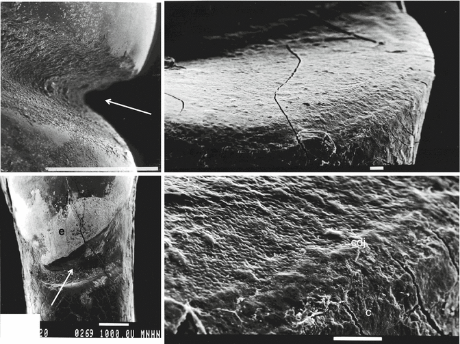
Fig. 8.3
Cervical erosion (arrows); C cementum, cdj cemento-dentinal junction
8.2.1.2 Dentin
In the superficial part of the outer dentin, two different layers have been identified. The thin (8–15 μm thick) Tomes’ granular layer is composed by calcospheritic (oval or round) structures. This zone contains a few minute tubules. The thin curved tubules are scarce and bent around the calcospheritic structures. Between the calcospherites, interglobular spaces are hypomineralized.
The subjacent layer (Hopewell-Smith hyaline layer, 8–15 μm thick) displays a small number of tubules. These tubules are straight, aligned, and at right angles to the tooth surface. The bulk of inner dentin, located around the dental pulp, is implicated in circumpulpal dentin formation. Usually, cells are alive and tubules are filled with odontoblast processes and their lateral branching. The zone of sclerotic dentin is thinner in these areas compared with normal dentin, and the changes occurring near the dentin surface are due to abrasion, abfraction, erosion, and wedge-shaped lesions. The carious decay contributes to the occlusion of the tubules by intratubular mineralizations (whitlockite crystallites, tricalcium phosphate) (Goldberg 2014; Tay and Pashley 2004) (Figs. 8.3 and 8.4).


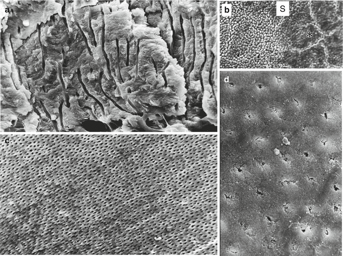
Fig. 8.4
(a) Outer globular Tomes’ granular structure. (b) Cementum covering the root surface (S). Left part of the figure: acid-etched enamel. (c) Circumpulpal dentin. (d) Sclerotic dentin
AFM-based nanoindentations found the DEJ to only be 11.8 μm across. Micro-Raman indicated a DEJ width of 7.0 μm, while dynamic modulus mapping indicated it was less than 1 μm across. The variations based on enamel-dentin phase intermixing suggest that DEJ is a biomimetic model for interfaces joining dissimilar materials (Marshall et al. 2003).
8.2.1.3 Cervical Lesions
Cracks generated in enamel stop at the DEJ, preventing catastrophic failure of the tooth. The major emerging question concerns the effects of organic matrix and water on the structural organization and how tooth microhardness and fracture toughness are affected. The removal of organic matrix resulted in 23 % increase in microhardness and 46 % decrease in fracture toughness. In contrast, water does not seem to influence these parameters. Moreover, the removal of organic matrix weakened the DEJ, leading to the formation of longer and more numerous cracks. Delamination of dentin and enamel along the DEJ suggests a strong physical bond between dentin and enamel crystals at the interface.
Retzius lines in the human cervical enamel display a staircase configuration. The Retzius lines have a curvilinear configuration, and crystallites are deficient in composition. Finally, direct contact is occurring between lattice fringes of dentin and enamel crystals, fusion being observed between enamel and dentin crystals. The DEJ is weak in mechanical and/or chemical attacks (Fig. 8.3).
8.2.2 Mechanical and Tribological Properties of the DEJ
The DEJ is a complex structure and poorly defined, bridging enamel to the bulk dentin. This structure constitutes a biomimetic model of a structure uniting dissimilar materials. Cracks cannot traverse the DEJ and/or produce cracks in dentin. The fracture toughness values for enamel were evaluated as 0.6–0.9 MPa.m1/2 (Marshall et al. 2001) (Figs. 8.3 and 8.4).
Many reports suggest that abfraction lesion formation is caused by the physical overloading of enamel (Rees and Hammadeh 2004). In the crowns of human teeth, beneath enamel, a 200–300 μm zone of resilient (less mineralized and elastic) dentin has been found in the DEJ (Zaslansky et al. 2006). The strain distribution in the 200 μm thick zone in dentin beneath the DEJ is a structural adaptation for transferring and minimizing stress (Wang and Weiner 1997). Root dentin is highly anisotropic in fracture behavior. Coronal dentin has a typical brittle fracture behavior along peritubular dentin, and this should be taken into consideration that dentin is not homogeneous with respect to fracture properties (Wang 2005).
Peritubular dentin located at some distance from the DEJ gradually thickens with increasing depth in the bulk dentin. A significant reduced stiffness of the superficial zone has been reported compared to bulk dentin. In mid-buccal regions of teeth, the average was 3.5GPa, compared with the 9.7 GPa in the mid-lingual regions. It was concluded that the superficial layer behaves as a stress-relieving layer between enamel and bulk dentin.
Microhardness and elastic modulus are higher in the head region of enamel rods. They decrease from the enamel surface toward the DEJ. Head and tail area are questionable structures, and many ultrastructural studies deny the reality of this organization. The mechanical and nanotribological properties of enamel rods depend on HAp orientation inside each rod. The wear rate increases with an increasing distance from the outer enamel surface along the longitudinal axis of the enamel rod reaching the DEJ (Jeng et al. 2011).
The DEJ exhibits a scalloped appearance. The scalloped model has higher maximum tensile stresses than the straight model, but axial pressures would push the two tissue apart, leading to delamination of the DEJ during loading (mastication). There is a direct correlation between prism decussation and scallop magnitude. The scallops are linked with the response to high bite forces. Exaptation is used for a function other than what is developed by natural selection.
8.2.3 Specific Composition of the DEJ
The enamel layer has a rich content in enamel proteins (amelogenins, enamelin, tuftelin, and other molecules). The proteins are characterized by the presence of high serine, glutamic acid, and glycine content. Components rich in proline and histidine are lost during enamel development and maturation (Glimcher et al. 1964).
These extracellular matrix proteins contribute to initiate the formation of the large and elongated enamel crystallites. By contrast, the ECM dentin molecules are composed by 90 % collagen, namely, type I collagen (Lin et al. 1993). The dentinoenamel junction is a fibril-reinforced bond, which is mineralized to a moderate degree.
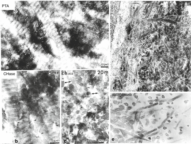
Other types of collagen are scarce (type III and V collagens), but actually present. Deposition of dentin crystallites occurs (1) within the collagen gap regions (due to the quarter stagger structure of collagen fibrils), (2) along the collagen fibrils network, and (3) bridging inter-collagen spaces. The nucleation and crystal growth of dentin crystals are promoted and developed by a series of non-collagenous proteins, namely, phosphorylated proteins (SIBLINGs) (Fig. 8.5a, b). Five of them play crucial roles in the mineralization process: the dentin phosphoprotein, dentin matrix protein-1, bone sialoprotein, osteopontin, and MEPE.


