Introduction
The objective of this study was to compare the degrees of skeletal and dental asymmetry between subjects with Class II subdivision malocclusions and subjects with normal occlusions by using cone-beam computed tomography.
Methods
Thirty subjects with Angle Class II subdivision malocclusions (mean age, 13.99 years) and 30 subjects with normal occlusions (mean age, 14.32 years) were assessed with 3-dimensional cone-beam computed tomography scans. Independent t tests were used to compare orthogonal, linear, and angular measurements between sides and between groups.
Results
Total mandibular length and ramus height were shorter on the Class II side. Pogonion, menton, and the mandibular dental midline were deviated toward the Class II side. Gonion and the anterior condyle landmark were positioned more posteriorly on the Class II side. The mandibular dental landmarks were located more latero-postero-superiorly, and the maxillary dental landmarks more latero-antero-superiorly on the Class II side. There was loss of maxillary arch length, and the mandibular molar was closer to the ramus on the Class II side.
Conclusions
The etiology of Class II subdivision malocclusions is primarily due to an asymmetric mandible that is shorter and positioned posteriorly on the Class II side. A mesially positioned maxillary molar and a distally positioned mandibular molar on the Class II side are also minor contributing factors.
Class II subdivisions are estimated to account for up to 50% of all Class II malocclusions and are among the most common dental asymmetries in the orthodontic population. Radiographic evaluation of Class II subdivision malocclusions have traditionally included submentovertex (SMV), posteroanterior (PA) cephalograms, and pantomographs. These 2-dimensional (2D) radiographs can be misleading, since complex 3-dimensional (3D) structures are projected onto flat 2D surfaces, creating distortion and subsequent magnification errors. Several studies have suggested that PA cephalograms have limitations in methodology and reliability, including lack of reproducibility and errors in landmark identification. In addition, slight asymmetries of the external auditory meatus can lead to rotations of the head and cause erroneous results. Panoramic imaging has also been used to assess mandibular asymmetries and evaluate Class II subdivision malocclusions, but many authors advocate caution in making absolute measurements or relative comparisons because of image distortion and positioning errors. SMV images have similar pitfalls, since maxillary structures are not easily detected and there is significant distortion in mandibular structures.
Case-controlled studies of Class II subdivision malocclusions with 2D radiographs failed to show any significant skeletal asymmetries between the groups. Alavi et al were the first to determine that Class II subdivisions result mainly from asymmetry in the mandibular first molars. Rose et al confirmed those results and concluded that Class II subdivisions occur from distal positioning of the mandibular first molars on the Class II side. Janson et al showed similar results and concluded that subdivision malocclusions were primarily due to dentoalveolar asymmetry without unusual skeletal or positional asymmetries. Most recently, Azevedo et al evaluated Class II subdivision patients with apparent facial asymmetry and concluded that the subdivision was primarily dentoalveolar with minimal skeletal involvement.
Cone-beam computed tomography (CBCT) and other 3D imaging modalities have an advantage in diagnosing skeletal and dental asymmetries, since true anatomic form can be visualized in 3 dimensions and without magnification. CBCT images can also be compared with accuracy and precision since they are true 1-to-1 measurements of craniofacial anatomy. The objective of this study was to compare the degrees of skeletal and dental asymmetry between subjects with Angle Class II subdivision malocclusions and subjects with normal occlusions based on true 3D analysis with CBCT.
Material and methods
In accordance with an institution review board protocol approved by the University of Connecticut, patient records were obtained from 2 orthodontic offices (Dr. Paul Rigali, Wallingford, CT and Dr. Carl Roy, Virginia Beach, VA) that routinely use cone-beam (CB) imaging for diagnostic purposes. Over 3000 patient records (clinical examination records, dental models, photographs, and CBCT scans) were reviewed to select the study sample based on the following inclusion criteria for the subdivision group: (1) complete Class I molar relationship on 1 side of the dental arch with a full-step Class II relationship on the other side; (2) all permanent teeth erupted, including second molars; (3) no malformed or missing teeth, or teeth with extensive restorations or gross decay; (4) no previous orthodontic treatment; (5) no crossbites; (6) no history of facial trauma or medical conditions that might have altered growth; (7) symmetrical spacing or crowding up to 3 mm per arch; (8) age between 10 and 18 years; and (9) no mandibular shift based on the clinical history. The inclusion criteria for the normal occlusion group were as follows: (1) Class I molar relationship on both sides of the dental arch, (2) coincident dental and facial midlines, (3) no clinical dental or skeletal asymmetries, and (4) inclusion criteria 2 through 9 from the subdivision group.
The subdivision and control groups were selected by 2 investigators (P.H.R. and D.S.). The subdivision group consisted of 30 consecutive patients (14 boys, 16 girls), with an average age of 13.99 years (SD, 1.81 years), who came for comprehensive orthodontic treatment with Class II subdivision malocclusions. The control group consisted of 30 consecutive patients (16 boys, 14 girls), with an average age of 14.32 years (SD, 1.67 years), who came for comprehensive orthodontic treatment with normal occlusions.
The 3D scans were acquired before any appliance placement with the patients in maximum intercuspation by using a CBCT unit capable of a full head scan (i-CAT Classic, Imaging Sciences International, Hatfield, Pa). Each volumetric data set was acquired with a 20-second scan time with a 16.0 (diameter) × 22.0 (height) cm field of view at a resolution of 0.4 mm voxels. All images were collected at 120 kVp and 5 mA based on the manufacturer’s specifications. Xoran (i-CAT software, version 2.1.22) was used to reconstruct and export the raw data as a 12-bit-depth digital imaging and communications in medicine (DICOM) file. The DICOM files were imported into Dolphin 3D (version 10.5, Dolphin Imaging, Chatsworth, Calif) for further analysis.
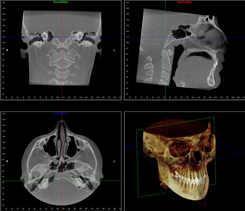
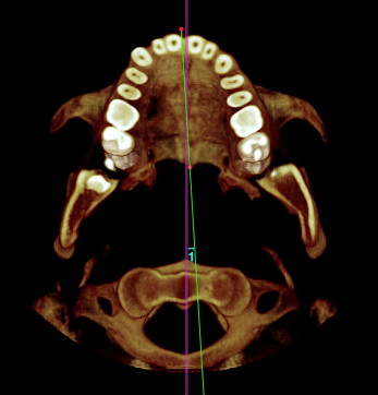
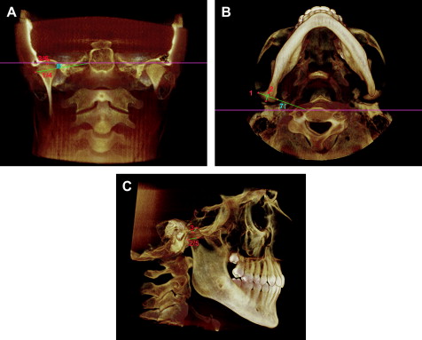
With the Dolphin 3D, the orientation of each 3D volumetric data set was standardized by using the Frankfort horizontal line as the x-axis, the transporionic line as the y-axis, and the midsagittal line as the z-axis ( Fig 1 ). The reference planes were defined by using the volumetric rendering view along with the multiple planar views. A custom analysis was developed, and a total of 34 anatomic landmarks modified from orthodontic cephalometric points were digitized ( Fig 2 , Table I ). The copy-and-paste function of the software was used to export the coordinate data for each defined landmark. The data were then exported into a database (Excel 2007, Microsoft, Redmond, Wash), and orthogonal, linear, and angular measurements were calculated. After the measurements were completed, statistical analyses were calculated with SPSS software (version 14.0.1, SPSS, Chicago, Ill). For convenience of comparison among the Class II subdivision patients, the Class II was set on the right side, which was simply performed by changing the x-coordinates.

| Landmark | Definition |
|---|---|
| Dental | |
| Mx1 | Incisal contact point of the maxillary central incisors |
| Md1 | Incisal contact point of the mandibular central incisors |
| Mx3R and Mx3L | Cusp tip of the maxillary canine |
| Md3R and Mx3L | Cusp tip of the mandibular canine |
| Mx6R and Mx6L | Mesiobuccal cusp tip of the maxillary first molar |
| Md6R and Mx6L | Buccal groove of the mandibular first molar |
| Midface | |
| ANS | The most anterior midpoint of the anterior nasal spine of the maxilla |
| PNS | The most posteror midpoint of the posterior nasal spine of the palatine bone |
| OrR and OrL | The most inferior point on the infraorbital rim of the maxilla |
| Mandible | |
| Me | The most inferior midpoint of the chin on the outline of the mandibular symphysis |
| Pg | The most anterior midpoint of the chin on the outline of the mandibular symphysis |
| GoR and GoL | The midpoint on the angle of the mandible, halfway between the corpus and ramus |
| CdSR and CdSL | The most superior point of the condylar head |
| CdLR and CdLL | The most lateral point of the condylar head |
| CdMR and CdML | The most medial point of the condylar head |
| CdAR and CdAL | The most anterior point of the condylar head |
| CdPR and CdAL | The most posterior point of the condylar head |
| Temporal bone | |
| GlSR and GlSL | The most superior point of the glenoid fossa of the temporal bone |
| GlAR and GlAL | The most inferior point of the articular eminence of the temporal bone |
| PoR and PoL | The most superior point of the external acoustic meatus |
| Reference planes | |
| Axial plane | Frankfort horizontal: a plane that connects the most superior point of the extrernal acoustic meatus (porion) with the most inferior point of the infraorbital rim (orbitale) on the right and left sides. |
| Sagittal plane | A plane constructed with paired midfacial anatomic structures (eg, orbits, frontal process of the maxilla, frontozygomatic suture) |
| Coronal plane | A plane constructed from the transporionic line |
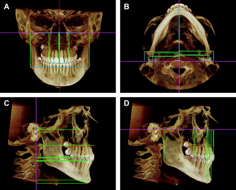
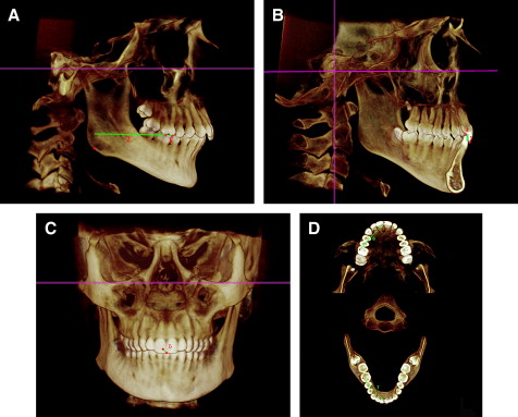
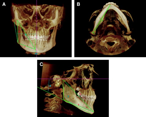
Orthogonal measurements from the volumetric image, defined as perpendicular measurements from each 3D landmark to each of the 3D reference planes, were expressed in millimeters ( Fig 3 ). Linear and angular measurements were expressed in millimeters and degrees for the evaluation of dental and dentoalveolar asymmetry ( Fig 4 , Table II ), mandibular morphologic asymmetry ( Fig 5 , Table III ), maxillary angular asymmetry ( Fig 6 , Table III ), and condylar morphologic asymmetry ( Fig 7 , Table IV ). All measurements were made by 1 examiner (D.S.), who was blinded when digitizing the CB images.
| Variable | Definition |
|---|---|
| Molar differential (mm) | The difference of Md6(z) and Mx6(z) |
| Midline difference (mm) | The absolute difference of Mx1(x) and Md1(x) |
| Overbite (mm) | Mx1(y) minus Md1(y) |
| Overjet (mm) | Mx1(z) minus Md1(z) |
| Mandibular molar position (mm) | Distance (z) between Go and Md6 |
| Maxillary arch length (mm) | Distance (x,y,z) between Mx6 and Mx1 |
| Mandibular arch length (mm) | Distance (x,y,z) between Md6 and Mx1 |
| Variable | Definition |
|---|---|
| Palatal plane to the sagittal plane (°) | Angulation (x,z) between ANS-PNS and the sagittal plane |
| Mandibular length in 3D (mm) | Distance (x,y,z) between CdS and Pg |
| Mandibular length in 2D (mm) | Distance (y,z) between CdS and Pg |
| Ramus height in 3D (mm) | Distance (x,y,z) between CdS and Go |
| Ramus height in 2D (mm) | Distance (y,z) between CdS and Go |
| Corpus length in 3D (mm) | Distance (x,y,z) between Go and Pg |
| Corpus length in 2D (mm) | Distance (y,z) between Go and Pg |
| Gonial angle ( o ) | Angulation (y,z) between CdS, Go, and Pg |
| Mandibular plane angle ( o ) | Angulation (y,z) between Go-Pg and Frankfort horizontal |
| Ramus inclination to the sagittal plane ( o ) | Angulation (x,y) between CdL-Go and the sagittal plane |
| Dental and chin inclination ( o ) | Angulation (x,y) between Md1-Me and the sagittal plane |
| Variable | Definition |
|---|---|
| Medio-lateral diameter of the condylar head in 3D (mm) | Distance (x,y,z) between CdL and CdM |
| Medio-lateral diameter of the condylar head in 2D (mm) | Distance (x,y) between CdL and CdM |
| Anteroposterior diameter of the condylar head in 3D (mm) | Distance (x,y,z) between CdA and CdP |
| Anteroposterior diameter of the condylar head in 2D (mm) | Distance (y,z) between CdA and CdP |
| Superior joint space in 3D (mm) | Distance (x,y,z) between CdS and GlS |
| Superior joint space in 2D (mm) | Distance (x,y) between CdS and GlS |
| Condylar head inclination to the coronal plane ( o ) | Angulation (x,z) between CdL-CdM and the coronal plane |
| Condylar head inclination to the axial plane ( o ) | Angulation (x,y) between CdL-CdM and the axial plane |
Statistical analysis
Before the main analyses of interest, intrarater reliability was assessed by using the intraclass correlation coefficient (ICC) on a randomly selected sample of 10 patients during a 2-week interval. ICC values were computed separately for measurements taken in the axial, coronal, and sagittal planes ( Table V ). After determining the reliability of the measurements, the data were then analyzed with the Student t test for independent groups, with a significance of P <0.05.
| Intrarater reliability | |||
|---|---|---|---|
| Variable | x | y | z |
| Maxillary central incisors | 0.966 | 0.962 | 0.520 |
| Mandibular central incisors | 0.972 | 0.967 | 0.489 |
| Maxillary right first molar | 0.899 | 0.878 | 0.441 |
| Mandibular right first molar | 0.823 | 0.867 | 0.409 |
| Maxillary right canine | 0.760 | 0.910 | 0.417 |
| Mandibular right canine | 0.934 | 0.967 | 0.551 |
| Maxillary left first molar | 0.822 | 0.873 | 0.380 |
| Mandibular left first molar | 0.843 | 0.883 | 0.386 |
| Maxillary left canine | 0.837 | 0.943 | 0.323 |
| Mandibular left canine | 0.906 | 0.983 | 0.447 |
| Anterior nasal spine | 0.947 | 0.942 | 0.855 |
| Posterior nasal spine | 0.941 | 0.755 | 0.627 |
| Menton | 0.695 | 0.940 | 0.535 |
| Pogonion | 0.932 | 0.910 | 0.582 |
| Gonion (R) | 0.975 | 0.709 | 0.967 |
| Gonion (L) | 0.944 | 0.680 | 0.971 |
| Condyle superior (R) | 0.907 | 0.979 | 0.910 |
| Condyle lateral pole (R) | 0.931 | 0.602 | 0.940 |
| Condyle medial pole (R) | 0.959 | 0.904 | 0.887 |
| Condyle posterior (R) | 0.896 | 0.647 | 0.985 |
| Condyle anterior (R) | 0.936 | 0.499 | 0.989 |
| Condyle superior (L) | 0.979 | 0.985 | 0.903 |
| Condyle lateral pole (L) | 0.931 | 0.866 | 0.921 |
| Condyle medial pole (L) | 0.971 | 0.933 | 0.964 |
| Condyle posterior (L) | 0.862 | 0.902 | 0.981 |
| Condyle anterior (L) | 0.924 | 0.822 | 0.958 |
| Glenoid fossa superior (R) | 0.884 | 0.941 | 0.951 |
| Glenoid fossa anterior (R) | 0.955 | 0.445 | 0.846 |
| Glenoid fossa superior (L) | 0.983 | 0.968 | 0.852 |
| Glenoid fossa anterior (L) | 0.975 | 0.811 | 0.808 |
| Porion (L) | 0.972 | 0.925 | 0.909 |
| Orbital (L) | 0.824 | 0.834 | 0.909 |
| Orbital (R) | 0.895 | 0.643 | 0.909 |
| Porion (R) | 0.985 | 0.811 | 0.909 |
The Angle molar classification was the primary difference between the 2 groups, and this variable (molar differential) was used as the main criterion to define each group in this study. To quantify the subdivision, the molar differential was calculated by using the left-right difference of the maxillary molar position minus the mandibular molar position from the coronal plane. The Pearson correlation coefficient (r) was calculated for each measurement against the molar differential to determine whether the variables were linearly associated with each other. For bilateral structures, comparisons were made between groups and between sides.
Results
In accordance with procedures validated by de Oliveira et al, intrarater reliability for the landmarks used in this study was established by using ICC values. Results, presented in Table V , indicated that the reliability for the landmarks used in this study was generally high for each of the 3 coordinate planes. Intrarater reliability was slightly reduced in the z-plane for the dental measurements.
In the comparison of dental measurements between groups and between sides ( Table VI ), there was a significant anteroposterior molar discrepancy on both sides of the dental arch between groups. The left side (Class I side) of the subdivision group was significantly more Class II by 0.43 mm ( P <0.05) than the left side of the control group. The right side (Class II side) of the subdivision group was significantly more Class II by 4.64 mm ( P <0.01) than the right side of the control group. Similarly, the average molar difference when comparing the differences between the sides of subdivision group and the control group was 4.21 mm ( P <0.01). This measurement was defined as the molar differential, which quantifies the asymmetric molar relationship of the subdivision group, and thus all other results were tested against this variable for correlation.
| Variable | Normal occlusion (n = 30) | Class II subdivision (n = 30) | P | Mean difference | Pearson correlation with molar differential | P | ||
|---|---|---|---|---|---|---|---|---|
| Mean | SD | Mean | SD | |||||
| Anteroposterior molar difference (mm) | ||||||||
| Left side normal/Class I side subdivision | 0.19 | 0.37 | 0.62 | 0.61 | 0.002 ∗ | –0.43 | 0.304 | 0.018 ∗ |
| Right side normal/Class II side subdivision | 0.15 | 0.42 | 4.79 | 0.89 | 0.000 † | –4.64 | 0.977 | 0.000 † |
| Difference (left – right) | 0.04 | 0.28 | –4.17 | 0.85 | 0.000 † | 4.21 | –1.000 | 0.000 † |
| Molar differential (mm) | −0.04 | 0.28 | 4.17 | 0.86 | 0.000 † | –4.21 | 1.000 | 0.000 † |
| Midline difference (mm) | 0.15 | 0.20 | 2.16 | 0.80 | 0.000 † | –2.01 | 0.877 | 0.000 † |
| Overbite (mm) | 3.07 | 1.00 | 3.28 | 1.26 | 0.478 | –0.21 | 0.133 | 0.310 |
| Overjet (mm) | 3.16 | 0.92 | 4.23 | 1.64 | 0.003 † | –1.06 | 0.356 | 0.005 † |
| Mandibular molar position (mm) | ||||||||
| Left side normal/Class I side subdivision | 45.78 | 4.06 | 45.88 | 4.01 | 0.924 | –0.10 | 0.017 | 0.897 |
| Right side normal/Class II side subdivision | 45.84 | 4.07 | 44.99 | 4.42 | 0.442 | 0.85 | –0.112 | 0.393 |
| Difference (left – right) | –0.07 | 1.44 | 0.88 | 1.64 | 0.02 ∗ | –0.95 | 0.340 | 0.008 † |
| Maxillary arch length (mm) | ||||||||
| Left side normal/Class I side subdivision | 39.11 | 1.94 | 38.59 | 2.23 | 0.339 | 0.52 | –0.041 | 0.757 |
| Right side normal/Class II side subdivision | 38.83 | 1.79 | 37.34 | 2.28 | 0.007 † | 1.49 | –0.288 | 0.026 ∗ |
| Difference (left – right) | 0.28 | 0.66 | 1.26 | 0.91 | 0.000 † | –0.97 | 0.579 | 0.000 † |
| Mandibular arch length (mm) | ||||||||
| Left side normal/Class I side subdivision | 35.12 | 1.61 | 35.51 | 1.71 | 0.363 | –0.39 | 0.220 | 0.092 |
| Right side normal/Class II side subdivision | 34.79 | 1.50 | 35.53 | 2.08 | 0.116 | –0.75 | 0.292 | 0.024 ∗ |
| Difference (left – right) | 0.33 | 0.68 | –0.02 | 1.00 | 0.115 | 0.35 | –0.198 | 0.129 |
There was a significant midline difference between the groups ( P <0.01), and the subdivision group showed a significant increase in overjet ( P <0.01). The mandibular molar position was not significantly different when comparing 1 side of the control group to the same side of the subdivision group; however, it was significantly different when comparing the difference between the sides of the control and subdivision groups. Maxillary arch length was decreased on the Class II side in relation to the same side of the control group ( P <0.01), and the difference between the sides of the subdivision and control groups was significant ( P <0.01).
The following dental measurement variables were correlated with the molar differential: midline difference ( P <0.01), overjet ( P <0.01), mandibular molar position difference ( P <0.01), maxillary arch length on the right side ( P <0.05), maxillary arch length difference ( P <0.01), and mandibular arch length on the right side ( P <0.05).
In the comparison of mandibular measurements between groups and between sides ( Table VII ), total mandibular length in 3 dimensions was significantly shorter on the Class II side compared with the same side of the control group ( P <0.05) and when comparing the difference between the sides of the Class II group and the control group ( P <0.01). The difference between right and left ramus heights in the 3D and 2D views was significant between the subject and the control groups ( P <0.01). The difference in corpus length in 2 dimensions between the left and right sides was significant between the subject group and the control group ( P <0.01). The gonial angle was significantly shorter on the Class I and Class II sides of the subdivision group when compared with the same sides of the control group ( P <0.05 and P <0.01). The difference of ramus inclination to the sagittal plane between sides was significant when comparing the subject and control groups. The dental and chin inclinations were also significantly greater in the subdivision group than in the control group ( P <0.05).
| Variable | Normal occlusion (n = 30) | Class II subdivision (n = 30) | P | Mean difference | Pearson correlation with molar differential | P | ||
|---|---|---|---|---|---|---|---|---|
| Mean | SD | Mean | SD | |||||
| Palatal plane to the sagittal plane (°) | 0.50 | 0.48 | 0.77 | 0.96 | 0.173 | –0.27 | 0.197 | 0.131 |
| Mandibular length in 3D (mm) | ||||||||
| Left side normal/Class I side subdivision | 118.00 | 6.73 | 116.65 | 5.37 | 0.397 | 1.34 | –0.105 | 0.424 |
| Right side normal/Class II side subdivision | 118.43 | 6.67 | 115.14 | 4.97 | 0.035 ∗ | 3.29 | –0.263 | 0.042 ∗ |
| Difference (left – right) | –0.43 | 1.53 | 1.51 | 2.04 | 0.000 † | –1.94 | 0.470 | 0.000 † |
| Mandibular length in 2D (mm) | ||||||||
| Left side normal/Class I side subdivision | 108.01 | 6.68 | 105.52 | 4.48 | 0.095 | 2.49 | –0.197 | 0.132 |
| Right side normal/Class II side subdivision | 108.22 | 6.64 | 105.92 | 4.69 | 0.126 | 2.30 | –0.194 | 0.138 |
| Difference (left – right) | –0.21 | 1.53 | –0.40 | 1.52 | 0.632 | 0.19 | –0.060 | 0.965 |
| Ramus height in 3D (mm) | ||||||||
| Left side normal/Class I side subdivision | 54.16 | 4.66 | 55.77 | 3.72 | 0.146 | –1.61 | 0.164 | 0.209 |
| Right side normal/Class II side subdivision | 54.97 | 4.60 | 55.15 | 3.72 | 0.866 | –0.18 | 0.025 | 0.850 |
| Difference (left – right) | –0.81 | 1.65 | 0.62 | 1.87 | 0.003 † | –1.42 | 0.316 | 0.014 ∗ |
| Ramus height in 2D (mm) | ||||||||
| Left side normal/Class I side subdivision | 53.98 | 4.62 | 55.47 | 3.72 | 0.175 | –1.49 | 0.156 | 0.233 |
| Right side normal/Class II side subdivision | 54.72 | 4.59 | 54.92 | 3.77 | 0.858 | –0.19 | 0.027 | 0.836 |
| Difference (left – right) | –0.75 | 1.60 | 0.55 | 1.79 | 0.005 † | –1.29 | 0.303 | .018 ∗ |
| Corpus length in 3D (mm) | ||||||||
| Left side normal/Class I side subdivision | 85.27 | 4.15 | 84.01 | 3.80 | 0.227 | 1.25 | –0.140 | 0.284 |
| Right side normal/Class II side subdivision | 85.17 | 4.18 | 83.40 | 3.76 | 0.089 | 1.77 | –0.219 | 0.093 |
| Difference (left – right) | 0.09 | 1.27 | 0.61 | 1.46 | 0.147 | –0.52 | 0.235 | 0.071 |
| Corpus length in 2D (mm) | ||||||||
| Left side normal/Class I side subdivision | 73.27 | 4.44 | 71.24 | 3.45 | 0.054 | 2.02 | –0.230 | 0.077 |
| Right side normal/Class II side subdivision | 73.19 | 4.42 | 72.75 | 4.08 | 0.692 | 0.44 | –0.057 | 0.666 |
| Difference (left – right) | 0.08 | 1.67 | –1.51 | 1.51 | 0.000 † | 1.59 | –0.393 | 0.002 † |
| Gonial angle (°) | ||||||||
| Left side normal/Class I side subdivision | 115.45 | 3.90 | 112.38 | 4.99 | 0.010 ∗ | 3.07 | –0.287 | 0.026 ∗ |
| Right side normal/Class II side subdivision | 114.97 | 4.52 | 111.54 | 4.70 | 0.006 † | 3.43 | –0.339 | 0.008 † |
| Difference (left – right) | 0.48 | 2.48 | 0.83 | 2.60 | 0.592 | –0.35 | 0.121 | 0.357 |
| Mandibular plane angle (°) | ||||||||
| Left side normal/Class I sides ubdivision | 25.85 | 5.49 | 24.38 | 5.79 | 0.319 | 1.46 | –0.145 | 0.269 |
| Right side normal/Class II side subdivision | 25.28 | 5.49 | 24.08 | 6.07 | 0.425 | 1.20 | –0.138 | 0.292 |
| Difference (left – right) | 0.57 | 1.66 | 0.30 | 2.07 | 0.587 | 0.26 | –0.011 | 0.934 |
| Ramus inclination to the sagittal plane (°) | ||||||||
| Left side normal/Class I side subdivision | 14.97 | 3.03 | 16.14 | 4.95 | 0.273 | –1.17 | 0.131 | 0.320 |
| Right side normal/Class II side subdivision | 16.19 | 3.64 | 14.99 | 4.85 | 0.282 | 1.20 | –0.112 | 0.394 |
| Difference (left – right) | –1.22 | 2.92 | 1.15 | 3.43 | 0.006 † | –2.37 | 0.302 | 0.019 ∗ |
| Dental and chin inclination ( o ) | 0.71 | 0.61 | 1.21 | 1.10 | 0.033 ∗ | –0.50 | 0.238 | 0.067 |
The following mandibular variables were correlated with the molar differential: mandibular length in 3 dimensions on the right side ( P <0.05), mandibular length in 3D difference ( P <0.01), ramus height in 3D difference ( P <0.05), ramus height in 2D difference ( P <0.05), corpus length in 2D difference ( P <0.01), gonial angle on the left side ( P <0.05), gonial angle on the right side ( P <0.01), and ramus inclination to the sagittal plane difference ( P <0.05).
In the comparison of maxillary measurements between groups ( Table VII ), there were no statistically significant differences in the angle between the palatal and sagittal planes between the groups. No variables were correlated with the molar differential.
In the comparison of condylar measurements between groups and between sides ( Table VIII ), there were no statistically significant differences with any condylar measurements between groups or between sides. No variables were correlated with the molar differential.
| Variable | Normal occlusion (n = 30) | Class II subdivision (n = 30) | P | Mean difference | Pearson correlation with molar differential | P | ||
|---|---|---|---|---|---|---|---|---|
| Mean | SD | Mean | SD | |||||
| Medio-lateral diameter of the condylar head in 3D (mm) | ||||||||
| Left side normal/Class I side subdivision | 19.09 | 2.10 | 18.29 | 2.15 | 0.147 | 0.80 | –0.137 | 0.296 |
| Right side normal/Class II side subdivision | 19.03 | 2.27 | 18.20 | 2.08 | 0.145 | 0.83 | –0.145 | 0.269 |
| Difference (left – right) | 0.06 | 1.22 | 0.09 | 0.92 | 0.925 | –0.03 | 0.023 | 0.859 |
| Medio-lateral diameter of the condylar head in 2D (mm) | ||||||||
| Left side normal/Class I side subdivision | 19.03 | 2.12 | 18.18 | 2.18 | 0.134 | 0.84 | –0.143 | 0.276 |
| Right side normal/Class II side subdivision | 18.96 | 2.26 | 18.05 | 2.22 | 0.121 | 0.91 | –0.154 | 0.240 |
| Difference (left – right) | 0.07 | 1.22 | 0.14 | 0.99 | 0.818 | –0.07 | 0.035 | 0.793 |
| Anteroposterior diameter of the condylar head in 3D (mm) | ||||||||
| Left side normal/Class I side subdivision | 6.93 | 1.08 | 7.35 | 1.20 | 0.160 | –0.42 | 0.150 | 0.254 |
| Right side normal/Class II side subdivision | 7.17 | 1.13 | 7.42 | 1.44 | 0.457 | –0.25 | 0.088 | 0.505 |
| Difference (left – right) | –0.24 | 0.85 | –0.07 | 0.92 | 0.461 | –0.17 | 0.067 | 0.609 |
| Anteroposterior diameter of the condylar head in 2D (mm) | ||||||||
| Left side normal/Class I side subdivision | 6.90 | 1.06 | 7.34 | 1.21 | 0.142 | –0.44 | 0.156 | 0.234 |
| Right side normal/Class II side subdivision | 7.14 | 1.13 | 7.40 | 1.44 | 0.423 | –0.27 | 0.094 | 0.473 |
| Difference (left – right) | –0.23 | 0.86 | –0.07 | 0.93 | 0.471 | –0.17 | 0.064 | 0.625 |
| Superior joint space in 3D (mm) | ||||||||
| Left side normal/Class I side subdivision | 3.32 | 0.71 | 3.39 | 0.87 | 0.750 | –0.07 | 0.047 | 0.719 |
| Right side normal/Class II side subdivision | 3.36 | 0.90 | 3.40 | 1.01 | 0.851 | –0.05 | 0.038 | 0.773 |
| Difference (left – right) | –0.03 | 0.78 | –0.01 | 0.76 | 0.923 | –0.02 | 0.002 | 0.988 |
| Superior joint space in 2D (mm) | ||||||||
| Left side normal/Class I side subdivision | 3.04 | 0.74 | 3.18 | 0.73 | 0.469 | –0.14 | 0.083 | 0.526 |
| Right side normal/Class II side subdivision | 2.96 | 0.79 | 3.07 | 0.84 | 0.621 | –0.11 | 0.078 | 0.555 |
| Difference (left – right) | 0.08 | 0.63 | 0.11 | 0.85 | 0.862 | –0.03 | –0.003 | 0.983 |
| Condylar head inclination to the coronal plane (°) | ||||||||
| Left side normal/Class I side subdivision | 26.85 | 14.24 | 23.64 | 12.41 | 0.357 | 3.20 | –0.160 | 0.223 |
| Right side normal/Class II side subdivision | 26.43 | 8.20 | 21.62 | 12.40 | 0.082 | 4.80 | –0.211 | 0.106 |
| Difference (left – right) | 0.42 | 10.15 | 2.02 | 9.74 | 0.536 | –1.60 | 0.013 | 0.921 |
| Condylar head inclination to the axial plane (°) | ||||||||
| Left side normal/Class I side subdivision | 2.10 | 4.87 | 1.89 | 6.66 | 0.891 | 0.21 | –0.026 | 0.845 |
| Right side normal/Class II side subdivision | 1.57 | 5.27 | 2.35 | 9.04 | 0.683 | –0.78 | 0.043 | 0.746 |
| Difference (left – right) | 0.53 | 3.99 | –0.46 | 6.49 | 0.479 | 0.99 | –0.086 | 0.511 |
In the comparison of orthogonal measurements with the sagittal plane between groups and between sides ( Table IX ), the maxillary dental midline between the groups was not significantly deviated from the midsagittal plane. The mandibular dental midline was significantly deviated toward the Class II side of the subdivision group when compared with the control group ( P <0.01). The maxillary canine on the Class I side of the subdivision group was significantly more medial than the maxillary canine on the same side of the control group ( P <0.01). The difference in medio-lateral position of the maxillary canine between sides was significant when comparing the Class II and the control groups ( P <0.01). The mandibular canine on the Class I side of the subdivision group was significantly more medial, and the mandibular canine on the Class II side of the subdivision group was significantly more lateral than the mandibular canines on the same side of the control group ( P <0.01). Likewise, the difference in medio-lateral position of the mandibular canines between sides was significant when comparing the Class II and Class I groups ( P <0.01). The maxillary first molar on the Class I side of the subdivision group was significantly more medial than the maxillary first molar on the same side of the control group ( P <0.01). The difference in medio-lateral position of the maxillary first molars between sides was significant when comparing the Class II and Class I groups ( P <0.01). The mandibular first molar on the Class I side of the subdivision group was significantly more medial, and the mandibular first molar on the Class II side of the subdivision group was significantly more lateral than the mandibular first molars on the same side of the control group ( P <0.01). Likewise, the difference in medio-lateral position of the mandibular first molars between the right and left sides was significantly different between the Class II and control groups ( P <0.01).
| Variable | Normal occlusion (n = 30) | Class II subdivision (n = 30) | P | Mean difference | Pearson correlation with molar differential | P | ||
|---|---|---|---|---|---|---|---|---|
| Mean | SD | Mean | SD | |||||
| Dental | ||||||||
| Mx1 (x) to the sagittal plane (mm) | –0.11 | 0.51 | –0.29 | 0.88 | 0.322 | 0.19 | –0.108 | 0.410 |
| Md1 (x) to the sagittal plane (mm) | –0.11 | 0.56 | –2.45 | 1.17 | 0.000 † | 2.34 | –0.784 | 0.000 † |
| Mx3 (x) to the sagittal plane (mm) | ||||||||
| Left side normal/Class I side subdivision | 16.94 | 0.98 | 15.96 | 1.17 | 0.009 † | 0.98 | –0.302 | 0.019 ∗ |
| Right side normal/Class II side subdivision | 16.96 | 1.22 | 17.36 | 1.39 | 0.238 | –0.40 | 0.212 | 0.104 |
| Difference (left – right) | –0.02 | 1.11 | –1.40 | 2.07 | 0.002 † | 1.38 | –0.403 | 0.001 † |
| Md3 (x) to the sagittal plane (mm) | ||||||||
| Left side normal/Class I side subdivision | 12.23 | 0.92 | 10.54 | 1.48 | 0.000 † | 1.69 | –0.522 | 0.000 † |
| Right side normal/Class II side subdivision | 12.48 | 1.08 | 14.23 | 1.56 | 0.000 † | –1.75 | 0.591 | 0.000 † |
| Difference (left – right) | –0.25 | 1.46 | –3.69 | 2.16 | 0.000 † | 3.44 | –0.683 | 0.000 † |
| Mx6 (x) to the sagittal plane (mm) | ||||||||
| Left side normal/Class I side subdivision | 25.41 | 1.21 | 24.34 | 1.69 | 0.007 † | 1.07 | –0.254 | 0.050 |
| Right side normal/Class II side subdivision | 25.16 | 1.41 | 25.05 | 1.70 | 0.786 | 0.11 | 0.063 | 0.632 |
| Difference (left – right) | 0.25 | 1.13 | –0.71 | 1.54 | 0.008 † | 0.96 | –0.346 | 0.007 † |
| Md6 (x) to the sagittal plane (mm) | ||||||||
| Left side normal/Class I side subdivision | 23.01 | 1.01 | 21.81 | 1.82 | 0.003 † | 1.20 | –0.289 | 0.000 † |
| Right side normal/Class II side subdivision | 22.73 | 1.26 | 23.97 | 1.63 | 0.002 † | –1.24 | 0.476 | 0.000 † |
| Difference (left – right) | 0.29 | 1.07 | –2.15 | 1.86 | 0.000 † | 2.44 | –0.620 | 0.000 † |
| Midface | ||||||||
| ANS (x) to the sagittal plane (mm) | –0.16 | 0.48 | –0.40 | 0.89 | 0.193 | 0.24 | –0.244 | 0.060 |
| PNS (x) to the sagittal plane (mm) | –0.22 | 0.54 | –0.13 | 0.43 | 0.447 | –0.10 | 0.104 | 0.428 |
| Or (x) to the sagittal plane (mm) | ||||||||
| Left side normal/Class I side subdivision | 33.61 | 2.20 | 33.90 | 2.09 | 0.595 | –0.30 | 0.089 | 0.499 |
| Right side normal/Class II side subdivision | 33.66 | 2.67 | 34.11 | 2.49 | 0.500 | –0.45 | 0.072 | 0.585 |
| Difference (left – right) | –0.05 | 1.70 | –0.21 | 1.85 | 0.734 | 0.16 | 0.003 | 0.983 |
| Mandible | ||||||||
| Me (x) to the sagittal plane (mm) | 0.11 | 0.68 | –2.31 | 1.60 | 0.000 † | 2.42 | –0.653 | 0.000 † |
| Pg (x) to the sagittal plane (mm) | 0.20 | 0.67 | –2.25 | 1.58 | 0.000 † | 2.45 | –0.663 | 0.000 † |
| Go (x) to the axial plane (mm) | ||||||||
| Left side normal/Class I side subdivision | 43.72 | 2.49 | 42.17 | 3.26 | 0.043 ∗ | 1.55 | –0.229 | 0.079 |
| Right side normal/Class II side subdivision | 43.24 | 2.97 | 42.90 | 2.62 | 0.643 | 0.34 | –0.072 | 0.583 |
| Difference (left – right) | 0.48 | 1.89 | –0.73 | 2.35 | 0.032 ∗ | 1.21 | –0.218 | 0.095 |
| CdS (x) to the sagittal plane (mm) | ||||||||
| Left side normal/Class I side subdivision | 47.64 | 2.72 | 47.40 | 3.63 | 0.779 | 0.23 | –0.058 | 0.658 |
| Right side normal/Class II side subdivision | 47.82 | 2.38 | 47.32 | 2.93 | 0.471 | 0.50 | –0.099 | 0.453 |
| Difference (left – right) | –0.19 | 1.81 | 0.08 | 2.54 | 0.642 | –0.27 | 0.035 | 0.791 |
| CdL (x) to the sagittal plane (mm) | ||||||||
| Left side normal/Class I side subdivision | 55.88 | 2.84 | 55.46 | 3.54 | 0.614 | 0.42 | –0.060 | 0.649 |
| Right side normal/Class II side subdivision | 56.43 | 3.15 | 55.20 | 3.15 | 0.134 | 1.24 | –0.175 | 0.180 |
| Difference (left – right) | –0.55 | 1.56 | 0.26 | 2.19 | 0.102 | –0.82 | 0.190 | 0.146 |
| CdM (x) to the sagittal plane (mm) | ||||||||
| Left side normal/Class I side subdivision | 38.41 | 1.74 | 38.57 | 2.92 | 0.797 | –0.16 | –0.019 | 0.885 |
| Right side normal/Class II side subdivision | 39.04 | 2.00 | 38.40 | 2.32 | 0.257 | 0.64 | –0.165 | 0.206 |
| Difference (left – right) | –0.62 | 1.28 | 0.18 | 2.16 | 0.087 | –0.80 | 0.173 | 0.146 |
| CdA (x) to the sagittal plane (mm) | ||||||||
| Left side normal/Class I side subdivision | 45.69 | 2.21 | 45.84 | 3.12 | 0.831 | –0.15 | 0.009 | 0.943 |
| Right side normal/Class II side subdivision | 47.03 | 2.58 | 46.16 | 2.66 | 0.201 | 0.87 | –0.171 | 0.192 |
| Difference (left – right) | –1.34 | 1.47 | –0.32 | 2.25 | 0.041 ∗ | –1.02 | 0.243 | 0.061 |
| CdP (x) to the sagittal plane (mm) | ||||||||
| Left side normal/Class I side subdivision | 48.77 | 2.22 | 48.58 | 3.54 | 0.804 | 0.19 | –0.049 | 0.707 |
| Right side normal/Class II side subdivision | 49.68 | 2.63 | 48.36 | 3.11 | 0.083 | 1.31 | –0.223 | 0.087 |
| Difference (left – right) | –0.90 | 1.51 | 0.22 | 2.23 | 0.026 ∗ | –1.12 | 0.257 | 0.047 ∗ |
| Temporal | ||||||||
| GlS (x) to the sagittal plane (mm) | ||||||||
| Left side normal/Class I side subdivision | 47.04 | 2.42 | 47.14 | 3.49 | 0.901 | –0.10 | –0.024 | 0.856 |
| Right side normal/Class II side subdivision | 46.80 | 2.05 | 46.96 | 2.78 | 0.801 | –0.16 | 0.000 | 0.998 |
| Difference (left – right) | 0.24 | 1.54 | 0.18 | 2.22 | 0.898 | 0.06 | –0.037 | 0.777 |
| GlA (x) to the sagittal plane (mm) | ||||||||
| Left side normal/Class I side subdivision | 43.04 | 2.38 | 43.59 | 2.74 | 0.413 | –0.55 | 0.091 | 0.492 |
| Right side normal/Class II side subdivision | 44.77 | 2.55 | 44.00 | 2.57 | 0.249 | 0.77 | –0.159 | 0.225 |
| Difference (left – right) | –1.73 | 1.52 | –0.42 | 2.65 | 0.022 ∗ | –1.32 | 0.285 | 0.027 ∗ |
| Po (x) to the sagittal plane (mm) | ||||||||
| Left side normal/Class I side subdivision | 45.15 | 2.44 | 45.24 | 2.73 | 0.893 | –0.09 | –0.029 | 0.826 |
| Right side normal/Class II side subdivision | 45.50 | 2.45 | 45.51 | 2.27 | 0.991 | –0.01 | –0.017 | 0.896 |
| Difference (left – right) | –0.35 | 1.66 | –0.27 | 2.20 | 0.869 | –0.83 | –0.018 | 0.893 |
Stay updated, free dental videos. Join our Telegram channel

VIDEdental - Online dental courses


