Fig. 26.1
(a–e) Surgical technique: (a) Incision lines. (b) Schematic drawing of the raised flaps. (c) The alveolar cleft denuded. All soft tissue has been removed. (d) The alveolar cleft packed with cancellous chips. (e) The posterior flap transferred to cover the graft site. Only attached gingiva covers the inferior part of the alveolar defect
During the exposure of the cleft, every effort is made to avoid traumatizing the thin bone lamellae that cover the dental roots adjacent to the cleft. The nasal floor is reconstructed, if necessary, and pushed upward. On the palatal side, the mucoperiosteal flaps are sutured together with everting mattress sutures. This leaves a well-defined cavity, whose walls are periosteum and denuded bone.
While the surgeon is exposing the cleft, an assistant has harvested cancellous bone from the anterior iliac crest through a small incision. A trap door of cortical bone is raised, hinged on the inner edge of the iliac crest. Chips of cancellous bone are removed by a sharp spoon, leaving the inner and outer cortex of the iliac bone intact. The donor cavity is filled with hemostatic felt (collagen). The lid of cortical bone is replaced and fastened with sutures.
The alveolar cleft is completely filled with cancellous bone chips. The alveolar crest must be formed up to the normal height and thickness. To improve nasal symmetry, sufficient chips must be placed under the alar base.
The lateral mucoperiosteal flap is advanced to cover the cleft and is sutured to the smaller medial flap and to the palatal flaps. In this way, only attached gingiva will cover the marginal area of the cleft site where the canine later will erupt.
In bilateral clefts, both sides are operated at the same time using the same operative technique.
26.2 Orthodontic Management
26.2.1 Preparation for Bone Grafting
Orthodontic treatment is initiated in the early mixed dentition. Fixed appliance therapy alone has been used.
The maxillary incisors, which often erupt rotated, retroclined, and in anterior crossbite, are corrected for aesthetic reasons and to facilitate oral hygiene. Treatment generally lasts 3–4 months.
Approximately 25 % of our patients with complete clefts of lip and palate have buccal crossbites attributable to segmental displacement.
Segmental repositioning is done just prior to the bone grafting procedure, and the orthodontic appliance is worn for 3 months postoperatively to retain the arch form. After this time, the bone seems able to retain the transverse dimension of the basal bone (any dentoalveolar relapse is corrected later). In patients with bilateral clefts, a mobile premaxilla is stabilized with a heavy rectangular arch wire for 3 months postoperatively.
26.2.2 Permanent Dentition Management
When the cleft-side canine has erupted spontaneously, orthodontic treatment is resumed. The alignment of the permanent teeth follows different principles, depending on whether orthodontic or prosthodontic space closure is planned. If at all possible, we prefer orthodontic space closure because we consider bridgework in young adults to be undesirable for a number of reasons. In all patients with a missing lateral incisor in whom orthodontic space closure seems to be possible, every effort is made to move the posterior teeth forward. For these patients, we have found the Delaire-style protraction head gear to be useful. We regard this face mask as an adjunct to orthodontics and not as a means of achieving clinically significant change in skeletal relationships.
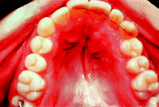
In most patients, the treatment period in the permanent dentition lasts 2 years and is completed at the age of 15. A bonded palatal retainer extending to two teeth on either side of the cleft is placed. The retainer is kept in place as long as the patient will accept it. Such retainers are unobtrusive and do not interfere with oral hygiene (Fig. 26.2).


Fig. 26.2
When the orthodontic treatment is finished, a bonded palatal retainer is placed. This retainer is unobtrusive and does not interfere with oral hygiene
26.3 Optimal Age for Secondary Bone Grafting
In the discussion of this subject, two factors are of particular importance: (a) the curve of growth of the various growth sites in the maxillary complex and (b) the clinical goal of the bone grafting.
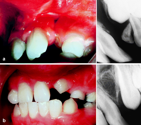
Possible interference with postnatal maxillary growth must be considered in identifying an optimum or ideal age for secondary bone grafting. Sagittal and transverse growth of the anterior maxilla has virtually ceased by the age of 8–9 years (Bjørk and Skiller 1976; Sillman 1964). The vertical growth of the maxilla occurs mainly as deposition of additional alveolar bone at the alveolar crest (Bjørk and Skiller 1974, 1976). The continuous eruption of teeth is thought to be the agent that stimulates the formation of alveolar bone. With a viable donor tissue such as autologous cancellous chips, the graft is rapidly transformed into functional alveolar bone responding physiologically to erupting teeth (Fig. 26.3). By spontaneous or orthodontically guided eruption of the canine, the capacity of erupting teeth to generate alveolar bone can be utilized to maintain the general growth in maxillary height.


Fig. 26.3
(a, b) The grafted bone responds physiologically to the erupting canine: (a) Alveolar cleft prior to bone grafting. (b) The canine erupting normally through the grafted bone
Cephalometric studies of the Oslo cases have shown that bone grafting of the alveolar cleft following the principles stated earlier and performed between the ages of 8 and 11 years had no adverse effect on anteroposterior or vertical maxillary growth (Semb 1988).
Some authors advocate bone grafting at the age of 5–6 years in order to give any lateral incisor an opportunity to migrate into and erupt through the bone graft. This is a strong argument from a dental point of view. However, further studies are necessary to prove that bone grafting at this age does not interfere with maxillary growth.
A primary goal of secondary bone grafting is achieving orthodontic closure of the cleft space. Orthodontic space closure has proven to be easier when bone grafting is performed prior to the eruption of the cleft-side canine. In our studies, in the group in which bone grafting was performed before the full eruption of the canine, orthodontic closure of the gap was possible in 93 % of the cases (335 out of 360). In the group in which the bone graft was performed in the permanent dentition, the success rate was 72 % (107 out of 149) (Abyholm et al. 1981; Åbyholm and Semb 1992; Bergland et al. 1986a).
An evaluation of the height of the interalveolar bony septum also showed a difference between the two groups with a better height of the interalveolar septum in patients who had the bone grafting performed before the full eruption of the cleft-side canine (Abyholm et al. 1981; Åbyholm and Semb 1992; Bergland et al. 1986a).
26.3.1 Secondary Bone Grafting
After 25 years of bone grafting in the transitional dentition, we have analyzed 1,070 patients, of whom 232 had bilateral clefts, making the total number of grafted cleft sites 1,302.
Of these, the teeth in the cleft region were in the final position in 992 cleft sites, and thus, the interdental septum could be evaluated. Good bony interdental septum height (3/4 or more of normal septum height) was found in 96 % when the operation was performed before the eruption of the canine and in 85 % when the operation was performed after the eruption of the cleft-side canine. Failure rate: no bone formation in 1.2 %. A complete dental rehabilitation without prosthodontics was achieved in 93 % of the patients when the bone grafting was performed before the eruption of the cleft-side canine.
In order to investigate the fate of the grafted bone, we have examined 18 consecutive patients with unilateral complete CLP with 3D CT scan 20 years after bone grafting (Kolbenstvedt et al. 2002). We found good bony support of the teeth adjacent to the cleft in all cases, but there was less bone in the cleft area than on the normal side. The apertura piriformis was lower on the cleft side in all cases, and there was hypoplasia of the maxilla on the cleft side.
26.4 The Grafting Tissue of Choice
When autogenous cancellous bone is transplanted under optimal conditions, osteogenic cells in the graft will survive, and new bone formation will start within a matter of days. Cancellous grafts subjected to minimal traumatic surgery undergo a rapid revascularization which is important for the long-term result (Albrektsson 1979). Fresh autologous cancellous bone transforms very rapidly into alveolar bone, which responds normally to tooth migration and orthodontic movement of teeth.
A cortical bone graft undergoes a slow transformation. The early establishment of nutrition to the cortical bone cells requires restoration of flow through existing vessels or canaliculi and ingrowth of capillaries. This is a very slow process, and the cortical bone will usually die and be replaced by invasion of bone cells originating from the recipient site. This is a fundamental difference between cortical and cancellous bone grafts in terms of both cell survival and vascularization. The slow transformation of a cortical graft makes the reestablishment of the tooth-bearing function of the alveolar process unfeasible.
26.4.1 Donor Sites
For autotransplantation of bone in cleft patients, several donor sites have been exploited: rib, tibia, skull, chin, and iliac crest. All of these sources have been used successfully. We have, however, found the iliac crest to be the most suitable donor site.
26.4.2 Complications
Serious complications are rare. Failures (i.e., no continuous bone bridge across the cleft) are probably due to poor surgical technique or infection. External cervical root resorption can occur if the root cementum is mechanically injured during the bone grafting procedure. This can be avoided by delicate surgical technique and by performing the operation at an age when the cervical region of the canine is still covered with bone (Bergland et al. 1986a).
26.5 Important Surgical Details
To achieve optimal results, we have found some details to be of great importance.
26.5.1 Flap Design
It is important that the mucoperiosteal flaps are designed in such a way that only attached gingiva will cover the marginal part of the graft. It is our clinical observation that a completely normal periodontium seems to occur mainly in cases where the flap design has ensured an orthotopic transfer of attached gingiva to cover the marginal part of the graft.
26.5.2 Good Exposure
The cleft must be widely exposed so that all scar tissue in the cleft area can be removed. The nasal floor has to be reconstructed if a fistula is present. It is important to elevate the nasal floor to achieve a good height of the alveolar crest so that the canine can be brought into a vertical position in the cleft area.
26.5.3 Cancellous Bone Only
Only autologous cancellous bone creates bone that responds normally to eruption and orthodontic movement of teeth.
26.6 Conclusion
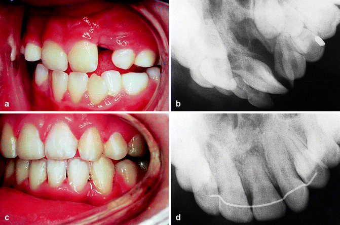
Autologous cancellous bone grafted to the alveolar cleft between the ages of 8 and 11 years creates alveolar bone which responds normally to eruption and orthodontic movement of teeth. A full dental rehabilitation is possible in the vast majority of cases when bone grafting is performed prior to the cleft-side canine eruption, rendering bridgework unnecessary in cleft patients (Fig. 26.4).


Fig. 26.4
(a–d) Unilateral complete cleft bone grafted prior to the eruption of the canine: (a) The dental arch before the bone grafting. (b) Radiograph of the cleft prior to bone grafting. (c, d) Dental arch and radiograph after the completion of the orthodontic treatment
Bone grafting performed between the ages of 8 and 11 years does not restrict maxillary growth.
This combined surgical/orthodontic treatment plan requires close cooperation between plastic surgeon and orthodontist. Fringe benefits include closure of oronasal fistulae and elimination of mucosal recesses, stabilization of maxillary segments, and improvement of nasal symmetry.
26.7 Optimal Timing for Secondary Alveolar Bone Grafting
Sayuri Otaki2, 3 and Masatomo Yorimoto4
26.7.1 Introduction
Timing for secondary alveolar bone grafting is critical to create an area of regenerated bone in the cleft site so that the adjacent teeth (canine and sometimes lateral incisor) can erupt spontaneously or can be moved orthodontically into it. Bone grafting before the eruption of these teeth causes their eruption through the newly grafted bone (the canine proceeds to lateral incisor position when it is missing), inducing additional bone generation and achieve higher interdental septum height. Thereafter, the presence and close approximation of these teeth prevent bone resorption. In addition, bone grafting before the eruption of these teeth allows the surrounding bone to protect the cervical region of their roots and thus prevent resorption. Maxillary growth is not disturbed when bone grafting is performed to facilitate canine eruption. Careful planning is necessary when bone grafting is performed to facilitate lateral incisor eruption because there is a possibility of causing a maxillary growth disturbance.
Secondary alveolar bone grafting using autogenous marrow and cancellous bone graft was introduced as a method for restoring tooth-bearing function (Boyne and Sands 1972, 1976). Thereafter, the central purpose for secondary alveolar bone grafting is to create an area of regenerated bone in the cleft site that is continuous with the rest of alveolus so that adjacent teeth (canine and sometimes lateral incisor) can erupt spontaneously or can be moved orthodontically into it. The timing for secondary alveolar bone grafting is critical to attain this purpose. There is a margin of time for bone grafting for other purposes, such as the stabilization of the maxillary segments, improving the soft tissue configuration to facilitate prosthetic restoration, preparing bed for the dental endosseous implant insertion, allowing closure of the oronasal fistula, and adding bony support to the alar base.
The timing of secondary alveolar bone grafting will affect the height of interdental septum at the former cleft site; tooth eruption and movement into former cleft site, which in turn affect the type of closure of the space in dental arch; root resorption; and maxillary growth. The optimal timing is going to be discussed with regard to these factors. Although various authors have presented the optimal timing based on their experience and/or investigations (Table 26.1), most practitioners so far accept that the optimal timing for secondary bone grafting is when the patient is 9–10 years old, before eruption of the canine, and the canine root is one-half to two-thirds formed. The canine eruption seems to be accelerated when canine root is two-thirds formed (Vig 1999).
Table 26.1
Optimal timing for secondary alveolar bone grafting
|
Author(s)
|
Age (years)
|
Eruption stage
|
Root development
|
|---|---|---|---|
|
Boyne and Sands (1972)
|
9–11
|
Before the full eruption of the canine
|
–
|
|
Boyne and Sands (1976)
|
7
|
Before the eruption of the lateral incisor
|
–
|
|
Abyholm et al. (1981)
|
–
|
Before the eruption of the caninea
|
–
|
|
El Deeb et al. (1982)
|
9–12
|
–
|
When 1/4–1/2 of the canine root has formed
|
|
Turvey et al. (1984)
|
8–10
|
Before the eruption of the canine
|
When 1/2–2/3 of the canine root has formed
|
|
Bergland et al. (1986a)
|
9–11
|
Before the full eruption of the caninea
|
–
|
|
Enemark et al. (1987)
|
–
|
Before the eruption of the caninea
|
–
|
|
Brattstrom and McWilliam (1989)
|
–
|
After the eruption of the incisors and before the eruption of the caninesa
|
–
|
|
Rune and Jacobsson (1989)
|
–
|
When the canine (or lateral incisor) is in an early stage of eruption
|
–
|
|
Kortebein et al. (1991)
|
Stay updated, free dental videos. Join our Telegram channel

VIDEdental - Online dental courses


