Introduction
Correct tooth position in the 3 planes of space is a major objective of orthodontic treatment. The aim of this study was to determine whether a panoramic radiograph (OPT) provides a true assessment of the mesiodistal root relationship of adjacent teeth.
Methods
OPTs of 22 patients near the end of treatment with fixed appliances in both arches were taken before debonding. When the roots of adjacent teeth were touching on the OPT, cone-beam computed tomography (CBCT) was used to show the true root relationships.
Results
We evaluated 235 interdental sites by OPT and CBCT; 47 areas showed contact between adjacent roots in the OPT images. However, the CBCT images showed true contact in only 5 of these areas; ie, 11% of the diagnoses based on OPT images were true positive, whereas the rest (89%) was false positive. One hundred eighty-eight sites showed no contact in the OPT images; this was confirmed by CBCT.
Conclusions
OPT has high sensitivity and relatively high specificity to detect adjacent roots touching each other. Root contacts are overestimated when evaluated by OPT.
Correct tooth position in the 3 planes of space is a major objective of orthodontic treatment. Appropriate axial inclination of the roots with near-parallel roots has been discussed often in the orthodontic literature and is a criterion in the examinations of the American Board of Orthodontics and the European Board of Orthodontics. Root proximity is the term used when the roots of adjacent teeth are 1.0 mm or less apart, as measured radiographically. Furthermore, by dividing the root into 3 sections—apical, middle, and cervical—different severities of root proximity might be obtained.
The clinical diagnosis of root proximity is based mainly on routine radiographic procedures, such as panoramic radiography (OPT). However, OPT provides a distorted and 2-dimensional representation of a 3-dimensional (3D) object. There are large discrepancies between optimal and actual beam directions, resulting in overlapping of teeth, especially in the premolar region.
Cone-beam computed tomography (CBCT) is a new radiographic method with applications in several diagnostic areas such as implant treatment, oral surgery, endodontic treatment, and tempomandibular joint imaging. Compared with conventional computed tomography, CBCT technology in clinical practice provides important advantages such as minimal radiation dose, image accuracy, rapid scan time, fewer image artefacts, and opportunity to use chair-side image display and real-time analysis. In orthodontics, the use of CBCT imaging has been restricted to impacted teeth, tempomandibular joint visualization, and determination of bone volume conducible to orthodontic tooth movement, and for cleft patients. Recently, Peck et al evaluated the accuracy of OPT and CBCT for determing mesiodistal root angulations. Using plaster models of 5 patients as the gold standard, they reported that CBCT scans can produce accurate measurements of root angulations, and OPT cannot provide reliable data.
By using CBCT as the gold standard, the purpose of this study was to determine whether OPT provides a true assessment of the mesiodistal root relationship of adjacent teeth in subjects approaching the end of orthodontic treatment.
Material and methods
From 119 consecutive patients at a private orthodontic practice in Winterthur, Switzerland, 22 patients (9 female, 13 male; mean age, 16.7 years; range, 12.8-37.3 years) in the permanent dentition were included in this study. They were approaching the end of orthodontic treatment with fixed appliances in both dental arches and had root contacts in the OPT images taken at that time. The patients were further referred to study the proximity of neighboring roots by CBCT.
The images were acquired with an OPT device (Cranex Excel, Soredex, Tuusula, Finland). The CBCT images were obtained with the 3D Accuitomo FPD (J. Morita, Kyoto, Japan). Two sizes of areas (40 x 40 and 60 x 60 mm) were imaged with super-high resolution (2.0 line pairs per millimeter; voxel size, 0.125 mm). The plane of primary reconstruction was aligned parallel to the long axis of the examined tooth by using iDixel software, as suggested by the manufacturer (J. Morita, Kyoto, Japan). Depending on the region of interest, either 1 or 2 CBCT images were taken of each patient.
Two calibrated examiners (M.L. and A.D.) assessed the OPT and CBCT images separately and blindly for root contacts. In case of disagreement between the examiners, a new evaluation was made, and this consensus was used for the final evaluation.
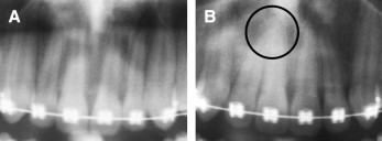
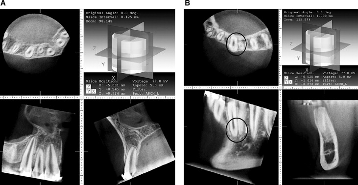
Figures 1 and 2 show the root contacts evaluated by OPT and CBCT, respectively. Root contact was determined when no periodontal space between adjacent roots was visible, resulting in “touching” roots.


Interrater agreement for both methods was assessed with Cohen’s kappa. The CBCT (value, 0.89) and the OPT (value, 0.83) methods had good agreement between the 2 observers.
The Pearson chi-square test was used to test the null hypothesis of no difference between the OPT and CBCT methods for evaluating root contacts. The validity of the OPT to detect the true root relationship was assessed by using the CBCT images as the gold standard. The frequency of root contact, the sensitivity and specificity, the positive and negative predictive values, and the likelihood ratio were calculated. The statistical analysis was done with SPSS for Windows (release 13.0, standard version; SPSS, Chicago, Ill).
Results
A total of 235 interdental sites were evaluated for root contacts. The number and percentages of teeth with root contact evaluated by OPT and CBCT are shown in the Table . Root contact was observed in 47 sites (20%) in the OPT images and in only 5 (2.1%) with the CBCT images. The hypothesis of no difference in evaluating root contact on OPT and CBCT images was rejected ( P <0.001). The OPT method had excellent sensitivity of 100% (95% CI, 46%-100%), combined with high specificity of 81% (95% CI, 76%-86%); this means that 81% of the areas without contact were diagnosed correctly by the OPT. The positive predictive value of the OPT was found to be quite low (10%), whereas the negative predictive value was 100%. The likelihood ratio for a positive test result was 5.4; this indicates that a positive result is 5 times as likely in an area with root contact than in one without root contact.
| CBCT | |||
|---|---|---|---|
| No contact | Contact | Total | |
| OPT | |||
| No contact | 188 (80.0) | 0 (0.0) | 188 (80.0) |
| Contact | 42 (17.9) | 5 (2.1) | 47 (20.0) |
| Total | 230 (97.3) | 5 (2.1) | 235 (100.0) |
Stay updated, free dental videos. Join our Telegram channel

VIDEdental - Online dental courses


