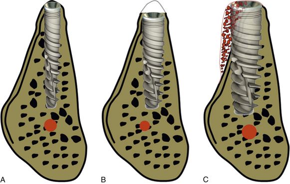
Fig 6.1 (A) Panoramic view of the edentulous mandibular molar site showing adequate bone length availability (15 mm) to place widest implant of any system (B) but the cross-sectional view of the same edentulous region shows inadequate bone availability at the ridge crest (5 mm), which cannot allow placement of even the narrowest implant of any system. The height of the bone above the mandibular canal is adequate (16.83 mm) to place an adequately long implant (14–15 mm long). (C) The implant with 4 mm x 15 mm dimensions can be placed with its platform at the crest level with simultaneous lateral bone augmentation to cover the facially exposed implant threads near the crest. (D) The shorter length (4 mm x 12 mm) implant is placed after 3 mm of osteoplasty, so that the implant platform can be placed at the level of wider subcrestal ridge.
Misch and Judy classification of bone availability
In 1985, Misch and Judy presented a classification of available bone for dental implant insertion, which is similar in both arches (< ?xml:namespace prefix = "mbp" />

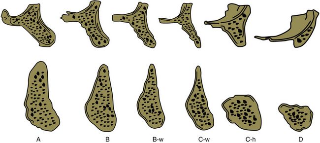
Fig 6.2 Misch and Judy classification of bone availability (Divisions A, B, C and D): Division A (abundant bone), Division B (barely sufficient bone), Division C (compromised bone), Division D (deficient bone), w (width), h (height).
Division A (abundant bone)
Division A bone is three-dimensionally abundant for the ideal implant insertion (

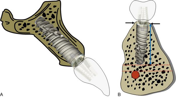
Fig 6.3 (A and B) Abundant bone in the maxilla and mandible suitable for inserting an implant with ideal dimensions.
Prosthetic options for Division A bone
a. is the best bone for any prosthetic option (
b. may need osteoplasty for implant overdentures, to achieve more vertical height space and accommodate the implant suprastructures (ball/bar) under the denture.

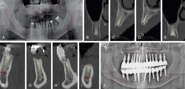
Fig 6.4 (A) Panoramic radiograph and (B–I) CT scan showing cross-sectional views of the maxilla and the mandible, showing the Division A bone (adequate bone) for implant placement without any ridge modification or grafting. (J) The implant-supported, full mouth fixed prosthesis can be seen in the radiograph.
Division B (barely adequate) bone
Bone in this category should be:
Prosthetic options for Division B bone
The bony ridge with Division B bone may be modified to division A bone by osteoplasty to achieve the wide ridge crest required to insert a regular diameter implant (
Stay updated, free dental videos. Join our Telegram channel

VIDEdental - Online dental courses


