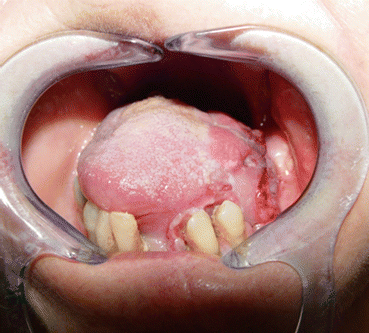
Fig. 4.1
Floor of the mouth and ventral tongue squamous cell carcinoma
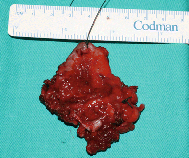
Fig. 4.2
Surgical specimen
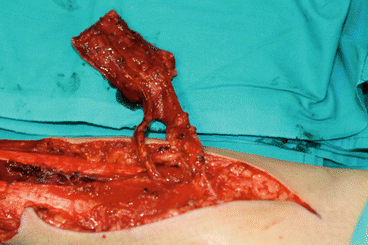
Fig. 4.3
Left antebrachial radial fasciocutaneous flap dissection
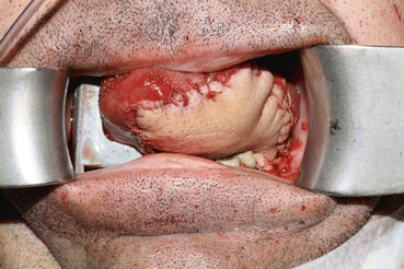
Fig. 4.4
Intraoperative clinical result
An alternative to this flap is the ulnar forearm flap, which is based on the ulnar artery and its venae comitantes. It can be used in cases where the radial artery is dominant in the supply to the forearm. According to Van Cann and Koole [6] and Antony et al. [7], the advantages of the radial forearm flap are a less severe cosmetic defect and better scarring at the donor site.
4.3.2 Anterolateral Thigh Flap (ALT)
First described by Song et al. [8], the ALT flap is a very useful perforator flap that is widely used in reconstruction procedures of the head and neck. The popularity of ALT flap led several authors, including Huang et al. [9], to reconsider continuing with the radial forearm flap. However, other authors use it to cover some of the indications of the myocutaneous abdominal rectus flap.
4.3.2.1 Indications
In our opinion, this flap should be used in cases where the radial forearm flap cannot provide sufficient volume for reconstruction of intraoral defects. It is a good option for reconstructing defects of the tongue stretching back to the oropharynx and cheek mucosa, extended hemiglossectomy, and full-thickness cheek defects where, in addition to reconstructing the skin and mucosa, the object of the procedure is to provide volume. As for extraoral defects, the ALT flap could be indicated in skin defects requiring large, bulkier flaps, such as those affecting the side of the face, the temporal region, and large defects of the scalp. According to Jiang et al. [10], the ALT can be harvested as a chimeric flap with multiple skin paddles.
4.3.2.2 Vascular Pedicle
The vascular pedicle of the ALT flap comprises the descending branch of the lateral femoral circumflex artery and its venae comitantes (approximately 2 mm in diameter), which are found in the septum between the vastus lateralis and femoris muscles. The perforators (approximately 0.5 mm in diameter) are mostly (85 %) musculocutaneous, whereas the remainder (15 %) are septocutaneous. The pedicle is dissected until the point where it leaves the lateral femoral circumflex artery. Lengths of up to 10 cm can be obtained.
4.3.2.3 Presurgical Planning
Doppler or echo Doppler is useful for locating the perforators. Lakhiani et al. [11] showed that most of the perforators are found in the middle third of the thigh, from the midpoint of a line that connects the anterior superior iliac spine and the supero-external part of the patella.
4.3.2.4 Tissue to Be Transferred
The ALT flap is harvested with the skin and subcutaneous cell tissue. The fascia is optional, although it is generally included. Suprafascial dissection is more cumbersome and complicated, although it spares the local anatomy and preserves muscle coverage. A sensory flap can be harvested by including branches of the lateral cutaneous nerve.
4.3.2.5 Morbidity
Harvesting the ALT flap has few complications, thus giving it an advantage over other flaps such as the rectus abdominis myocutaneous flap or the radial forearm flap, since it does not damage the abdominal wall or require major vascular axes to be sacrificed. It generally does not require free grafts, since it enables direct closure of defects measuring 8–9 cm. Leg extension and mobility are barely affected because they are compensated by the remaining extensor muscles. However, there is a possibility of loss of sensation in the thigh because of the damage to the lateral cutaneous femoral nerve. The unsightly scar left on the thigh is also a potential drawback.
4.3.2.6 Postsurgical Care
It is important to monitor for development of compartment syndrome (Figs. 4.5, 4.6, 4.7, 4.8, and 4.9).
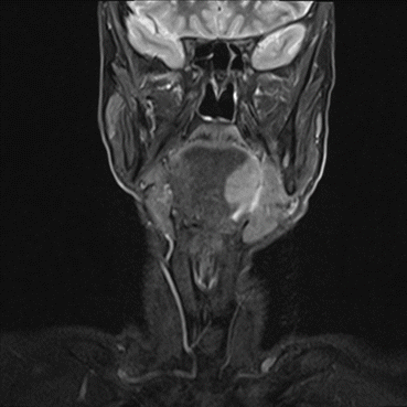
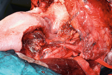
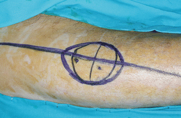
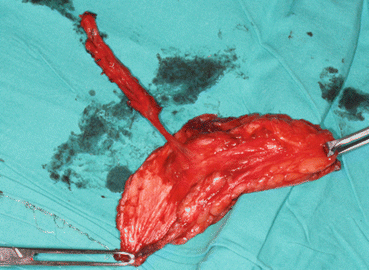
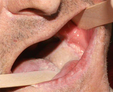
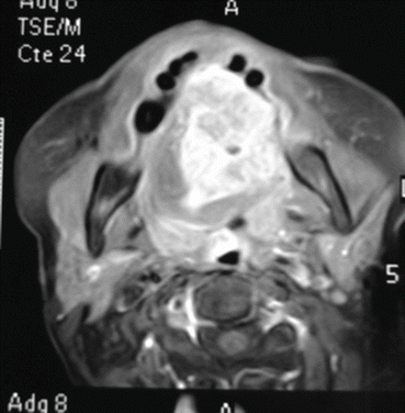
Fig. 4.5
MRI. Base of the tongue squamous cell carcinoma
Fig. 4.6
Surgical defect after resection. Pull through cervical approach
Fig. 4.7
ALT perforator flap design
Fig. 4.8
Septocutaneous perforator flap
Fig. 4.9
One year postsurgery
The vastus lateralis flap is a variant of the ALT perforator flap. Dissection differs from that of the ALT, since it is necessary to locate the vascular pedicle without locating and isolating the perforators. It is necessary to follow the principles for dissection of the myocutaneous flaps, in which the skin and subcutaneous tissue are not separated from the underlying muscle. This flap is easier to harvest than the ALT flap, although it does require part of the vastus lateralis muscle to be sacrificed. Therefore, it is used for the reconstruction of three-dimensional defects requiring greater bulk, as reported by Engel et al. [12] after total glossectomy or to isolate the base of the skull or central nervous system. The vastus lateralis myocutaneous flap is a good alternative to the rectus abdominis myocutaneous flap (Figs. 4.10, 4.11, 4.12, 4.13, and 4.14).
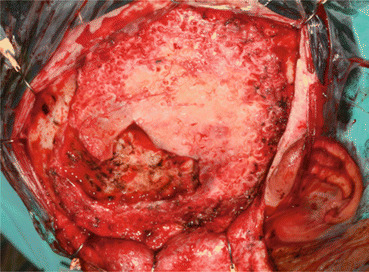
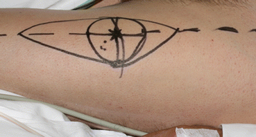
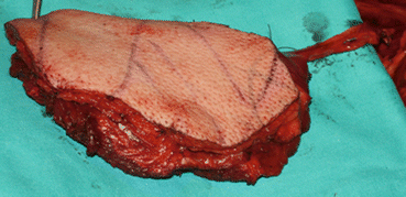
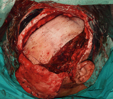
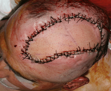
Fig. 4.10
Temporal defect associated to cerebrospinal fluid leak
Fig. 4.11
Design of the myocutaneous vastus lateralis flap similar to the ALT perforator flap
Fig. 4.12
Flap with muscular component
Fig. 4.13
Flap inset using the muscle to seal the leak
Fig. 4.14
Final intraoperative result
Another variant of this flap is the adipofascial ALT, which is indicated for the reconstruction of defects of the intraoral and nasal mucosa [13].
4.3.3 Rectus Abdominis Myocutaneous Flap
Pennington and Pelly [14] were the first to describe this flap, which is based on the deep inferior epigastric artery and vein. Together with the radial forearm flap, the rectus abdominis myocutaneous flap has been used for many years in the reconstruction of head and neck defects requiring considerable bulk. The use of perforator flaps and the possibility of including muscle in this type of flap mean that it has been less widely used.
4.3.3.1 Indications
The reconstruction of large intraoral defects, such as total glossectomy or type IV maxillectomy [15], that include orbital exenteration. It can be used in combination with the temporal myofascial flap. The rectus abdominis flap provides bulk to fill the middle third of the face, and the temporal myofascial flap is used to reconstruct the palate. As for extraoral reconstruction, the rectus abdominis myocutaneous flap is used mainly to seal bone defects at the base of the skull, which is isolated from the upper aerodigestive tract [16].
4.3.3.2 Vascular Pedicle
Deep inferior epigastric vessels. The caliber is usually around 3 mm. The greatest density of musculocutaneous perforators is found in the periumbilical region.
4.3.3.3 Presurgical Planning
This flap does not require specific assessment of the vasculature. However, it is important to determine whether the patient has previously undergone surgery of the abdominal region, especially in the iliac fossa, hypogastrium, and mesogastrium, since the pedicle could be damaged.
4.3.3.4 Tissue to Be Transferred
The rectus abdominis flap is generally a musculocutaneous flap. The skin paddle can be placed in several ways, although it is normally placed vertically over the longitudinal axis of the muscle. The whole muscle component can be transferred. It is important to respect the anterior fascia below the arcuate line.
4.3.3.5 Morbidity
The rectus abdominis flap should only be harvested after careful deliberation, since it is necessary to remove a basic muscle of the abdominal wall, whose flexor activity would therefore be compromised. When harvesting the flap, extra care must be taken not to accidentally open the peritoneal cavity. Abdominal hernia is another complication at the donor site. The use of synthetic mesh reduces the likelihood of hernia.
4.3.3.6 Postsurgical Care
Stay updated, free dental videos. Join our Telegram channel
VIDEdental - Online dental courses


