Oral pigmentation may be focal, multifocal, or diffuse. The lesions may be blue, purple, brown, gray, or black. They may be macular or tumefactive. Some are localized harmless accumulations of melanin, hemosiderin, or exogenous metal; others are harbingers of systemic or genetic disease; and some can be associated with life-threatening medical conditions that require immediate intervention. The differential diagnosis for any pigmented lesion is extensive, and can include examples of endogenous and exogenous pigmentation. Although biopsy is a helpful and necessary aid in the diagnosis of focally pigmented lesions, with diffuse presentations lesions require a thorough history and laboratory studies to establish a definitive diagnosis.
Key points
- •
Pigmented lesions within the oral cavity may present a diagnostic dilemma for the clinician.
- •
A differential diagnosis for a pigmented lesion may include traumatic, reactive, neoplastic, and systemic pathologies.
- •
Mucosal melanomas have no distinctive appearance.
- •
A clinicopathologic correlation is often required to ensure accurate diagnosis of systemic causes of diffuse pigmentation.
The identification of pigmented tissue within the oral cavity may present a diagnostic dilemma for the clinician. The manifestation of mucosal pigment is variable and can range from focal to diffuse macular coloration or from a small nodular growth to a large mass. Is the pigmentation of physiologic origin? Is it pathologic in etiology? Does it represent a malignant process? These are questions that typically come to mind when a patient presents with oral mucosal pigmentation. The color, location, duration, distribution, and appearance of the pigmented lesions may be of diagnostic importance. A thorough investigation of dental, medical, family, and social histories is also necessary to ensure accurate diagnosis. The presence of cutaneous pigmentation or other systemic signs and symptoms may be helpful in formulating a differential diagnosis. Clinical laboratory testing may also be beneficial. However, if the underlying cause of the pigmentation cannot be readily identified, a tissue biopsy is essential for definitive diagnosis.
A differential diagnosis for a clinically pigmented lesion may include an array of traumatic, reactive, and neoplastic pathologies and pigmentation associated with systemic disease. Lesions or conditions of melanocytic, vascular, hematopoietic, and hemosiderotic origin may be given consideration. Genetic dysfunction may also manifest as oral pigmentation; Peutz-Jeghers syndrome is the prototypical example. Where appropriate, exogenous substances including chemical coloring agents, amalgam, graphite, drug metabolites, and chromogenic bacteria could also be considered. To this end, relatively common causes of mucosal coloration, including petechiae, purpura, ecchymoses, hematomas, vascular tumors, and exogenous substances are not considered true pigmented lesions. In contrast, melanin, which is synthesized by melanocytes, is a true pigment and usually imparts a brown, blue, or black appearance to the mucosa.
Melanin pigmentation may be focal, multifocal, or diffuse in its presentation. Melanin is the pigment derivative of tyrosine and is synthesized by melanocytes, which typically reside in the basal cell layer of the epithelium. In the skin, melanin is thought to be cytoprotective against the damaging effects of sunlight. The role of melanocytes in oral epithelium remains unclear. Unless a patient is of a race or ethnicity in which mucocutaneous pigmentation is physiologic, melanocytes are uncommonly observed in routine oral mucosal biopsies. Thus, in a white patient, oral melanocytic pigmentation may not always be of any significant clinical consequence but it is always considered pathologic in origin.
Pathologic melanin production within the oral mucosa may be associated with an array of etiologies. The most concerning of these are malignant melanoma and various systemic disorders, including adrenal insufficiency and Cushing disease (discussed later). Importantly, the oral manifestations of these potentially life-threatening disorders can mimic an array of idiopathic, reactive, and benign neoplastic lesions. Thus, an understanding of the various disorders and substances that can contribute to oral mucosal coloration is essential for the appropriate evaluation, diagnosis, and management of the patient. This article focuses on oral pathologies of melanocytic origin.
Reactive melanocytic pigmentation
Melanotic Macule
Melanotic macules are the most common oral mucosal lesions of melanocytic origin. These small, solitary, well-circumscribed and often uniformly pigmented lesions develop most commonly in adult female patients. Any mucosal site may be affected but the lower lip, gingiva, and palate are the most common areas. Localized trauma may be a potential cause but this remains uncertain.
Melanotic macules rarely present larger than 1 cm in diameter. Although these are innocuous lesions, a biopsy is usually warranted for diagnosis because mucosal melanoma can mimic the appearance of a melanotic macule. After a diagnosis is rendered, no additional treatment is required and the lesion typically does not recur. Because most melanotic macules do present on the lip, patients often request complete removal because of esthetic concerns.
Melanotic macules are caused by functional hyperactivity of the regional melanocytes (ie, there is increased melanin production). Histologically, this is evidenced by abundant melanin pigmentation within the basal epithelial cell layer with melanin incontinence in the superficial portions of the submucosa. Importantly, there is no increase in melanocyte number. If there is evidence of melanocytic hyperplasia, it should be diagnosed as such and the lesion would likely warrant complete surgical removal. It is possible that melanocytic hyperplasia may potentially herald the development of malignant melanoma.
The differential diagnosis for melanotic macule is often limited to entities that may present as focal, macular clinical pigmentation. In addition to melanoma, a melanocytic nevus, amalgam tattoo, and focal ecchymosis are usually given consideration. Oral mucosal melanomas have no defining clinical characteristics. Thus, a biopsy of any persistent solitary pigmented lesion is always warranted.
Oral Melanoacanthoma
Oral melanoacanthoma is a relatively uncommon melanocytic lesion that may cause rapid, diffuse, and dark pigmentation of a large mucosal area. However, this is an innocuous pathology that is often self-limiting and may spontaneously resolve, with or without surgical intervention. Acute regional trauma or a history of chronic irritation may precede the development of the lesion.
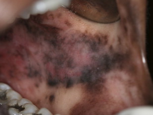
Oral melanoacanthoma usually presents as an asymptomatic, ill-defined, rapidly enlarging, macular pigmentation, commonly observed in a black female patient. Although most lesions are heavily pigmented, the coloration may or may not be uniform. Any mucosal site may be affected, but buccal mucosal involvement is most common ( Fig. 1 ). Although typically solitary, rare patients may present with multifocal lesions.
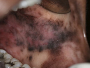
Oral melanoacanthoma is characterized by spongiotic epithelium containing dendritic, pigmented melanocytes extending through its full thickness. A mild inflammatory infiltrate composed of lymphocytes and occasional eosinophils is also seen.
Because of the ominous clinical presentation, melanoma is invariably considered in the differential diagnosis. After a histologic diagnosis of oral melanoacanthoma has been established, no further treatment is required. The biopsy procedure itself may lead to spontaneous regression of the lesion.
Smoker’s Melanosis
In addition to cancer and numerous other systemic complications, cigarette smoking can also induce oral mucosal pigmentation. Smoker’s melanosis is not considered a preneoplastic condition. Although the exact pathogenesis remains uncertain, melanin stimulation may represent a protective mucosal response to either the heat of the smoke or to an irritant within the cigarette. Females are most commonly affected. Smoker’s melanosis usually presents as diffuse but patchy and irregular pigmentation of the anterior facial maxillary and mandibular gingivae. Other mucosal sites are less commonly affected.
Histologically, the findings are nonspecific in the form of abundant melanin within the basal cell layer with melanin incontinence. Similar histologic findings can be seen in melanotic macule, and in an array of other conditions that can present as diffuse pigmentation. A clinicopathologic correlation is required to make an accurate diagnosis of smoker’s melanosis. Importantly, melanoma can present as diffuse patchy pigmentation. Thus, if only one mucosal site is affected, melanoma should be considered in the differential diagnosis. Melanoma is not typically considered in a patient with multifocal pigmentation affecting noncontiguous mucosal sites.
Inflammation-associated hyperpigmentation
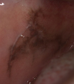
Inflammation-associated hyperpigmentation most commonly develops in dark-complexioned individuals. When it occurs in the skin, the pigmentation usually develops in an area previously subjected to trauma or inflammation, such as in acne-prone areas of the face. In the oral cavity, this form of reactive pigmentation is most commonly observed in patients with clinical evidence of lichenoid inflammation. The pigment may be focal or diffuse and patchy, but is often regional to the lichenoid lesion ( Fig. 2 ). The coloration is often light brown. However, in rare instances, the pigmentation may become so dark that it obscures the underlying lichenoid pathology. In most cases, the clinical manifestation of the lichenoid lesion is usually what prompts the biopsy. In addition to the typical lichenoid histologic features, there is melanin pigmentation in the basal cell layer with melanin incontinence. After a histologic diagnosis is rendered, treatment is aimed at resolution of the lichenoid inflammation, if the patient is symptomatic. The pigmentation may or may not disappear after treatment.
Drug-induced melanosis
Medications may induce mucocutaneous coloration by a variety of different mechanisms. In some instances, drug metabolites may be incorporated or deposited into the regional tissues. Although the mucosa may appear clinically pigmented, this is not true pigmentation. Drugs of the tetracycline family are representative examples of this phenomenon ( Fig. 3 ). In contrast, a host of medications, including antimalarial drugs, phenothiazines, oral contraceptives, and various cytotoxic medications, induce true mucocutaneous pigmentation by induction of melanin. The mechanisms by which drugs induce melanin synthesis remain unclear. However, the mechanisms may vary between different drug classes. Some drugs, including chloroquine and chlorpromazine, have been shown to physically bind melanin. This results in retention of the drug within melanocytes, which may contribute to the increased pigmentation. Alternatively, it is possible that the drugs or specific drug metabolites may stimulate melanin synthesis.
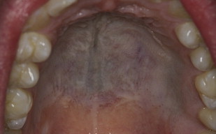
Drug-induced pigmentation may be diffuse but localized to one mucosal region, or it can be multifocal and involve multiple surfaces ( Fig. 4 ). The pigmentation is macular and may or may not be uniformly colored. Some drugs may be associated with a specific pattern of pigmentation. For example, hydroxychloroquine typically triggers pigmentation of the palatal mucosa.
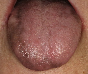
A differential diagnosis includes other causes of diffuse mucosal pigmentation. A good clinical history and understanding about known drug side effects may help in achieving an appropriate diagnosis. If the melanosis can be temporally associated with use of a specific medication that is known to induce pigmentation, then no further intervention is typically warranted. The discoloration may fade within several months after the medication is discontinued. Laboratory testing may be necessary to rule out an underlying endocrinopathy.
As with all other pigmented lesions, a biopsy is warranted if a diagnosis cannot be appropriately rendered. The histologic changes mimic those of melanotic macule; there is abundant melanin accumulation within the basal cell region with melanin incontinence. Thus, a clinicopathologic correlation is often necessary to ensure accurate diagnosis.
Reactive melanocytic pigmentation
Melanotic Macule
Melanotic macules are the most common oral mucosal lesions of melanocytic origin. These small, solitary, well-circumscribed and often uniformly pigmented lesions develop most commonly in adult female patients. Any mucosal site may be affected but the lower lip, gingiva, and palate are the most common areas. Localized trauma may be a potential cause but this remains uncertain.
Melanotic macules rarely present larger than 1 cm in diameter. Although these are innocuous lesions, a biopsy is usually warranted for diagnosis because mucosal melanoma can mimic the appearance of a melanotic macule. After a diagnosis is rendered, no additional treatment is required and the lesion typically does not recur. Because most melanotic macules do present on the lip, patients often request complete removal because of esthetic concerns.
Melanotic macules are caused by functional hyperactivity of the regional melanocytes (ie, there is increased melanin production). Histologically, this is evidenced by abundant melanin pigmentation within the basal epithelial cell layer with melanin incontinence in the superficial portions of the submucosa. Importantly, there is no increase in melanocyte number. If there is evidence of melanocytic hyperplasia, it should be diagnosed as such and the lesion would likely warrant complete surgical removal. It is possible that melanocytic hyperplasia may potentially herald the development of malignant melanoma.
The differential diagnosis for melanotic macule is often limited to entities that may present as focal, macular clinical pigmentation. In addition to melanoma, a melanocytic nevus, amalgam tattoo, and focal ecchymosis are usually given consideration. Oral mucosal melanomas have no defining clinical characteristics. Thus, a biopsy of any persistent solitary pigmented lesion is always warranted.
Oral Melanoacanthoma
Oral melanoacanthoma is a relatively uncommon melanocytic lesion that may cause rapid, diffuse, and dark pigmentation of a large mucosal area. However, this is an innocuous pathology that is often self-limiting and may spontaneously resolve, with or without surgical intervention. Acute regional trauma or a history of chronic irritation may precede the development of the lesion.
Oral melanoacanthoma usually presents as an asymptomatic, ill-defined, rapidly enlarging, macular pigmentation, commonly observed in a black female patient. Although most lesions are heavily pigmented, the coloration may or may not be uniform. Any mucosal site may be affected, but buccal mucosal involvement is most common ( Fig. 1 ). Although typically solitary, rare patients may present with multifocal lesions.

