Introduction
Previous studies have used the right and left sides of the same jaw to compare different force levels, types of movement, and durations of forces. However, the amounts of root resorption have not been compared between the right and left sides after applying the same amount of force. The aims of the study were to quantitatively compare the volumes of the root resorption lacunae between the right and left first premolars to determine whether 1 side can serve as a control to the other and to compare the volumes of root resorption lacunae of the first premolars between the maxilla and the mandible.
Methods
Forty-four first premolars, orthodontically indicated for extraction from 11 patients (left and right maxillary and mandibular first premolars from each) were moved buccally by using beta-titanium-molybdenum alloy 0.017 × 0.025-in cantilever springs with continuous heavy (225 g) force. After the experimental period, the teeth were extracted under a strict protocol to prevent root cementum damage and then analyzed by using a microcomputed tomography scan x-ray system (1172; SkyScan, Aartselaar, Belgium) and specially designed software (Convex Hull 2D, University of Sydney, Sydney, Australia) for direct volumetric measurements.
Results
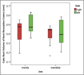
There were no statistically significant differences in the mean cube root volumes of root resorption craters between the right and left sides ( P = 0.18) or between the maxillary and mandibular jaws ( P = 0.10). There was also no statistical significance for the interception ( P = 0.41), which indicated that the jaw and the side had independent effects.
Conclusions
The amount of root resorption on the left and right sides of the jaw were similar in both the maxilla and the mandible. Therefore, for future root resorption studies, it is justifiable to use the split-mouth technique so that teeth from 1 side of the jaw can serve as the controls.
Numerous studies have analyzed the amount of root resorption after applying different magnitudes of forces. These studies often involved applying a certain amount of force on 1 side and leaving the other side as the control. It was assumed that there was symmetry between the right and left sides of the body. As shown in human chromosome and embryogenesis, clearly symmetry exists and carries onto the structural organization. Human dentition is thought to be a form of matching symmetry, with the right side as a mirror image of the left with identical genetic determination. Previous research has shown no differences in the cementum’s hardness between the buccal and lingual root surfaces or between the maxillary and mandibular premolars. In addition, Chutimanutskul et al analyzed the hardness and elasticity of premolar cementum hardness and found no significant differences between the right and left premolars.
Sameshima and Sinclair studied the pretreatment and posttreatment radiographs of 868 patients and found no significant differences in root resorption between the right and left sides or between the maxillary and the mandibular teeth. In contrast, McFadden et al reported a difference in root shortening between the maxillary and mandibular incisors, and suggested that it might be related to less intrusion in 1 arch or differences in the rate of bone turnover. Various factors can contribute to the differing amounts of root resorption in the maxilla and the mandible, including bone density, turnover of bone, and anatomic relationships in the maxilla and the mandible. In terms of anatomic relationships, there seems to be no single pretreatment characteristic of facial or dentoalveolar apparatus that is responsible for apical root shortening.
No previous study has investigated the quantitative amounts of root resorption between the right and left sides of the jaws after applying the same amount of orthodontic force. Moreover, few studies have compared the amounts of root resorption between teeth in the maxilla and the mandible. Therefore, the aims of this study were to quantitatively compare the volumes of the root resorption lacunae between the maxillary right and left, and mandibular right and left, first premolars and to see whether 1 side or jaw can serve as a control for the other.
Material and methods
Forty-four first premolars were collected from 11 patients (3 boys, 8 girls; mean age, 14.9 years; range, 12.2-18.8 years) who required 4 premolar extractions for orthodontic treatment purposes. Ethics approvals were obtained from the Medical Faculty Ethics Committee of the University of Ondokuz Mayis in Turkey and the Human Research Ethics Committee of the University of Sydney in Australia. The patients were selected according to the selection criteria described previously. Informed consent and questionnaires about fluoride intake, birthplace, and previous living places were obtained from the subjects. They received a heavy continuous buccal orthodontic force (225 g) on all 4 first premolars for 4 weeks. Each experimental premolar from each quadrant of the mouth was allocated to groups maxillary right, maxillary left, mandibular right, and mandibular left.
The 0.022-in slot SPEED brackets (Strite Industries, Cambridge, Ontario, Canada) were bonded on the buccal surfaces of the first permanent molars and first premolars. Self-ligating brackets were selected to allow for standardized ligation of the experimental teeth. Heavy buccally directed forces of 225 g were induced by a 0.017-in × 0.025-in cantilever spring (Beta III Titanium, 3M Unitek, Monrovia, Calif). The force magnitude was measured with a strain gauge (Dentaurum, Ispringen, Germany). Light-cured band cement (Transbond Plus, 3M Unitek) was bonded onto the occlusal surfaces of the mandibular first permanent molars to minimize occlusal disturbance to the first premolars during the experimental period. The experimental teeth were extracted 28 days after initial force application by 2 oral surgeons who followed an established protocol to prevent any forceps contact on the cervical cementum. Immediately after extraction, the teeth were individually stored in marked containers of sterilized deionized water (Milli Q, Millipore, Bedford, Mass). The teeth were then placed in an ultrasonic bath for 10 minutes to remove any residual periodontal ligament and soft-tissue fragments. The remaining periodontal ligament was removed with damp gauze until all visible signs of it were removed. The teeth were then bench-dried for a minimum of 48 hours.
The teeth were scanned by a high-resolution desktop x-ray microcomputed tomography system (1172; SkyScan, Aartselaar, Belgium) to assist in 3-dimensional visualization of the microstructure. All teeth were scanned from the cementoenamel junction to the tooth apex with resolutions of 15.07 to 17.33 μm pixel sizes. Scanning of the teeth was done with a 360° rotation around the vertical axis and a single rotation step of 0.2°. At each rotation step, an x-ray absorption radiograph was acquired. A total of 1800 x-ray absorption radiographs were acquired for each tooth and saved as 16-bit tagged image file format (TIFF) files.
After acquisition, an axial slice-by-slice reconstruction was performed with specific software (Nrecon, version 1.4.2; SkyScan). Two-dimensional images were generated as 1024 × 1024 pixel bite-map images having an 8-bit gray-scale dynamic range. Then, Studio Max software (version 1.2, Volume Graphics, Heidelberg, Germany) was used to collate the axial 2-dimensional slices to form a 3-dimensional reconstruction of the images. Each resorption crater was isolated, and its axial slices were exported to the Convex Hull 2D software (University of Sydney, Sydney, Australia), which measured the volume of each resorption crater.
Statistical analysis
Univariate analysis of variance (ANOVA) was used to determine the significant differences between the 4 groups (maxillary right and left, mandibular right and left). The fixed factors used in the model were side and jaw. The significance level was P = 0.05. During statistical evaluation, the raw data were transformed for the residual plots to conform to normality. The cube root of the volumetric reading was used to create a model for statistical analysis that replaces each volume by the radius of an equivalent hemispheric crater. This approach was also used in previous studies. The Statistical Package for Social Sciences (SPSS for Windows, version 16, SPSS, Chicago, Ill) was used for all statistical calculations.
Results
There was no statistically significant difference in the mean cube root volumes of the root resorption craters between the left and right sides ( P = 0.18) or the maxillary and mandibular jaws ( P = 0.10) ( Fig ). There was also no statistical significance for the interception ( P = 0.41), which indicated that the jaw and the side had independent effects. The Table gives the means and standard errors of each group.