Introduction
Detection of mandibular second molar (MM2) impaction is imperative for orthodontic diagnosis and treatment. In this study, we examined a possible genetic trait in MM2 impaction in 2 populations and defined distinctive characteristics.
Methods
Initial panoramic radiographs of patients of Israeli (n = 3500) and Chinese-American (n = 3000) origin, aged 11 to 15 years, were examined. Twelve distinctive characteristics were compared between the unilateral impacted and the nonimpacted sides.
Results
A total of 120 subjects with MM2 impaction were found (1.8%). The Chinese-American population had a higher prevalence (n = 71, 2.3%) of MM2 impaction compared with the Israeli population (n = 49, 1.4%; P = 0.004). For the subjects with MM2 impaction, the Israelis had significantly ( P = 0.039) fewer bilateral impactions (27%) than did the Chinese-Americans (45%). Mesially inclined impacted MM2s were more common (88% and 89%) in the Israeli and Chinese-American populations, respectively. The unilateral impacted side demonstrated reductions in the distance between the mandibular first molar and the ramus ( P <0.001), the length of the mesial root of the MM2 ( P <0.001), and the height between the MM2 and the mandibular third molar, and increases in the angulations of the MM2 ( P <0.001) and the mandibular third molar ( P <0.003).
Conclusions
An autosomal genetic trait is present in MM2 impaction with greater penetrance in the Chinese-American population. Within developmental impediments, the deficient mesial root length of the MM2 is the primary impaction factor.
Tooth eruption is defined as the axial or occlusal movement of a tooth from its developmental position in the jaw toward its functional position in the occlusal plane. A permanent tooth usually emerges when approximately two thirds of its roots are developed. It has been reported that the mandibular permanent second molar (MM2) emerges 1.3 years after three quarters of its roots have been formed. The axial inclination of the MM2 was found to maintain almost the same angle during its development.
A tooth is impacted when it fails to emerge in a timely fashion and its eruption is arrested because of an obstacle such as a supernumerary tooth or an odontoma, an abnormal eruption path, lack of space, or with no apparent etiology (idiopatic). Tooth impaction is not a rare developmental dental anomaly and can involve any tooth in the dentition. MM2 impaction is relatively rare, with a reported incidence of 3 in 1000 (0.3%). Unilateral impaction of MM2 is more common than bilateral impaction, and MM2 impaction occurs more frequently in the maxilla. Impacted MM2s are found more often in men than in women and more frequently on the right side than on the left.
MM2s have been reported to be impacted in 3 forms of angulations: mesially or distally inclined, or vertically positioned. Most commonly, they are found in mesial angulation, and less so in distal angulation or in a vertical position.
Crowding and arch-length deficiency in the posterior region of the mandibular arch or in the anterior segment of the mandibular arch were suggested as the main local factors causing MM2 impaction. Therefore, correcting anterior crowding by distal movement of the mandibular first molar (MM1) (eg, with a lip bumper) might result in MM2 impaction. However, occasionally, a normally developing MM2 suddenly changes its angulation for unknown reasons and become impacted. This was demonstrated in an 8-year-old boy who had more than enough space available for the developing MM2. The tooth changed its normal position within a 3.5-year interval for unknown reasons to become horizontally impacted. A possible explanation for this phenomenon is the “guidance” theory, first suggested for the maxillary canine, which requires the guidance of the lateral incisor root for its normal eruption path. Similarly, it could well be that the MM2 requires the guidance of the roots of the MM1 during its eruption. Moreover, unlike reports suggesting a close association between arch-length deficiency and MM2 impaction, excess space between the developing MM2 and MM1 might allow a more mesial inclination of the MM2, resulting in impaction under the distal bulge or the height of the contour of the MM1 crown. The MM2 might occasionally upright itself spontaneously, if the third molar (MM3) bud is not developing on top of, or pressing against, the erupting MM2. In contrast, the MM2 might further incline mesially, resulting in oblique or horizontal impaction.
A 3-fold increase in the prevalence of MM2 impaction during a 10-year study period was reported by Evans. She suggested that, in extraction patients, change in the previous extraction pattern of MM1 (larger teeth) in Bristol, England, to the extraction pattern of the first premolars (smaller teeth) resulted in a shortage of space for the MM2 and probably contributed to this impaction increase. Compared with nonextraction treatment, extraction of a premolar in each buccal segment and closure of the extraction spaces might allow the eruption of the otherwise unerupted MM2s. However, if an MM2 is strongly mesially angulated, it might not erupt even if space is provided. Interestingly, a close association was found between unilateral impaction of MM2 and a mandibular midline shift toward the impacted tooth. The suggested explanation was an asymmetric space deficiency for the erupting MM2 by the side affected by the shift.
Early detection of the arrested eruption of an MM2 is imperative, because corrective measures might eliminate its potential impaction and reduce the need for complicated orthodontic treatment.
The objectives of this study were to evaluate genetic traits by comparing the prevalence of MM2 impaction in Israeli and Chinese-American populations, and to define distinctive linear and angular characteristics of the impacted side by comparing measurements of the impacted side with those of the nonimpacted side.
The null hypotheses of this study were that the prevalence rates of MM2 impactions in Israelis and Chinese-Americans are similar, and there is no difference in dentoskeletal parameters between the impacted and the nonimpacted sides.
Material and methods
For the first objective, initial panoramic radiographs were screened from 3000 consecutively treated Israeli patients from the Department of Orthodontics at Tel Aviv University as well as 500 consecutively treated Israeli patients from a private orthodontic practice (Y.S., Jerusalem, Israel), and 3000 consecutively treated patients of Chinese-American origin from a private orthodontic practice (Y.H.L., New York). All patients were aged 11 to 15 years (mean ages, 12.48 and 13.13 years for Israelis and Chinese-Americans, respectively). The total group’s mean age was 12.8 years ( P = 0.21). There was no significant difference in the patients’ ages between the 2 groups. The study was approved by the Helsinki committee of Tel Aviv University.
For the second objective, 50 subjects with unilateral mesial impaction of MM2 were selected, 36 from the initial pool at Tel Aviv University and 14 from private practices of the faculty members.
The panoramic radiographs were scanned into the computer with 300-dpi resolution by using a scanner (PowerLook-1000, UMAX, Dallas, Tex). The digitized images were measured in pixels by using the Java image processing program (Image-J 1.34s, National Institutes of Health, Bethesda, Md). Only radiographs with similar right and left mesiodistal crown sizes of the MM1s were selected for the study. Since enlargement can differ between panoramic radiographs, comparisons were made between contralateral paired measurements in the same patient. All measurements were taken by the same examiner (T.F.) twice, and the average was calculated. The pixels were then converted to millimeters according to a calculated calibration where 1 mm = 7.874 pixels. Thus, the measurements in this study represent the actual sizes in millimeters as viewed on the panoramic radiographs.
MM2 impaction was defined according to the following criteria: (1) full eruption of the MM2 was observed on 1 side, but the contralateral MM2 had not emerged even though more than three quarters of 1 root was formed; and (2) the mesial cusps of the impacted MM2 were angulated and often locked in tight contact with the distal wall undercut of the MM1.
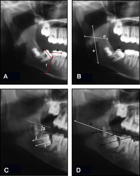
Table I and Figure 1 depict the 12 measurements made on the impacted and nonimpacted sides in each panoramic radiograph.
| a | MM1 width | Mesiodistal width of the mandibular first molar crown. |
| b | MM2 width | Mesiodistal width of the mandibular second molar crown. |
| c | MM1-ramus ant | Distance from the distal height of the contour of the MM1 to the ramus parallel to the occlusal plane. (The occlusal plane was determined by a line drawn from the tip of the mesial cusp of the MM1 to the tip of its distal cusp.) |
| d | Ramus width | The narrowest width of the ramus parallel to the occlusal plane. |
| e | Ramus height | The distance from the mandibular notch to the antegonial notch. |
| f | Mand height | Mandibular corpus height (in the MM1 area) |
| g | MM2-MM3 vert | The vertical distance between the coronal surface of the MM2 and the coronal surface of the MM3, perpendicular to the occlusal plane. |
| h | MM1-ramus post | Distance between the distal height of the contour of the MM1 to the distal end of the ramus, parallel to the occlusal plane. |
| k | MM2-angulation | The angle between the long axis of the MM2 and the occlusal plane. |
| l | MM3-angulation | The angle between the long axis of the MM3 and the occlusal plane. |
| i | MM2-mesial root | Length of the mesial root of the MM2 measured from the tip of the mesial cusp to the developing apex. |
| j | MM2-distal root | Length of the distal root of the MM2 measured from the tip of the distal cusp to the developing apex. |
| i-j | MM2 roots diff | The difference between the length of the mesial root of the MM2 measured from the tip of the mesial cusp to the developing apex to the length of its distal root measured from the tip of the distal cusp to the developing apex. |
The statistical analysis included the Pearson chi-square test, used to compare the prevalence of MM2 impaction in the Israeli and Chinese-American populations (first objective). A paired t test was used to compare the linear and angular measurements of the impacted and the nonimpacted sides at a significance level of 0.05.
Results
A total of 120 subjects with MM2 impaction were found (1.8%) of whom 49 (1.4%) were Israelis and 71 (2.3%) were Chinese-Americans ( Table II ).
| MM 2 | Israelis | Chinese-Americans | P | |||
|---|---|---|---|---|---|---|
| n | Percentage | n | Percentage | |||
| 1 | Prevalence | 49 | 1.4% | 71 | 2.3% | 0.004 ∗ |
| 2 | Unilateral impaction | 36 | 73% | 39 | 55% | 0.039 ∗ |
| Bilateral impaction | 13 | 27% | 32 | 45% | ||
| 3 | Mesial impaction | 43 | 88% | 63 | 89% | 0.964 |
| Vertical impaction | 4 | 8% | 6 | 8% | ||
| Distal impaction | 2 | 4% | 2 | 3% | ||
| 4 | Male | 23 | 47% | 41 | 58% | 0.611 |
| Female | 26 | 53% | 30 | 42% | ||
| 5 | Right unilateral impaction | 20 | 55.6% | 13 | 33% | 0.053 ∗ |
| Left unilateral impaction | 16 | 44.4% | 26 | 67% | ||
Stay updated, free dental videos. Join our Telegram channel
VIDEdental - Online dental courses


