Multiple treatment options are available for patients who have impacted canines in addition to congenitally missing lateral incisors. This article describes the treatment of a 13-year-old postpubertal girl with bilaterally impacted permanent maxillary canines, missing lateral incisors, retained deciduous canines, and a midline diastema. The orthodontic treatment plan included extraction of the deciduous canines. A lingual and labial approach (1-couple force system) was used to move the permanent canines into the arch. Through a collaborative team effort, including an orthodontist and a periodontist, the outcome was excellent esthetically and functionally.
Maxillary canines are important both esthetically and functionally. They are also the second most frequently impacted teeth, with a prevalence of 1% to 4%. Palatally impacted canines occur twice as frequently in females as in males, and bilateral occurrence has been reported to be in the range of 8% to 45%. Jacoby stated that 85% of the impacted canines are palatal, and they usually occur in patients with adequate arch length. Patients with impacted maxillary canines are perceived to be more difficult and time-consuming to treat than those with a routine malocclusion.
From a treatment perspective, congenitally missing maxillary lateral incisors also pose significant problems for orthodontists. They are the third most commonly missing teeth after third molars and mandibular second premolars, and they account for 20% of all congenitally missing teeth. Jena and Duggal stated that there is a high probability of palatal canine impaction when adjacent lateral incisors are anomalous or missing. Becker et al found that 5.5% of patients with a palatally impacted canine had a congenitally missing lateral incisor. Anic-Milosevic et al reported that 16% of patients with palatal canine impaction have peg-shaped lateral incisors, missing lateral incisors, or missing second premolars, suggesting a significant association between palatally impacted canines and missing lateral incisors. Mossey et al stated that smaller than average crown width of lateral incisors is an important factor for palatal canine impaction; however, no statistical evidence has yet given credibility to the association between the crown width of lateral incisors and palatal canine impaction. Nevertheless, when it does happen, careful treatment planning is imperative. Strang stated that in patients with missing lateral incisors and impacted canines, the best option is to create space for the lateral incisors followed by prosthetic replacement for the missing lateral incisors. On the other hand, Carlson emphasized canine substitution of the lateral incisors followed by selective recontouring of the canines.
A maxillary midline diastema is a malocclusion characterized by spaces between the maxillary central incisors; it has functional and esthetic consequences. It is more prevalent in the African American population than in white people. The most common etiology reported in the literature is tooth size or jaw size discrepancy including missing lateral incisors.
Retained deciduous canines are sometimes present with palatally impacted canines. Studies have shown that the removal of a deciduous canine results in successful eruption; in some cases, it has been shown to be successful in inducing eruption and even uprighting the impacted canines. Similarly, several authors have proposed the extraction of deciduous canines to return ectopically erupting canines to normal eruption pathways, especially if the persistence of the deciduous teeth would be a mechanical obstacle for the emergence of the permanent teeth.
In this article, we report on a patient with bilaterally impacted canines, congenitally missing lateral incisors, hypertrophic and incisively placed labial frenum, and retained deciduous canines.
Case report
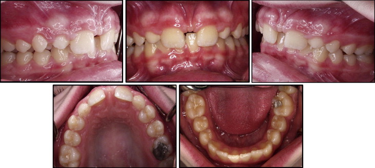
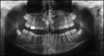
A 13-year-old postpubertal girl came to our clinic with the chief complaint of spacing between her maxillary central incisors ( Fig 1 ). She had no significant medical history, but her dental history included an amalgam restoration and sealants on the posterior teeth. She reported a history of crepitus in the left temporomandibular joint, but no limitations of mouth opening or pain were reported at the initial examination. The pretreatment records showed a straight to mild tendency toward a concave facial profile. She had a Class II (end on) molar relationship bilaterally. Clinical examination showed an 80% deepbite, 3 mm of overjet, U-shaped maxillary and mandibular arches, and the mandibular midline shifted toward the right by 2 mm. The maxillary labial frenum was attached to the incisive papilla. The patient had retained deciduous canines and congenitally missing lateral permanent incisors in the maxillary arch. The orthopantomogram ( Fig 2 ) showed that both maxillary canines were bilaterally impacted, and the occlusal and periapical radiographs confirmed that the impactions were palatal. Tomographic images showed that the crowns of the canines were close to the roots of the central incisors, and the roots of the impacted canines were close to the roots of the premolars ( Fig 3 ).

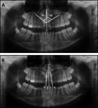
The position of the impacted canines was evaluated with the orthopantomogram, periapical and occlusal radiographs, and tomograms. According to the method suggested by Ericson and Kurol, the right and left canines were impacted at 55° and 58°, respectively ( Fig 4 , A ). Mesiodistal evaluation according to the method of Baccetti at al showed a sector 5 impaction, where the impacted canine completely overlapped the central incisor crown ( Fig 4 , B ).
The etiology of palatally impacted canines is obscure and multifactorial. In this patient, we speculated on 2 possible reasons for the impaction of the maxillary canines: genetics and congenitally missing lateral incisors. Because neither her sibling nor her parents had impacted canines, heredity was ruled out as a potential factor for impaction. Therefore, the missing lateral incisors might have been responsible for the bilateral canine impaction. Previously, it was speculated that the root of the lateral incisor might play an important role in the eruption of the permanent canine by providing adequate guidance for its eruption.
The patient was diagnosed with an Angle Class II malocclusion, congenitally missing maxillary lateral incisors, palatally impacted maxillary canines, midline diastema, incisively placed frenum, and 80% deepbite.
Treatment plan
The treatment objectives for this patient were to achieve an optimal soft-tissue profile and establish an ideal occlusal relationship after the eruption of the impacted canines and the attainment of normal overjet and overbite.
The treatment plan to achieve these objectives was established as follows. The impacted canines would be surgically exposed on an outpatient basis under local anesthesia and brought into the arch using cantilever mechanics; the retained deciduous canines would be extracted. The congenitally missing lateral incisors and impacted canines would be replaced with the canines and the premolars, respectively.
The midline diastema would be closed; to prevent the relapse, the labial frenum would be surgically resected. The deepbite would be corrected and an ideal occlusal relationship established.
Treatment alternatives
The second option was to distalize the molars bilaterally to achieve an Angle Class I molar relationship and to restore the space for prosthetic implants in place of the congenitally missing lateral incisors, but this option was rejected because the patient was only 13 years old, and we must wait at least 5 years before placing implants. Also, the parents opted out of it for economic reasons.
The other option was to convert the deciduous canines into lateral incisors, but this option was also rejected because more than half of the roots of the canines were resorbed, and we were not sure about the longevity of the deciduous canines.
Stay updated, free dental videos. Join our Telegram channel
VIDEdental - Online dental courses


