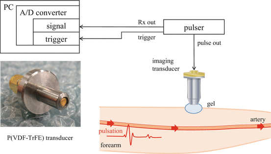
Measurement of Skin Elasticity Using High Frequency Ultrasound Elastography with Intrinsic Deformation Induced by Arterial Pulsation

Fig. 21.1

Block diagram of 3D ultrasound microscope system
A322452_1_En_21_Fig2_HTML.gif
Fig. 21.2

Schematic of elasticity measurement

21.2.2 Subject

Subject is one 24-year-old healthy male. A measurement area is skin in his forearm.

21.2.3 Velocity Measurement
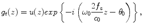
The velocity induced by the pulsation was measured from the RF echo data of the fixed position by implementing a 1-D cross correlation method. RF signal of time t at depth z was defined as r t (z). Analytical signal g t (z) was obtained by applying the Hilbert transform to the RF signal r t (z). Pulse waves with angular frequency ω 0 = 2πf 0 were transmitted at a time interval of ΔT. Analytical signal of time t and t + ΔT at depth z can be modeled as


$$ {g}_t(z)=u(z) exp\left\{-i\left({\omega}_0\frac{2{f}_s}{c_0}z-{\theta}_0\right)\right\}, $$
(21.1)

$$ {g}_{t+\Delta \mathrm{T}}(z)=u(z) exp\left[-i\left\{{\omega}_0\frac{2{f}_s}{c_0}\left(z-{z}_{\tau}\right)-{\theta}_0\right\}\right], $$
(21.2)

where u(z) was the envelope of the analytical signal, f s was the sampling frequency, c 0 was the sound speed, θ 0 was the initial phase, and z τ was the true displacement induced by the pulsation. The complex cross-correlation function γ(z lag ) at lag z lag was defined as


$$ \upgamma \left({z}_{lag}\right)=\frac{\sum_{z=-\frac{N}{2}}^{z=\frac{N}{2}}{g}_t^{*}(z){g}_{t+\Delta \mathrm{T}}\left(z+{z}_{lag}\right)}{\left|{\displaystyle {\sum}_{z=-\frac{N}{2}}^{z=\frac{N}{2}}}{g}_t^{*}(z)\right|\left|{\displaystyle {\sum}_{z=-\frac{N}{2}}^{z=\frac{N}{2}}}{g}_{t+\Delta \mathrm{T}}\left(z+{z}_{lag}\right)\right|} $$
(21.3)

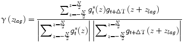
where N was 1-D cross correlation window of 256 pixels, corresponding to 195 μm depth window with 91 % overlap using hamming window. An index at maximum value of the real part of the Eq. (21.3) corresponded to the index of 
$$ {\hat{z}}_{lag} $$
. In this paper, f s was 1 GHz, and the temporal resolution was enough high to observe the deformation with HFUS of 100 MHz. Additionally, the received RF signal was up-sampled to 4 GHz before implementing the 1-D cross-correlation. Because of these reasons, the estimated displacement 
$$ {\hat{z}}_{lag} $$
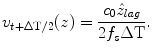
was almost equal to the true displacement z lag . The velocity, denoted by v t + ΔT/2(z), of the skin in his forearm between the interval was given as follows:


$$ {v}_{t+\Delta \mathrm{T}/2}(z)=\frac{c_0{\hat{z}}_{lag}}{2{f}_s\Delta \mathrm{T}}. $$
(21.4)

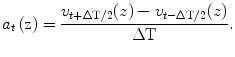
The acceleration was calculated from the measured velocity. The acceleration was calculated by differentiating the measured velocity as


$$ {a}_t\left(\mathrm{z}\right)=\frac{v_{t+\Delta \mathrm{T}/2}(z)-{v}_{t-\Delta \mathrm{T}/2}(z)}{\Delta \mathrm{T}}. $$
(21.5)

21.2.4 Shear Wave Measurement

By comparing an acceleration at reference depth with an acceleration at interest depth, a velocity of shear wave propagating from an artery toward a skin surface can be calculated from a relationship between the depth and the arrival time of shear wave at the depth. The relationship φ(z) can be expressed as


$$ \upvarphi (z)=\frac{{\displaystyle {\sum}_{t=-\frac{N_{\alpha }}{2}}^{t=\frac{N_{\alpha }}{2}}}{a}_t^{*}\left({z}_0\right)\cdot {a}_t(z)}{\left|{\displaystyle {\sum}_{t=-\frac{N_{\alpha }}{2}}^{t=\frac{N_{\alpha }}{2}}}{a}_t^{*}\left({z}_0\right)\right|\left|{\displaystyle {\sum}_{t=-\frac{N_{\alpha }}{2}}^{t=\frac{N_{\alpha }}{2}}}{a}_t(z)\right|}, $$
(21.6)

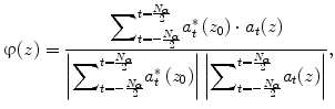
where N α was an estimation window of 50 pixels, corresponding to a 50 ms time. A hamming window was used for the estimation. A variable z 0 was a reference depth, and a variable z was an interest depth. A time lag between the reference depth and the interest depth was expressed as follows:


$$ \updelta \uptau (z)=\frac{\varphi (z)}{2\pi {f}_c} $$
(21.7)
The center frequency f c was obtained from the power spectrum calculated by applying the Fourier transform to the acceleration signals. Figure 21.3 shows the relationship between the depth z and the time lag. A regression line can be obtained by applying the least square method to the relationship. A slope a 1 of the regression line can be obtained by minimizing a least mean square error e as follows:

A322452_1_En_21_Fig3_HTML.gif
Fig. 21.3

Relationship between depth and time lag. Dots: experiments data, and line: regression line calculated by least square method

$$ \mathrm{e}={\displaystyle \sum_{z=0}^{z={Z}_N}}{\left|\updelta \uptau (z)-\left({a}_1z+{a}_2\right)\right|}^2 $$
(21.8)
A variable Z N is a total number of the region of interest. The velocity C s of shear wave was given by dividing the depth distance Δz by the estimated slope value 
$$ {\hat{a}}_1 $$

Only gold members can continue reading. Log In or Register to continue

Stay updated, free dental videos. Join our Telegram channel

Sep 17, 2015 | Posted by in General Dentistry | Comments Off on Measurement of Skin Elasticity Using High Frequency Ultrasound Elastography with Intrinsic Deformation Induced by Arterial Pulsation

VIDEdental - Online dental courses

Get VIDEdental app for watching clinical videos