The need to segment the maxilla to achieve esthetic and functional objectives has been recognized for at least 80 years. Initially, segmental maxillary osteotomy procedures were performed as isolated and definitive treatments. As orthognathic surgical expertise evolved, the ease and predictability of segmenting the entire maxilla have been realized; thus, the indications for these isolated procedures are rare. The focus of this chapter is total maxillary segmental osteotomy.
HISTORY
Although segmental mandibular osteotomies were described by Hullihen in 1849, the segmental maxillary osteotomy was not introduced until 1921, by Cohn-Stock. Wassmund described a single-stage labial approach in 1926, followed by Axhausen, who described a palatal tunneling technique in 1937. Schuchart developed a two-stage approach with the palate operated initially followed 4 weeks later by a labial approach, to complete the access to the lateral maxilla. In 1963, Wunderer published a modification of Wassmund’s procedure. He raised a palatal flap and left a broad buccal pedicle to section the anterior maxilla. Köle’s article on segmental surgery was published in 1959. This article, which was written in English, stimulated much interest in segmental osteotomy, especially among British and American surgeons. A complete generation of surgeons used these techniques routinely until Bell’s revascularization studies revolutionized the performance of surgery performed to correct maxillary deformities. Studies indicated that the entire maxilla could be mobilized and segmented with only transient ischemic effects and little adverse effect on wound healing. Beginning in the 1970s, a number of surgical techniques were described for mobilizing the entire maxilla and segmenting it to improve outcomes. Epker et al made significant contributions to the design of total maxillary segmental osteotomies conducted through multiple approaches. Today, most segmental maxillary surgery is performed via Le Fort I down-fracture; this procedure is the topic of this chapter.
INDICATIONS
Although transverse and vertical maxillary discrepancies have been recognized for decades, they have not received the same attention as sagittal maxillary discrepancies. The reason for this is most likely the difficulty involved in managing these conditions. Vertical steps in the dental arch occur with a variety of malocclusions. Of particular concern are the vertical steps in the maxillary arch, which accompany anterior open bite. Leveling these steps orthodontically may be possible, but the stability of the correction when done orthodontically alone is questionable, especially when teeth are extruded. Surgical leveling is possible and requires an interdental osteotomy between the teeth involved. If an anterior open bite is present, and no vertical step is present in the maxillary arch, closing the bite without segmental surgery should be possible by rotating the maxilla superiorly in the posterior region or inferiorly in the anterior region, or performing a combination of both. If the anterior bite is closed by rotating the maxilla downward anteriorly and the posterior bite opens, segmenting the maxilla so that pieces move independently is the treatment of choice.
Transverse maxillary deficiency is commonly seen in dentofacial deformities. On many occasions, orthodontic preparation alone facilitates transverse arch compatibility when minor discrepancies exist. In other instances, the discrepancy is sufficient that it cannot be stably improved with conventional orthodontics; in these instances, surgery is indicated to improve the width of the maxilla. Although some advocate surgically assisted rapid maxillary expansion, this procedure can be problematic. Furthermore, in many situations, it does not address the entire occlusal issue, which commonly involves more than just the transverse dimension.
Transverse maxillary deficiency and a vertical step in the plane of occlusion may accompany each other; when this occurs, or when either occurs in isolation, surgery of the maxilla in segments should be considered.
An increasing number of adult patients are seeking surgery and orthodontics to correct their conditions. It is estimated that 30% of these patients have transverse discrepancies between the dental arches. The most common problems are sagittal and transverse maxillary deficiencies combined with mandibular prognathism. In North America, maxillary problems commonly occur in combination with mandibular deficiency. In South Americans and in other Hispanic populations, the maxillary problems seem to occur more commonly with mandibular prognathism.
An appropriate dental arch transverse relationship improves masticatory efficiency and long-term periodontal health and reduces trauma to the buccal mucosa during chewing. This relationship also reduces the need for tooth removal to achieve functional occlusion. It is important to note that appropriate transverse relationships increase buccal tooth surface exposure during smiling, and this enhances the facial appearance.
When transverse problems occur in adults, surgery designed to widen the maxilla or to narrow the mandible can be performed to deal with these problems. When transverse discrepancies exist, the following five options should be considered for correction:
- 1.
Surgically assisted rapid palatal expansion (SARPE)
- 2.
Multisegment Le Fort I osteotomy for expansion
- 3.
Mandibular narrowing via midline osteotomy
- 4.
Slow dentoalveolar expansion with orthodontic movement
- 5.
Orthopedic rapid maxillary expansion
Rapid palatal expansion was introduced in the early 1960s to address transverse maxillary discrepancies. In growing children and in adolescents, the procedure is more reliable than when used in adults. In adults, alveolar bending or lateral tooth movement causing periodontal defects has been observed. Presumably, this occurs because of fusion of the midpalatal and other articulations between the maxilla and the palatal bone. Surgically assisted rapid maxillary expansion was introduced to assist the correction of transverse problems in the adult.
Clinical examination of the face and occlusion and dental model evaluation provide the greatest amount of diagnostic material needed to develop a treatment plan. Lateral cephalometric and panoramic radiographs are more helpful for identifying vertical problems than transverse problems. They also are helpful for identifying potential interdental osteotomy sites. Posterior anterior cephalometric measurements are of limited value because of the landmark identification issue, lack of standardization of radiographs, and problems with image quality. This makes determination of the origin (dental or skeletal) more difficult. Cone beam computed tomography (CT) imaging may enhance the ability to distinguish skeletal from dental causes in that the angulation of teeth and bone can be clearly visualized.
TECHNIQUE
Although this technique is controversial, Xylocaine with 1:100,000 epinephrine can be injected into the tissues before the incision is made. Another controversy involves the efficacy of hypotensive anesthesia during this surgical procedure. Use of these blood-preserving techniques is common in practice, but in theory, they may adversely affect wound healing and perfusion of the segments. Surgeons must develop their own philosophy regarding the potential benefits of reduced blood loss versus potential adverse wound healing consequences.
An incision through the vestibular mucosa of the upper lip is designed to leave a broad vascular pedicle along the buccal surface of the maxilla. The incision is made high in the vestibule and begins at the second molar, continuing anteriorly to the midline. This incision is joined by an identical one that is placed on the opposite side. The subperiosteal dissection from the incision should be directed superiorly, anteriorly, and posteriorly but should not be conducted inferiorly. The intention is to leave as much buccal tissue pedicled to the tooth-bearing portion of the maxilla as possible. Once the rim of the nose has been dissected, the lateral nasal walls and the anterior nasal spine are dissected. At the zygomaticomaxillary buttress region, the tissue should be dissected posteriorly in a subperiosteal plane to the pterygomaxillary juncture. The Le Fort I osteotomy should be performed as described in Volume III, Chapter 7 .
Once the maxilla has been down-fractured and the soft tissues of the nasal floor have been dissected from the nasal surface and thoroughly mobilized, the segmental osteotomy is begun. A large retractor is placed that reflects the soft tissue lining of the nose cephalically to adequately expose the osseous floor of the nose. A rounded tip fissure burr (Steiger burr) is used in a rotary instrument to section the bone of the lateral nasal floor from the posterior hard palate to the anterior region at the cuspid area. A very thin bur 701, a thin saw .015, or a piezoelectric osteotome should be used when planing is completed, and a wide burr 703 should be used when the maxilla is going to be narrowed ( Figures 8-1, 8-2, 8-3 ).
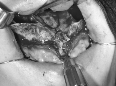
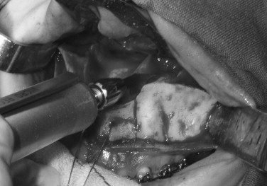
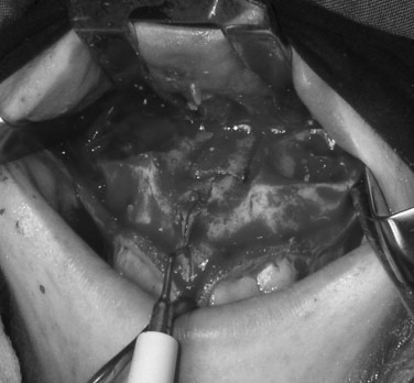
The bone is generally shallow posteriorly and thickens anteriorly. A similar osteotomy is conducted on the opposite side of the nasal floor. These two osteotomies are joined by a transpalatal osteotomy at the anterior extent of the two parallel osteotomies. The bone at the midline of the palate is thickest. The soft tissues are thinnest in this area, so care must be taken to prevent damage to the palatal mucosa.
Once the osteotomies have been completed, a periosteal elevator or a small bi-beveled osteotome is used to mobilize the free island of palatal bone that remains pedicled to the palatal mucosa. The interdental osteotomy site then is identified. A tooth forceps and a thin periosteal elevator are used to facilitate dissection of the buccal pedicle at the interdental osteotomy site. The root position should be determined from radiographs and by observation of the prominence of the buccal cortex over the roots. A tunnel is created, with care taken to leave a similar amount of buccal tissue pedicled on either side of the osteotomy site. A small fissure burr (#701) then is used to score the osteotomy through the cortical bone of the alveolus. The osteotomy should run from the alveolar crest superiorly to the horizontal osteotomy along the lateral maxillary wall. If the central or lateral incisor interspace is the interdental osteotomy site, the osteotomy is directed into the floor of the nose. Superiorly (above the tooth apices), the osteotomy can be deepened and should be connected to the nasal floor osteotomies. Care should be taken to avoid perforating the palatal tissues.
A thin osteotome is malleted to complete the interdental osteotomy by inserting the cutting end into the scored cortical bone. Conducting the osteotomy in this manner reduces the chance that bone may be damaged by heat from the drill; it also reduces the likelihood of damage to the roots ( Figures 8-4, 8-5, and 8-6
Stay updated, free dental videos. Join our Telegram channel
VIDEdental - Online dental courses


