Introduction
Our objective was to determine the perception of smile esthetics among orthodontists and laypeople with respect to asymmetries in the maxillary canines’ gingival margins in full-face and close-up smile analyses.
Methods
Full-face and close-up photographs of the frontal smiles of 4 subjects (2 women, 2 men) were used. The images were digitally altered to create a symmetrical image with the gingival margin levels of the maxillary canines matching the central incisors. From this new image, 5 stages of alterations were made in the gingival margin of the right canine in 0.5-mm increments. Final full-face and close-up images of the smiles were assessed by 50 orthodontists and 50 laypeople, who indicated the level of attractiveness of each smile on visual analog scales. The data collected were statistically analyzed by means of 1-way analysis of variance with the Tukey post-hoc test and the unpaired Student t test.
Results
In general, the most attractive smiles for the orthodontists were those without asymmetries and the one with a 0.5-mm asymmetry, whereas laypersons could not detect an asymmetry up to 1.5 mm ( P <0.05). For both groups of raters, the lowest scores were assigned for the smiles with asymmetries of 2.0 and 2.5 mm ( P <0.05). When opinions of orthodontists and laypersons were compared, in most situations a statistically significant difference was found, with orthodontists more sensitive in detecting deviations ( P <0.001). Moreover, there was no significant difference ( P >0.05) between the full-face and close-up assessments of the smiles.
Conclusions
It can be concluded that the perceptions of unilateral asymmetries in the gingival margin levels of the maxillary canines were 1.0 mm for orthodontists and 1.5 to 2.0 mm for laypersons.
In recent years, facial esthetics has become a major focus for the public worldwide. Having a beautiful, youthful smile is among patients’ main concerns, and esthetic improvements are routinely requested in dental offices. For this reason, orthodontists worldwide are working hard to incorporate into their clinical routine different tools to focus on improving smile esthetics.
An esthetically pleasing smile should include aspects such as symmetry and proportion between the central incisors, minimal gingival display, moderate to minimum buccal corridor spaces, ideal smile arc with the curvature of the maxillary anterior incisal edges parallel to the lower lip, and adequate design of the gingival margins in the esthetic zone.
With these considerations, studies have evaluated the perception of asymmetries in smile esthetics. Kokich et al conducted a study to evaluate the esthetic perceptions of general dentists, orthodontists, and laypersons regarding alterations in crown lengths of the maxillary central incisors and gingival margins of the maxillary lateral incisors. With regard to the crown size of the central incisors, they concluded that orthodontists could perceive a 1-mm discrepancy and that general dentists identified 1.5 mm, but the discrepancy was noticeable by laypersons only when the alteration was above 2 mm. In another study, Kokich et al investigated this aspect again using asymmetric increments and concluded that orthodontists were more sensitive and could detect a 0.5-mm discrepancy. General dentists and laypeople were less rigorous, and they only perceived alterations of 1.5 and 2 mm, respectively.
An interesting aspect is that in these studies, only the central and lateral incisors were assessed. It is debatable whether these data can be applied to the canines. Pinho et al investigated the perceptions of incisal-edge asymmetries between maxillary canines and found that discrepancies up to 2 mm were not detected by orthodontists, prosthodontists, and laypeople. On the other hand, we found no studies that considered the perception of smile esthetics with respect to gingival margin discrepancies between the maxillary canines. This information is of paramount importance because this is a common clinical finding. The establishment of this parameter is critical in some situations such as canine substitution cases where premolars substitute as canines. In these situations, a gingival asymmetry will be present, and some authors suggest dental intrusion followed by restoration to match the gingival margin level from the contralateral canine. This strategy might be questionable because if those asymmetries cannot be detected, it might be unnecessary to treat them.
Thus, does this discrepancy need to be corrected? In other words, if laypersons cannot recognize dental or gingival asymmetries as unesthetic, why should they be treated? Would the correction of some gingival discrepancies be an overtreatment more than an esthetic need?
Therefore, the aim of this study was to determine the perceptions of smile esthetics among orthodontists and laypeople with respect to asymmetries in the maxillary canines’ gingival margins in facial and close-up smile analyses. The null hypothesis was that these asymmetries would be rated as attractive equally by orthodontists and laypeople.
Material and methods
The study was approved by the research ethics committee of the Dental School, Federal University of Bahia in Brazil, under report number 24/12 and registered at CONEP: CAAE 03798512.9.0000.5024. All participants in the study signed a form of free and informed consent.
The sample size was determined using a pilot study and the Student unpaired t test with 80% power and a bilateral alpha level of 5%. It was determined that the sample should include at least 49 persons in each group of examiners.
Forty-eight images—24 full-face views and 24 close-up views—of the smile were used from 4 subjects, who were volunteer patients in the Section of Orthodontics at the Federal University of Bahia; the 2 women and 2 men were white, between 25 and 35 years of age, with no apparent facial asymmetries, and with attractive smiles. These smiles followed some principles of an ideal smile described in the literature: adequate width-to-length proportion of the esthetic zone, symmetry between maxillary central incisors, convex smile arc, gingival display less than 1.0 mm, and moderate buccal corridors.
The photographs were taken by the same operator (B.D.C.) using the digital Rebel camera with an MR-14EX ring flash and a 100-mm macro lens (Canon, Tokyo, Japan) mounted on a tripod.
The selected images were digitally altered using Photoshop (CS5.1; Adobe Photoshop, San Jose, Calif). The photos were altered to produce symmetrical images and retouched to adjust color, brightness, and contrast, as well as to remove any discolorations on the lips and skin. Each image was then condensed to achieve an image with measurements identical to those on the actual patient. Thus, for the close-up images, each millimeter measured on the digital and printed images was equivalent to each millimeter measured clinically on the patient, with the maxillary central incisor as the reference. For the full-face images, the magnification ratio was 1:2; ie, 1 mm on the printed image was equivalent to 2 mm in the real patient.
The gingival line of the central incisors was used as a reference, and the canines’ gingival margins were matched with this line, with the lateral incisor 0.5 mm below and an incisal step of 1 mm between the central and lateral incisors. All mandibular teeth were removed from the images.
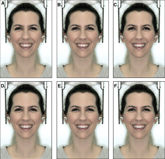
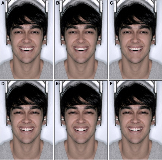
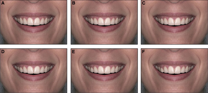
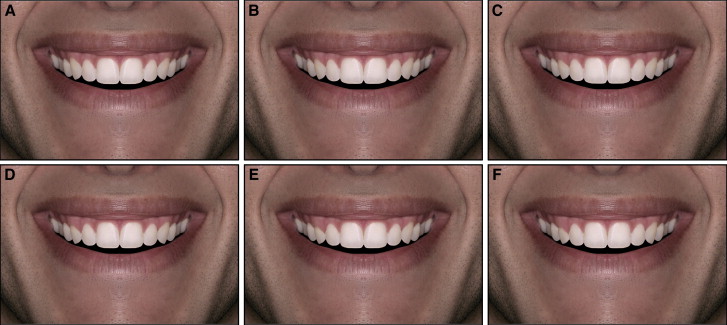
After these procedures, 5 alterations were created in the gingival margins of the canines on 1 side, with discrepancies of 0.5, 1, 1.5, 2, and 2.5 mm in relation to their contralateral teeth. Therefore, 6 images were produced for each subject. The upper limit of the full-face image ( Figs 1 and 2 ) was the region just above the top of the head, and the lower limit was the base of the neck. The upper limit of the close-up image of the smile ( Figs 3 and 4 ) was the base of the nose, and the lower limit was above the chin.
The final images were digital files with a resolution of 300 dpi. They were professionally printed on standard A4 size format (29.7 × 42 cm). Then a photo album was assembled containing all images in random order.
The album was given to the 100 raters (50 orthodontists, and 50 laypeople with a college education but no dental background). Each rater was given information about the study and asked to evaluate the attractiveness of the images. Along with the album, each rater received a form with 100-mm visual analog scales printed for each image, as in previous studies. The scale ranged from “very unattractive” on the far left to “very attractive” at the far right. A line was also printed at the midpoint of each scale as a reference line for an average level of attractiveness. All raters marked a point along the scale according to their perception of smile esthetics. The scores were then measured in millimeters by the first author (B.D.C.) with an electronic digital caliper (Starrett, Suzhou, China).
To test the reliability of the method, 10 examiners (5 laypeople, 5 orthodontists) were randomly selected to evaluate 2 pages of photographs (1 containing a full-face view and the other a close-up view of the smile) with 2 identical images on the same page. Then the scores corresponding to the 2 identical images were tabulated and examined by the intraclass correlation test with the BioEstat program (version 5.0; Mamirauá Institute, Belém, Pará, Brazil). The result (0.72) showed good reliability of the method.
Descriptive statistics were reported as means and standard deviations. Differences in the mean esthetic scores in the 6 levels of asymmetry were analyzed using 1-way analysis of variance with the Tukey post-hoc test. To compare the distributions of the mean scores between the full-face and close-up images and also between the orthodontists and laypersons, the Student t test was used. The level of significance was established at 5%.
Results
When comparing assessments of full-face images with close-up views, no statistically significant difference was found ( P >0.05) ( Tables I-IV ). After this result, the close-up view was used as the reference to address the results.
| Variable (mm) | Full-face view | Close-up view | P | ||
|---|---|---|---|---|---|
| Mean | SD | Mean | SD | ||
| 0.0 | 76.5 | 17.0 | 80.2 | 16.9 | 0.120 |
| 0.5 | 72.1 | 17.5 | 71.2 | 16.7 | 0.687 |
| 1.0 | 60.2 | 21.3 | 61.7 | 19.4 | 0.608 |
| 1.5 | 57.4 | 22.6 | 54.8 | 20.8 | 0.397 |
| 2.0 | 48.3 | 27.5 | 42.5 | 24.8 | 0.122 |
| 2.5 | 40.4 | 29.9 | 36.1 | 26.4 | 0.282 |
| Variable (mm) | Full-face view | Close-up view | P | ||
|---|---|---|---|---|---|
| Mean | SD | Mean | SD | ||
| 0.0 | 75.6 | 17.8 | 77.9 | 16.4 | 0.342 |
| 0.5 | 68.2 | 17.5 | 71.2 | 15.7 | 0.095 |
| 1.0 | 61.6 | 18.8 | 63.4 | 16.9 | 0.479 |
| 1.5 | 52.8 | 21.6 | 55.3 | 19.3 | 0.400 |
| 2.0 | 35.6 | 25.0 | 36.3 | 21.4 | 0.826 |
| 2.5 | 30.9 | 23.1 | 32.8 | 24.5 | 0.558 |
| Variable (mm) | Full-face view | Close-up view | P | ||
|---|---|---|---|---|---|
| Mean | SD | Mean | SD | ||
| 0.0 | 74.1 | 17.9 | 77.9 | 16.4 | 0.202 |
| 0.5 | 70.0 | 16.3 | 73.7 | 16.3 | 0.131 |
| 1.0 | 58.2 | 20.5 | 60.7 | 18.7 | 0.374 |
| 1.5 | 50.1 | 22.8 | 51.7 | 22.9 | 0.605 |
| 2.0 | 38.7 | 25.6 | 42.8 | 26.2 | 0.269 |
| 2.5 | 34.8 | 26.6 | 36.2 | 25.9 | 0.702 |
| Variable (mm) | Full-face view | Close-up view | P | ||
|---|---|---|---|---|---|
| Mean | SD | Mean | SD | ||
| 0.0 | 75.8 | 17.1 | 77.0 | 16.2 | 0.619 |
| 0.5 | 69.2 | 18.4 | 70.3 | 15.7 | 0.647 |
| 1.0 | 58.1 | 17.7 | 62.6 | 18.1 | 0.079 |
| 1.5 | 44.6 | 21.8 | 48.4 | 20.8 | 0.211 |
| 2.0 | 35.0 | 24.2 | 36.6 | 23.5 | 0.660 |
| 2.5 | 29.5 | 25.2 | 30.1 | 26.4 | 0.869 |
In Table V , according to the orthodontists, in the assessment of the close-up view of the smile for woman number 1, the highest scores were assigned to the smiles without asymmetries and with a 0.5-mm asymmetry. For the laypeople, the highest scores were for the smiles without asymmetries and with asymmetries of 0.5 and 1.0 mm. For both groups of raters, the lowest scores were assigned for the smiles with asymmetries of 2.0 and 2.5 mm ( P <0.05).
| Variable (mm) | Orthodontists | Laypersons | Orthodontists × laypersons | ||||
|---|---|---|---|---|---|---|---|
| Mean | SD | Results ∗ | Mean | SD | Results ∗ | ||
| 0.0 | 80.73 | 16.76 | A | 79.76 | 17.20 | A | |
| 0.5 | 73.24 | 12.56 | A | 73.08 | 19.96 | A, B | |
| 1.0 | 55.41 | 14.83 | B | 67.95 | 21.51 | A, B | † |
| 1.5 | 45.77 | 15.25 | C | 63.80 | 21.83 | B, C | † |
| 2.0 | 31.09 | 16.46 | D | 54.00 | 26.50 | C, D | † |
| 2.5 | 23.32 | 18.71 | D | 48.86 | 26.93 | D | † |
∗ Variables with the same letter do not differ statistically ( P <0.05).
† statistical difference between groups of examiners ( P <0.001).
Table VI shows the results for woman number 2. The orthodontists assigned the highest scores to the smile without asymmetries and the lowest scores to smiles with asymmetries of 2.0 and 2.5 mm. The laypeople assigned higher scores to the symmetrical smile and smiles with asymmetries of 0.5, 1.0, and 1.5 mm. The lowest scores were for discrepancies of 2.0 and 2.5 mm ( P <0.05).
| Variable (mm) | Orthodontists | Laypersons | Orthodontists × laypersons | ||||
|---|---|---|---|---|---|---|---|
| Mean | SD | Results ∗ | Mean | SD | Results ∗ | ||
| 0.0 | 81.61 | 13.33 | A | 74.17 | 18.36 | A | |
| 0.5 | 69.70 | 14.12 | B | 74.56 | 16.90 | A | |
| 1.0 | 57.97 | 15.30 | C | 68.82 | 16.83 | A | † |
| 1.5 | 46.84 | 13.29 | D | 63.70 | 20.75 | A | † |
| 2.0 | 28.31 | 15.91 | E | 44.30 | 23.34 | B | † |
| 2.5 | 21.69 | 17.06 | E | 43.98 | 25.92 | B | † |
Stay updated, free dental videos. Join our Telegram channel
VIDEdental - Online dental courses


