Introduction
The purpose of this study was to determine the prevalence of hypodontia, hyperdontia, and impacted teeth in children with various types of clefts.
Methods
This study sample consisted of 201 cleft patients including 131 male subjects with a mean age of 12.3 ± 4 years and 70 female subjects with a mean age of 12.6 ± 3.9 years. Charts, models, radiographs, and intraoral photographs were used for the study. t tests, chi-square tests, and binomial tests were used for assessment of the data.
Results and Conclusions
Hypodontia was found in 129 subjects (64.1%). The chi-square test showed no statistically significant difference between the type of cleft and hypodontia ( P <0.319). The binomial test showed that the frequencies of subjects with hypodontia were significantly higher in both unilateral and bilateral cleft lip and palate patients ( P <0.015 and P <0.001, respectively). Hyperdontia and impacted teeth were also found to occur mostly in the maxillary arch, and maxillary canines were the most commonly impacted teeth in both unilateral and bilateral cleft lip and palate patients.
Highlights
- •
There is no relationship between hypodontia and the type of cleft.
- •
No sex differences were found in regard to the prevalence of hypodontia.
- •
Both supernumerary and impacted teeth occur mostly in the maxillary arch.
- •
Maxillary canines are often impacted in unilateral and bilateral cleft lip and palate patients.
Cleft lip and palate (CLP) is among the most common congenital human malformations. Failure of fusion of the maxillary and medial nasal prominences or between the palatal processes results in clefts of varying extent, unilaterally or bilaterally. CLP affects between 1 and 7 of 1000 newborns. The frequency of clefts is higher in Asian people than in other races.
When compared with the general population, subjects with CLP have always been found to have a higher prevalence of dental anomalies, such as variations in tooth number and position and reduced tooth dimensions, most of which are localized in the area of the cleft defect. Akcam et al investigated the frequency of various dental anomalies in the maxillary dental arch in various cleft groups and found that a significant proportion (96.7%) of subjects with a cleft had at least 1 dental anomaly. In another study, a higher prevalence of enamel discoloration was found in children with CLP when compared with a control group. Shapira et al found that in the cleft area, most developmental dental irregularities are related to the maxillary lateral incisor in both the deciduous and permanent dentitions. Tooth agenesis, also known as hypodontia or congenital absence of teeth, is the most frequently observed developmental anomaly of the human dentition. Clefts of all types are often associated with congenitally missing teeth. Shapria et al found a 77% prevalence of hypodontia in a combined sample of children with cleft lip, cleft palate, and both. They also reported that hypodontia was found to be considerably more frequent on the left side in both the maxilla and the mandible, and clefts occurred more often on the left side, clearly indicating left-side predominance for these anomalies.
The literature contains little information regarding the prevalence of various dental anomalies in patients with different types of clefts. Therefore, the aim of this study was to conduct a comprehensive research on tooth anomalies of cleft patients according to the different cleft types.
Material and methods
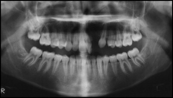
This study was carried out in accordance with the ethical standards of the 1964 Declaration of Helsinki. Informed written consent was obtained from each patient and a parent or guardian. In the study, we included 202 consecutive patients with cleft lip or cleft lip and palate who were referred to the orthodontic department of Shahid Beheshti University of Medical Sciences in Tehran, Iran, from 2009 to 2011. Except for 1 subject who was excluded from the study, no subjects had other known syndromes. The subjects’ distribution according to sex is given in Table I . The final sample of 201 included 131 male subjects with a mean age of 12.3 ± 4 years and 70 female subjects with a mean age of 12.6 ± 3.9 years. The youngest patient was 5 years old, and the eldest was 25 years old; however, only 1 subject was younger than 8 years old. The patients were divided into 8 groups according to the classifications proposed by Whitaker et al. The patient population was racially and ethnically similar. Lateral cephalograms, orthopantomograms, photos, and dental casts of the patients that were taken for treatment were used for the observational purposes of this study. Panoramic, periapical, and occlusal radiographs of the patients were used to determine the presence or absence of the teeth. A further clinical examination was done to confirm the absence ( Fig ).
| Sex | n (%) | Age (y), mean ± SD |
|---|---|---|
| Male | 131 (65.2) | 12.3 ± 4 |
| Female | 70 (34.8) | 12.6 ± 3.9 |
Statistical analysis
Two observers (A.D., R.H.) analyzed the records of the patients at the same time. The results of their observations were blinded to each other. Interobserver agreement was calculated through kappa analysis. A kappa value of 1 showed perfect agreement.
The anomalies of the subjects including hypodontia, hyperdontia, and impaction were investigated. Impaction was studied only in subjects with permanent dentition. Periapical radiographs were taken using paralleling techniques to diagnose hyperdontia. The Statistical Package for the Social Sciences (version 20; IBM, Armonk, NY) was used to analyze the data. We used t tests, chi-square tests, and binomial tests to determine the significance of the findings. Statistical significance was set at P <0.05.
Results
This study showed that unilateral and bilateral CLP followed by unilateral cleft lip and alveolus were more common than the other types of cleft in a Persian population ( Table II ). One hundred thirty-four of the subjects had permanent dentition. Hypodontia was found in 129 subjects (64.1%) ( Table III ). To assess the relationship between the frequency of hypodontia and the various types of cleft, the cleft types that were not related to bony defects were omitted, and chi-square tests showed no statistically significant difference between the remaining types of cleft and hypodontia ( P <0.319) ( Table IV ). In both the male and female subjects, no statistically significant differences were found between those who had hypodontia and those without it. Chi-square tests showed no statistically significant difference between the sexes ( P <0.740) ( Table V ).
| Sex | Unilateral cleft lip | Bilateral cleft lip | Unilateral cleft lip and alveolus | Bilateral cleft lip and alveolus | Cleft palate | Unilateral cleft lip and palate | Bilateral cleft lip and palate | Total |
|---|---|---|---|---|---|---|---|---|
| Male | 1 | 1 | 18 | 8 | 2 | 64 | 37 | 131 |
| Female | – | 1 | 11 | 4 | 7 | 27 | 20 | 70 |
| Total | 1 | 2 | 29 | 12 | 9 | 91 | 57 | 201 |
| Cleft type | Patients (n) | Sex | Patients with hypodontia | Patients without hypodontia | Total | |||
|---|---|---|---|---|---|---|---|---|
| n | % | n | % | n | % | |||
| Unilateral cleft lip | 1 | Male | 0 | 0 | 1 | 0.5 | 1 | 0.5 |
| Female | 0 | 0 | 0 | 0 | 0 | 0 | ||
| Unilateral cleft lip and alveolus | 29 | Male | 9 | 4.5 | 9 | 4.5 | 18 | 8.9 |
| Female | 6 | 3 | 5 | 2.5 | 11 | 5.4 | ||
| Unilateral cleft lip and palate | 91 | Male | 40 | 19.9 | 24 | 12 | 64 | 31.8 |
| Female | 18 | 8.9 | 9 | 4.5 | 27 | 13.5 | ||
| Bilateral cleft lip | 2 | Male | 1 | 0.5 | 0 | 0 | 1 | 0.5 |
| Female | 1 | 0.5 | 0 | 0 | 1 | 0.5 | ||
| Bilateral cleft lip and alveolus | 12 | Male | 5 | 2.5 | 3 | 1.5 | 8 | 4 |
| Female | 3 | 1.5 | 1 | 0.5 | 4 | 2 | ||
| Cleft palate | 9 | Male | 2 | 1 | 0 | 0 | 2 | 1 |
| Female | 3 | 1.5 | 4 | 2 | 7 | 3.5 | ||
| Bilateral cleft lip and palate | 57 | Male | 26 | 12.9 | 11 | 5.4 | 37 | 18.4 |
| Female | 15 | 7.4 | 5 | 2.5 | 20 | 10 | ||
| Total | 129 | 64.1 | 72 | 35.9 | 201 | 100 | ||
| Cleft type | Total patients (n) | Patients with hypodontia (n) | Patients without hypodontia (n) |
|---|---|---|---|
| Unilateral cleft lip and alveolus | 29 | 15 | 14 |
| Unilateral cleft lip and palate | 91 | 58 | 33 |
| Bilateral cleft lip and alveolus | 12 | 8 | 4 |
| Bilateral cleft lip and palate | 57 | 41 | 16 |
| Total | 189 | 122 | 67 |
| Sex | Patients | Patients with hypodontia | Patients without hypodontia | P value | |||
|---|---|---|---|---|---|---|---|
| n | % | n | % | n | % | ||
| Male | 131 | 65.2 | 83 | 41.3 | 48 | 23.9 | 0.466 |
| Female | 70 | 34.8 | 46 | 22.8 | 24 | 12 | 0.497 |
| Total | 201 | 100 | 129 | 64.1 | 72 | 35.9 | – |
Binomial tests were used for more detailed analysis of hypodontia according to cleft type. This test showed that the frequency of subjects with hypodontia was significantly higher in both unilateral and bilateral CLP patients ( Table VI ). Chi-square tests showed that hypodontia was significantly higher in the cleft side of the unilateral subjects and on both sides of the bilateral patients ( Table VII ). The binomial test also showed that missing teeth were significantly higher in the patients’ cleft side ( P <0.001) ( Table VIII ). Table IX shows that only 10 patients had hyperdontia. Only 9 of the bilateral CLP patients suffered from impacted teeth, followed by 7 unilateral CLP patients ( Table X ).
| Cleft type | Patients with hypodontia | Patients without hypodontia | Total | P value | |||
|---|---|---|---|---|---|---|---|
| n | % | n | % | n | % | ||
| Unilateral cleft lip | 0 | 0 | 1 | 0.5 | 1 | 0.5 | – |
| Unilateral cleft lip and alveolus | 15 | 7.5 | 14 | 7 | 29 | 14.5 | 1 |
| Unilateral cleft lip and palate | 58 | 28.8 | 33 | 16.5 | 91 | 45.3 | 0.015 ∗ |
| Bilateral cleft lip | 2 | 1 | 0 | 0 | 2 | 1 | 0.250 |
| Bilateral cleft lip and alveolus | 8 | 4 | 4 | 2 | 12 | 6 | 0.388 |
| Cleft palate | 5 | 2.5 | 4 | 2 | 9 | 4.5 | 1 |
| Bilateral cleft lip and palate | 41 | 20.3 | 16 | 7.9 | 57 | 28.2 | 0.001 ∗ |
| Total | 129 | 64.1 | 72 | 35.9 | 201 | 100 | – |
Stay updated, free dental videos. Join our Telegram channel
VIDEdental - Online dental courses


