Introduction
In this study, we aimed to investigate the effect of buccolingual inclinations of the maxillary canines and premolars on the perceived attractiveness of the smile when viewed from the frontal perspective.
Methods
A smiling frontal photograph was taken of a man. A 3-dimensional digital dental model of this subject was constructed within which the buccolingual inclinations of the canines and premolars could be altered relative to the occlusal plane. Three-dimensional models of the altered digital models were then printed in resin and mounted on articulators. Frontal photos of the mounted models were taken and transferred to the smile image. A series of images was produced with the canines and premolars inclined buccally or lingually by different degrees. The smile images were assessed by 2 panels, orthodontists and laypeople.
Results
There was a broad range of esthetic acceptability for the buccolingual inclinations of the maxillary canines and premolars. The range of preferred inclinations was not as broad. Smile esthetics was significantly compromised ( P <0.01) when the canines were lingually inclined more than −12°, or the premolars were lingually inclined more than −15°, as perceived by orthodontists and laypersons. Buccally tipping the canines more than 6° also made the smile esthetics less satisfying ( P <0.01).
Conclusions
It could be esthetically satisfying to position the teeth within the ranges of 0° to −7° of inclination for the canines and −3° to −11° of inclination for the premolars, as assessed by the orthodontists, or of 3° to −10° of inclination for the canines and 5° to −11° of inclination for the premolars, as assessed by the laypersons. Clinicians could exercise flexibility within this range, when compromising tooth positions for transverse jaw discrepancies.
Highlights
- •
We constructed and manipulated a series of 3-dimensional digital dental models.
- •
Altered inclinations of the teeth were transferred from mounted models to the smile images.
- •
Esthetic acceptability was wide for buccolingual inclination of maxillary canines and premolars.
- •
The range of preferred inclinations was not so broad.
An attractive smile is an important aspect of facial appearance. Researchers have conducted extensive studies on the impact of various aspects of smile attractiveness including buccal corridor spaces, arch widths, smile arcs, midline deviations, gingival displays, occlusal plane inclinations, and facial measurements. Recently, the buccolingual inclination of the posterior teeth has been recognized as an important aspect of both occlusion and smile attractiveness. A full, broad smile with minimal buccal corridor space has been deemed by some to be a preferred esthetic feature. Zachrisson stated that lingually inclined posterior teeth would increase the negative corridor and consequently decrease the smile fullness and attractiveness.
There are some deficiencies in studies regarding the impact of buccolingual inclinations of the maxillary posterior teeth on perceived attractiveness, resulting in conflicting findings. Previous researchers have failed to separately evaluate the esthetic effect of buccolingual inclinations of posterior teeth and the effect of negative buccal corridor spaces, since these 2 factors were coupled together in the study designs. Also, attention has been focused on the tilting of the posterior teeth in the frontal smile and the incisors for the profile smile, but the inclinations of the canines and premolars were omitted. The maxillary canine could be of prime importance in smile esthetics because of its location on the corner of the dental arch, joining the anterior and lateral segments of the teeth. Conclusions reached in previous studies were arrived at by digitally altering the positions of teeth on photographs, without knowing the exact angular inclination of the teeth relative to the occlusal plane in actual situations; thus, these studies could not provide detailed principles that can be systematically applied to clinical practice.
The term “inclination of teeth” was described in the 6 keys of normal occlusion, and ideal norms were set for buccolingual inclinations. In “Six elements of orofacial harmony” by Andrews, it is suggested that the root of each tooth should be centered between the faciolingual borders of the basal bone, and that each crown should be inclined so that it can interface fully and function gnathologically. In practical situations, dentoalveolar transverse compensation is often needed because of skeletal transverse discrepancies. The borders of the basal bone provide limitations for transverse tooth movement, in consideration of health and stability. In such conditions, the inclinations of the teeth should be a compromise from the published norms. For patients who seek orthodontic therapy, much attention is often paid to enhancing dentofacial esthetics rather than improving occlusal relationships. This calls for further research on the range of what might be considered esthetically acceptable regarding the buccolingual inclinations of teeth, as perceived by laypersons and dental professionals. Such research could help define the boundaries for transverse dental compensation when there are jaw discrepancies.
In this study, we hypothesized that there might be a range of esthetic acceptability for the buccolingual inclinations of the maxillary canines and premolars from a frontal view of the smile. We performed this study to investigate the effect of buccolingual inclinations of the maxillary canines and premolars on the perceived attractiveness of a frontal smile.
Material and methods
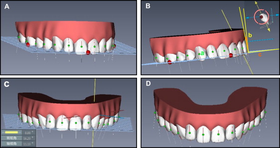
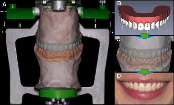
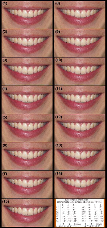
This study was carried out in 3 steps. In the first step, a 3-dimensional digital dental model was constructed, and alterations were made in the buccolingual inclinations of the selected maxillary teeth ( Fig 1 ). Second, photographic manipulations were performed so that the altered inclinations of the teeth could later be transferred from mounted maxillary models to the frontal view of the smile ( Fig 2 ). Finally, the 15 frontal smile images ( Fig 3 ) were assessed and rated by groups of orthodontists and laypeople.
A man considered to have an esthetic smile was selected. Informed consent was obtained. He met the following criteria: (1) well-aligned teeth and tooth-size symmetry, (2) Class I molar and canine relationships with normal overjet and overbite, (3) less than 2 mm of maxillary gingival display, (4) the dental midline coincided with the facial midline, (5) when adopting his natural head posture, both the interpupillary line and the Frankfort horizontal plane were parallel to the true horizontal, and (6) the angle between the occlusal plane and the sella-nasion plane was within a normal range.
A silicone rubber impression of his dentition was taken and then converted to digital data. The 3-dimensional digital dental models were constructed and manipulated using OrthoRX software (version 4.0; Henghui Technology Co, LTD, Xi’an, China). The facial axis of the clinical crown and its midpoint, the facial-axis point, as described by Andrews, were marked on the buccal surfaces of the teeth on the models. The occlusal plane was formed by the bisection of the vertical overlap of the incisal edges of the maxillary and mandibular incisors and the bisection of the vertical overlap of the anterior cusp tips of the mandibular and maxillary first molars. The buccolingual inclinations of the teeth were measured relative to this occlusal plane.
Alterations in the buccolingual inclinations of the maxillary teeth on the digital models were performed using OrthoRX software ( Fig 1 ). Before making the inclination alterations, a trial experiment was conducted using frontal smile images with the canines and premolars inclined in 10° increments between +20° and −30°. The inclinations outside the range between +10° and −20° were unacceptable by over 90% of the evaluators. There was less need to study unesthetic smiles, which would certainly be prevented by clinicians. The esthetic effect of inclinations between +10° and −20° was undefined and needs investigation using alterations in smaller increments of degrees.
We made the inclination alterations using a series of numbers deviating from the ideal norms. There were series 1, with inclinations deviating from −7°, −7°, −7° (norms proposed by Andrews for canines, first premolars, and second premolars, respectively), and series 2, with inclinations deviating from −3°, −7°, −7° (norms for Chinese people ). The inclinations of the canines were altered in 3° increments between +2° and −16° for series 1 and between +6° and −12° for series 2. The inclinations of the premolars were altered in 4 increments between +5° and −19° for both series. These alterations created 15 different groups of buccolingual inclinations of the teeth, including 7 groups for each series and the original smile photograph. A total of 15 digital maxillary models were constructed and printed tangibly in resin.
The frontal posed smile photograph of the subject was taken with a digital camera (D100; Nikon, Tokyo, Japan). When the smile was photographed, the lens of the camera was positioned perpendicular to the face in natural head position and simultaneously parallel to the interpupillary line, with the center point of the image superimposed on the labial interproximal contact area of the maxillary central incisors. The entire lower third of the face was captured in the image.
The printed resin maxillary models, representing 15 different groups of buccolingual inclinations of teeth, were each mounted on the same articulator. The wax-bite registration was made in the molar and incisor regions, and the same bite registration was used during each mounting. A frontal photograph was taken of each mounted cast with the same camera ( Fig 2 ). This principle was also applied so that the camera was held parallel to the true horizontal with the center point of the image superimposed on the labial interproximal contact area of the maxillary central incisors. Thus, a total of 15 images were created, each corresponding to a distinct resin model.
To transfer the altered buccolingual inclinations of the teeth from the articulator mounted models to the frontal view of the smile, the smile image was superimposed on 1 model photograph; then the inclinations of the teeth on the smile image were altered accordingly to match the model photograph using Photoshop software (version 11.0; Adobe, San Jose, Calif). To prevent any discrepancies in camera angle or magnification between the 2 superimposed images, the image scale was adjusted so that the mesiodistal crown widths of the maxillary right central incisors on the 2 images coincided. A total of 15 frontal smile images were created, representing 15 different groups of buccolingual inclinations of the teeth ( Fig 3 ).
For image assessment, a trial experiment was conducted, and a sample size calculation estimated that 52 subjects were required in each panel to detect a difference of 10 points in the smile ratings for multiple comparisons between groups, with power of 85% and 5% precision. The judges who evaluated the smile images belonged to 2 panels: orthodontic professionals (30 men, 30 women; mean ages, 38.5 and 36.2 years, respectively) and laypersons (30 men, 30 women; mean ages, 20.7 and 21.4 years, respectively). The professional panel consisted of orthodontists who practiced in Chengdu, China. The lay panel consisted of college students with no link to dentistry.
Each judge was shown a binder in which the 15 smile images were arranged in random order. The judges were asked to rate the attractiveness of the smiles using the visual analog scale in a quiet, nonclinical environment under good natural lighting conditions. There were 10 grades to choose from: 0 (least attractive), 10, 20, 30, 40, 50, 60, 70, 80, 90, and 100 (most attractive) scores. Specific instructions were given on the use of the visual analog scale. The judges were told to view all images before commencing the rating and not to return to previously rated images while progressing forward. The judges were not allowed to confer or discuss their ratings with others. The same observer gave instructions to all judges when necessary.
To test intrasubject reproducibility, repeated assessments were made 6 weeks after the initial ratings. The order of the 15 images was randomly changed between the first and second evaluations. Every image was examined twice by each judge, and the mean scores were used for statistical comparisons.
Statistical analysis
Data analysis was performed using software (version 13.0; SPSS, Chicago, Ill). The intrasubject reproducibility between the 2 assessments was tested with the intraclass correlation coefficient (ICC), with a 95% confidence interval. Means and standard deviations of the ratings for each image were calculated for each panel and sex group. Two-way analysis of variance (ANOVA) was used to evaluate the effects of the 2 independent variables (profession and sex) on the ratings for each image. One-way ANOVA was used to determine significant differences in the scores between the groups. For each panel, the least significant difference and the Student-Newman-Keuls methods were used for multiple comparisons to determine whether the judges scored differently between the images. The level of significance was set at 0.05.
Stay updated, free dental videos. Join our Telegram channel
VIDEdental - Online dental courses


