http://evolve.elsevier.com/Haveles/pharmacology
Oral disorders or diseases are among the most prevalent diseases in American society. Each year, dental disorders result in a loss of more than 164 million hours from work. Nearly one third of adult Americans have untreated tooth decay and one in every seven adults 35-44 years of age have gum disease. Severe gum disease affects 14% of Americans aged 45-54 years. The rate of gum disease increases to one in four Americans age 65 years and older. Less than 60% of adults older than 65 years visit an oral health care provider during a given year. Almost 25% of Americans 60 years and older have lost all of their permanent teeth. The growing number of older persons with their own natural teeth has many dental implications.1
Poor or improper oral hygiene is a direct cause of dental caries, gingivitis, and halitosis. Nonprescription products for preventing and treating hygiene-related oral disorders are available in pharmacies, food stores, and other retail outlets. Dental hygienists are in the forefront on educating the public about the proper use of these products and their role in preventing hygiene-related oral disorders.
Dental caries
Approximately 20% of the general population has experienced dental caries. The incidence of dental caries in children had decreased from the 1970s until the mid 1990s. This decrease has been attributed to fluoridation of public water supplies, dentifrices, and mouth rinses, not improved oral hygiene. Despite improvements in these areas, the incidence of dental caries in primary teeth of children 2-11 years of age appears to be on the rise. Currently, 42% of children 2-11 years of age have caries in their primary teeth. Of these children, 23% have untreated caries. Also, 21% of children 6-11 years of age have caries in their permanent teeth, and 8% have untreated decay. The incidence of tooth decay in adolescents aged 12-19 years is 59%. Patients at highest risk for caries are those with poor oral hygiene. Patients at increased risk include those with orthodontic appliances, xerostomia, or gum recession, those who are living at or below the poverty level, black non-Hispanic, or of Hispanic origin, and those who use tobacco. Box 13-1 gives a more detailed overview of the risk criteria for caries.
Dental caries is considered an infectious disease that affects the calcified tissue of the teeth. Certain plaque bacteria generate acid from dietary carbohydrates, causing acid demineralization of tooth enamel, which then leads to the formation of carious lesions. Plaque buildup is directly related to the incidence of oral disease. If left untreated, these lesions can destroy the tooth.
Carious lesions start slowly on the enamel surface and initially produce no clinical symptoms. Once demineralization of the tooth progresses through the enamel to the soft dentin, the destruction proceeds at a much faster pace. At this point, the patient becomes aware of the problem either by directly noticing the carious lesion or by experiencing sensitivity to hot and cold stimuli. If left untreated, the lesion can damage the dental pulp and lead to necrosis of vital pulp tissue.
Prevention
The key to preventing dental caries is good dental plaque control. Reduction in the amount and frequency of refined carbohydrate intake, plaque removal, and fluoride use can lower the incidence of dental caries. Antiplaque products aid in the mechanical removal of plaque and slow or inhibit its buildup on teeth. Two methods are available to remove plaque from the teeth: mechanical and chemical. Mechanical methods include brushing and flossing, and chemical methods include specific drug products to prevent or remove plaque buildup. The dental hygienist should teach the patient that the best way to ensure healthy teeth and gingival tissues is to mechanically remove plaque by brushing at least twice daily and by flossing at least once a day.
Nonpharmacologic Therapies
Dietary measures
One of the easiest ways, although in some ways the most difficult, to prevent caries is to avoid highly cariogenic foods. Foods with higher water content, those that stimulate saliva flow, and foods high in protein are less cariogenic. Proteins in dairy products raise pH levels and can inhibit bacterial growth (Figure 13-1). Noncariogenic sugar substitutes, such as sorbitol, xylitol, and aspartame, can help reduce the risk for development of caries.

Mechanical measures
Toothbrushes, floss, oral irrigating devices, and specialty aids are the primary types of plaque removal devices.
Toothbrushes
Both manual and electric toothbrushes are available for plaque removal. The proper frequency and method of brushing often vary from patient to patient. Although there are no definite guidelines as to how often patients should replace a toothbrush, it is recommended that the average life of a toothbrush is 3 months. Wear and tear and bacterial accumulation lead to increased plaque buildup instead of plaque removal. Box 13-2 describes the proper method of brushing.
Dental floss
Interdental plaque removal can help decrease the incidence of proximal caries, gingival inflammation, and periodontal pocketing. Proper flossing techniques require some finger dexterity and practice. Box 13-3 describes the proper method of flossing.
Pharmacologic Therapies
Pharmacologic management of plaque and calculus enhances the mechanical removal by either acting directly on plaque bacteria or disrupting plaque so that it can be removed mechanically.
Fluoride
Fluoride is the agent most commonly used to reduce demineralization and remineralize decalcified areas. The type and amount of fluoride that a person receives depend on his or her risk for development of caries (see Box 13-1). Those with a low risk for caries require only fluoridated dentifrices. Additional, professionally applied fluoride is not recommended in this group because of insufficient evidence for any benefit. Patients considered to have a moderate-to-high risk for caries benefit from professionally applied fluoride products. According to the American Dental Association (ADA), only adults who have had active caries in the last 3 years and have risk factors for caries should receive professionally applied fluoride products.
Mechanism of action
Fluoride is thought to work by two different means. Fluoride ions interact with mineralized tissue, including bones and teeth. Once incorporated into developing teeth, fluoride systemically reduces the solubility of dental enamel by enhancing the development of fluoridated hydroxyapatite, thereby forming the stable compound calcium fluoride at the enamel surface. This chemical structure facilitates the remineralization of early carious lesions during repeated cycles of demineralization and remineralization. This same action is thought to occur when topical fluoride is administered. The second action of the fluoride ion is thought to occur on the individual microorganisms in biofilm. Topically applied stannous fluoride (SnF) inhibits bacterial enzyme systems and alters the acid production that would result in demineralization of tooth structure.
Toxicity
As with any drug, side effects can occur with fluoride. Nausea and vomiting have been reported in children who have swallowed some of their fluoride treatment. Both acute and chronic toxicity can occur with fluoride use. Acute toxicity is a result of fluoride overdose and is a medical emergency. Chronic fluoride toxicity occurs over time and is treated medically.
Acute toxicity of fluoride occurs with a single overdose of fluoride. Signs and symptoms of acute toxicity are nausea, vomiting, diarrhea, intestinal cramping, profuse salivation, black stools, progressive hypotension, and cardiac abnormalities. Death can occur as the result of cardiovascular and respiratory collapse.
Immediate treatment is necessary; it involves giving the patient calcium. Milk should be given because it will bind to fluoride and prevent systemic absorption. A designated member of the oral health care team should call 911 for emergency medical treatment. Other team members should induce emesis to get the fluoride out of the stomach if the patient does not spontaneously vomit. Monitor patient vital signs and prepare for cardiopulmonary resuscitation (CPR) until emergency help arrives.
Drinking water with more than 2 ppm of fluoride can lead to fluorosis of tooth enamel during the period of tooth mineralization. Dental fluorosis or mottled tooth enamel is the most common sign of chronic fluoride toxicity during tooth development. The color changes in tooth enamel are a result of hypomineralization of the outer third of the tooth enamel. Children who drink water with at least 1 ppm of fluoride and ingest fluoride supplements are at risk for chronic toxicity. Table 13-1 reviews the current recommendations of the American Academy of Pediatrics, American Academy of Pediatric Dentistry, and the ADA Council on Access, Prevention, and Interpersonal Relations regarding fluoride supplementation and drinking water fluoridation. The treatment of chronic toxicity, which is one of esthetics, consists of bleaching the anterior teeth and covering the anterior teeth with porcelain restorations.
Table 13-1
Dosing Schedule for Fluoride Supplement Dependent on Water Fluoride Ion Concentrations In Drinking Water
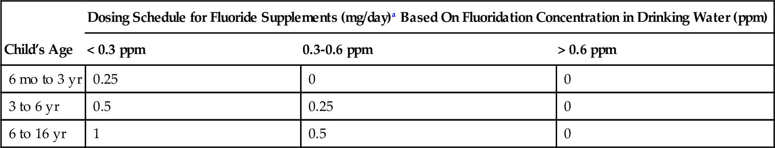
Data from Council on Scientific Affairs, American Dental Association: Intervention: fluoride supplementation. In ADA Council on Access, Prevention and Interprofessional Relations: Caries diagnosis and risk assessment, J Am Dent Assoc 126 (6 Suppl):19-S, 1995.
a
Stay updated, free dental videos. Join our Telegram channel
VIDEdental - Online dental courses


