Orthodontists have long been among the most progressive of the dental specialists, quick to embrace new technologies for enhancing clinical efficiencies and practice workflow. Orthodontic software innovations, whether for imaging and clinical applications or for managing the business side of a practice, have led the consistent need for more powerful computing requirements for more than 4 decades. This article recounts the history of how computers and orthodontic software have been used in America from their nascence to today and provides an outlook for the future.
Highlights
- •
Orthodontics has been harnessing computer and software technologies for more than 40 years.
- •
Orthodontists have historically been quick to embrace new technologies for enhancing workflow.
- •
Going forward, orthodontics advancements will be increasingly intertwined with technology.


It has been said that in life two things are inevitable: death and taxes. May I add a third (though not so vital): the application of the computer to orthodontic research and diagnosis.
These were the opening words of American Association of Orthodontists’ annual meeting in New Orleans in May 1971.
Orthodontics has long enjoyed a reputation for being among the most progressive of the dental specialties. Its practitioners were using computer technology for clinical pursuits as early as the late 1960s, although it remained primarily within the realm of researchers and academicians for the first decade or so. It was not until much later, in the early 1980s, that mainstream computer technology caught the attention of Main Street orthodontists—this time as a possible solution for the clerical tasks involved in running an orthodontic practice.
Harnessing computer technology to tackle the scheduling and financial tasks was the first pursuit in the early days of practice management software systems. An orthodontic practice is a business, and most orthodontists did not graduate from business school. They wanted—needed—to have an accurate idea of their financial situation. Clerical tasks such as accounts receivable, past-due control, basic patient history, and reporting were the minimum capabilities expected from the computers of the time. More progressive orthodontists were using computers to calculate staff salaries and fee schedules, and to perform sophisticated reporting. Tracking appointments and recalls were also part of the functions of those early systems. “The 1980s was definitely a period of early adopters,” recalled Todd Blankenbecler, manager of Dolphin Management Development (Chatsworth, Calif) and former national sales manager at Orthodontic Practice Management System (OPMS) and PracticeWorks. “Key features in those early systems were billing statements, insurance tracking, automated letter writing, and basic reporting.”
Dr Marc S. Lemchen, a New York City orthodontist, was a forerunner of the movement driving computer technology into the orthodontic practice. He was using an early form of a computerized treatment card while the rest of the crowd was still impressing itself with billing statements and “thank you” letters. “In the beginning, there were only 2 practice management systems on the scene,” remembered Lemchen. “There was no digital imaging, or digital x-rays, or any kind of image-management systems. But we had the treatment card, so we didn’t have to pull a chart for the patient unless we wanted to see the x-rays.” That was the early 1980s. At that time, any “state of the art” system had about 10 MB of memory and a footprint the size of a small refrigerator. For the sake of perspective, today’s entry level smartphones have 16,000 MB (16 GB) of memory and fit in the palm of your hand.
The 1980s: embracing technology and forging forward
In typical fashion, technology advanced at such a pace that by 1984, a dozen computer companies were hawking their wares to orthodontists. “The industry really took off in 1984,” remembered Reid Simmons, computer science professional and founder of OrthoTrac (Carestream Dental, Atlanta, Ga) and Cloud9Ortho (Kennesaw, Ga). That year, the American Association of Orthodontists held a winter meeting in Dallas, and handouts offered at the meeting reviewed the practice management systems that were currently available. By this time, orthodontic practice management systems could perform a variety of tasks, including registration, financials and accounting, appointments, insurance information, and treatment information.
Additional tasks available in programs at that time included payroll, general ledger, inventory, and more.
Most of the first practice management systems were installed on UNIX platforms. “In 1982 OrthoTrac ran on the Oasis operating system and was limited to 5 workstations,” said Simmons. “In 1983, we switched to UNIX, which could handle up to 11; later it was unlimited.” In the late 1980s, DOS-based systems began to appear, and by the mid-1990s, the first Windows-based systems (Microsoft, Redmond, Wash) were being introduced. By the 2000s, most of the platforms being sold worked with Windows.
Although orthodontic imaging systems joined the game later, their roots reach back far earlier to the onset of the computer technology in the late 1960s. That is when Robert M. Ricketts, affiliated with Rocky Mountain Orthodontics Data Services (Denver, Colo), announced a computer portal service for orthodontists. The idea was that a doctor would send a lateral head x-ray, a photocopy of the impressions, and a diagnostic sheet, and receive back a computed analysis of the x-ray along with a growth prognosis and treatment plan. The emerging computer technology of the era inspired others in the orthodontic community to consider the diagnostic possibilities. A few years later, Geoffrey F. Walker published a report in the American Journal of Orthodontics suggesting 177 cephalometric landmarks for computer analysis.
Digitization of cephalometrics
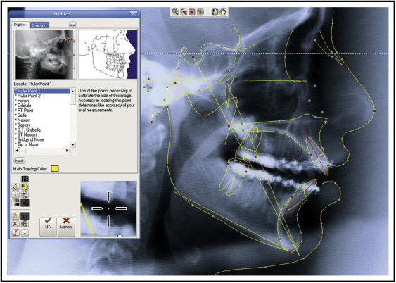
Introduced in 1931 by B. H. Broadbent, cephalometrics remains a vital diagnostic tool for orthodontists. Tracing of the cephalograph was done manually until the early 1970s, when Robert M. Ricketts created a computerized cephalometric tracing/visual treatment objective system. Today’s cephalometric tracing programs often contain hundreds of analyses, any number of which can be performed simultaneously by the doctor or skilled staff ( Figs 1 and 2 ).

In 1988, Dolphin introduced the DigiGraph, which used sonic echolocation—similar to what dolphins use—to gather cephalometric information, thereby eliminating radiation from the process. The DigiGraph’s components consisted of a cabinet with a digitizer probe, computer, monitor, head holder, microphone array, video cameras, and software. “Since the DigiGraph predated the general availability of digital cameras, the most practical way to get the patient’s lateral photo into a computer was to use an analog video camera signal fed into a frame-grabber circuit board in a computer,” explained Ken Gladstone, manager of Imaging Software Products at Dolphin and a member of the original DigiGraph team. “The image would then display the video on the computer screen, allowing the user to grab a single still image from the video feed.” The digitizing probe placed lightly on the patient’s face would send sonic signals to the microphone array positioned above the patient’s head. “Then the computer used the signals to triangulate the probe’s exact location in 3 dimensions,” said Gladstone. Combining the data sets allowed the practitioner to perform cephalometric tracings and analyses, superimpositions, and virtual treatment objectives ( Fig 3 ).

Whereas the concept of the DigiGraph proposed an interesting direction, before long it became apparent that the traditional method (x-rays) of gathering cephalometric imagery was more complementary to the other parts of the orthodontist’s tool set.
Digitization of cephalometrics
Introduced in 1931 by B. H. Broadbent, cephalometrics remains a vital diagnostic tool for orthodontists. Tracing of the cephalograph was done manually until the early 1970s, when Robert M. Ricketts created a computerized cephalometric tracing/visual treatment objective system. Today’s cephalometric tracing programs often contain hundreds of analyses, any number of which can be performed simultaneously by the doctor or skilled staff ( Figs 1 and 2 ).
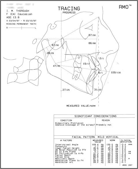
In 1988, Dolphin introduced the DigiGraph, which used sonic echolocation—similar to what dolphins use—to gather cephalometric information, thereby eliminating radiation from the process. The DigiGraph’s components consisted of a cabinet with a digitizer probe, computer, monitor, head holder, microphone array, video cameras, and software. “Since the DigiGraph predated the general availability of digital cameras, the most practical way to get the patient’s lateral photo into a computer was to use an analog video camera signal fed into a frame-grabber circuit board in a computer,” explained Ken Gladstone, manager of Imaging Software Products at Dolphin and a member of the original DigiGraph team. “The image would then display the video on the computer screen, allowing the user to grab a single still image from the video feed.” The digitizing probe placed lightly on the patient’s face would send sonic signals to the microphone array positioned above the patient’s head. “Then the computer used the signals to triangulate the probe’s exact location in 3 dimensions,” said Gladstone. Combining the data sets allowed the practitioner to perform cephalometric tracings and analyses, superimpositions, and virtual treatment objectives ( Fig 3 ).