Fig. 3.1
(a) Crystallite of a mouse incisor. Alignments of hydroxyapatite monocrystals form a lattice, with – 5 Å–6.8 Å spacing. (b) Axial dissolution of a crystallite. (c) The acidic dissolution starts on the basal face (A), then increases in depth (B), and dissolves the central part of the crystals along the c-axis (C). (d) View on the longitudinal and central dissolution of enamel crystallites
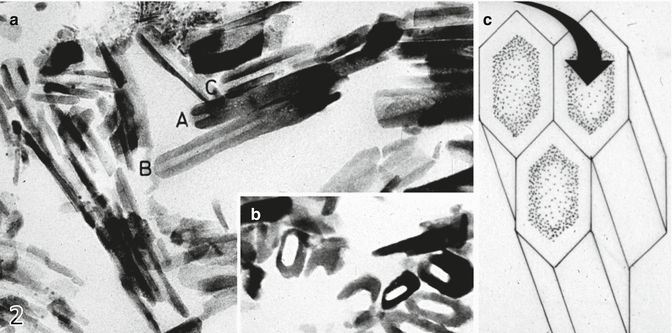
Fig. 3.2
Shows the successive steps of crystallite dissolution: (a) longitudinal dissolution; (b) dissolution in the central c-axis; (c) schematic drawing of the crystallite dissolution
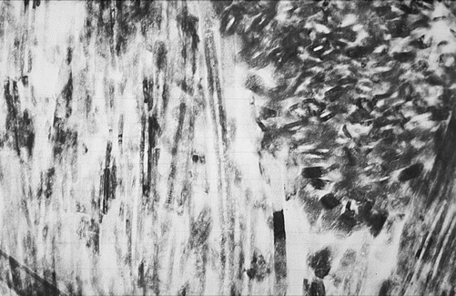
Fig. 3.3
Longitudinal (left part of the figure) and axial (right part of the figure) dissolution of enamel crystallites
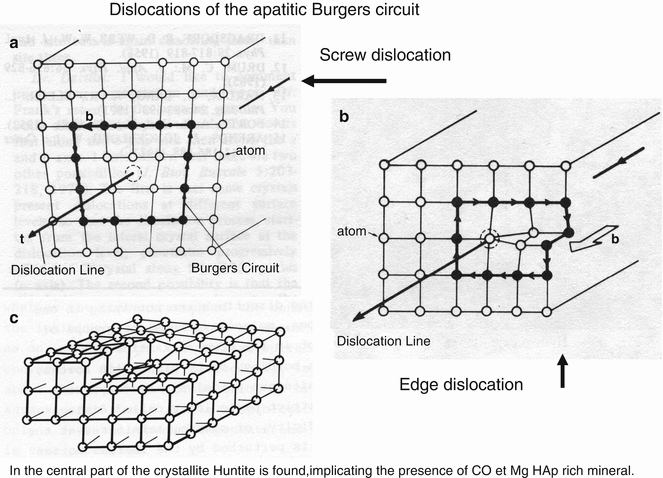
Fig. 3.4
Screw dislocation (a) and edge dislocation (b) of the apatitic Burgers circuit. In (c), huntite is found in the central part of crystallites. The huntite-rich central part implicates an elevated content of carbonate and magnesium
Measurements of the overall size of the crystallite on transverse sections have been reported to be 700 nm × 300 nm (mean value: L + l : 2 = 500 nm) (263 Å ± 22 x 683 Å ± 34- Daculsi et al. 1984). They appear as long ribbons with a length over a few millimeters, starting at the dentinoenamel junction and probably reaching at the surface the whole enamel thickness.
In the outer aprismatic layer, crystallites are parallel to each other and form a palisade-like structure. Acidic solutions or gels spread at the surface and dissolve the center of the crystal or enlarge the narrow intercrystalline structures. After a short period of time, the row of crystallites is reduced in height. The aprismatic layer disappears and the new surface created reaches the prismatic layer.
During enamel formation, ameloblast Tomes’ processes fill structures that will become rod (or prisms) and mineralize (Figs. 3.3, 3.4, 3.5, 3.6, 3.7, and 3.8). At the rod periphery, the interrod material forms a continuous network. The width of the rods is about 3 μm and includes more than 1000 crystallites/rod unit, whereas the thinner interrod displays a mean thickness of 0.5 μm and contains about 100–300 tightly packed crystallites. In rods, the crystals are bending from the head to the tail. A thin layer of enamel matrix proteins, the so-called enamel sheath, separates rods and interrods. Depending on the location of the etched area at the enamel surface, rods and interrod crystallites form a 60°angle.
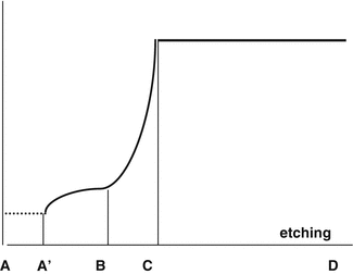
Fig. 3.5
(a) Human enamel surface cleaned with tape running water. Empty rods are open at the surface. (b) Human enamel surface cleaned with a chloroform/methanol, which dissolves extracellular matrix lipids. The organic material inside rods is eliminated and intercrystalline spaces are empty. (c) Untreated enamel surface. Rods (r) and interrods (ir) constitute irregularities at the surface. (d) Perikymata and prints of ameloblasts are visible on the enamel surface
Fig. 3.6


The surface enamel located between A and A’ has been totally dissolved and disappeared. The mineral percentage between A’ and B is slowly increasing, and a more accentuate rise in mineral content occurs between B and C. Between C and D mineralization is unchanged despite the effect of acid etching
Stay updated, free dental videos. Join our Telegram channel
VIDEdental - Online dental courses
