Introduction
Our aim was to prospectively assess in-vivo color alterations of natural teeth associated with fixed orthodontic treatment.
Methods
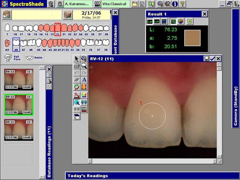
Twenty-six consecutive patients were treated with fixed appliances bonded with a chemically cured or a light-cured resin with a split-mouth design. The spectrophotometric data of a standardized labial circular area of all teeth were recorded, before bracket bonding and after debonding and cleaning procedures. The color parameters of the Commission Internationale de l’Eclairage—L∗, a∗, and b∗ (lightness, red/green, and blue/yellow)—were measured for each adhesive and type of tooth, and the corresponding color differences (ΔE) between the interval groups were calculated. The effect of these parameters on color was assessed with 3-way mixed analysis of variance (ANOVA) and the Bonferroni comparisons test (α <0.05).
Results
Orthodontic treatment was associated with changes in color parameters. The L∗ values decreased ( P <0.001), whereas the a∗ and b∗ values increased ( P <0.001) at the end of treatment. All measured types of teeth demonstrated significant color changes (ΔE); their mean differences ranged from 2.12 to 3.61 ΔE units. Chemically cured resin was associated with greater color changes than light-cured composite.
Conclusions
The color of natural teeth is changed in various ways after fixed orthodontic treatment.
Editor’s comment
This article examines an issue of importance, especially with the increased focus on esthetics. According to patients and parents, when the braces come off, teeth should be as white and perfect as the alignment. We now know that this is not always the case. It is normal for patients and parents to easily forget what was beneath the appliances to begin with. There are 2 common methods of analyzing in vivo the apparent tooth color: visual determination and instrumental measurement. The general demand for objective color matching in dentistry coupled with rapid advances in optical electronic sensors and computer technology has made instrumental measurement devices an adjunct to visual tooth-color evaluation. Various commercial systems—tristimulus colorimeters, spectroradiometers, spectrophotometers, and digital color analyzers—are used in clinical and research settings for objective color specification.
The aim of this prospective clinical trial was to assess in vivo with a reflectance spectrophotometer the color alterations of natural teeth associated with comprehensive orthodontic treatment.
Twenty-six patients completed this study; their treatment required the placement of a variety of fixed appliances on maxillary and mandibular teeth from first premolar to first premolar. Each tooth underwent spectrophotometric analysis on a standardized area in the middle third of its labial crown surface before placement of fixed appliances and immediately after removal and cleanup. The results suggest that the color of natural teeth undergoes significant changes. Current bracket debonding and adhesive removal protocols typically cause enamel morphologic alterations and texture modifications that cannot be restored with polishing media at postfinishing. These irreversible changes could affect the optical properties of enamel surface (gloss and luster) and, consequently, influence the color of natural teeth. The authors discussed other reasons for changes, including age, gingival inflammation, and variations of blood flow in the dental pulp. These findings emphasize the potential risk of tooth-color alterations of fixed orthodontic treatment.


