Introduction
The aim of this study was to assess the use of 3-dimensional facial averages for determining morphologic differences from various population groups.
Methods
We recruited 473 subjects from 5 populations. Three-dimensional images of the subjects were obtained in a reproducible and controlled environment with a commercially available stereo-photogrammetric camera capture system. Minolta 900VI (Konica Minolta, Tokyo, Japan) and 3dMDface (3dMD LLC, Atlanta, Ga) systems were used. Each image was obtained as a facial mesh and orientated along a triangulated axis. All faces were overlaid, one on top of the other, and a complex mathematical algorithm was performed until average composite faces of 1 man and 1 woman were achieved for each subgroup. These average facial composites were superimposed based on a previously validated superimposition method, and the facial differences were quantified.
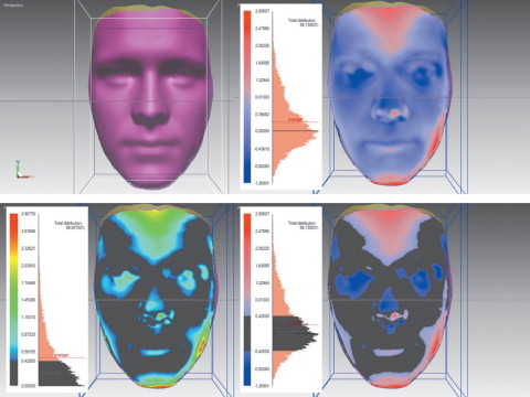
Results
Distinct facial differences were observed among the groups. The linear differences between surface shells ranged from 0.37 to 1.00 mm for the male groups. The linear differences ranged from 0.28 and 0.87 mm for the women. The color histograms showed that the similarities in facial shells between the subgroups by sex ranged from 26.70% to 70.39% for men and 36.09% to 79.83% for women. The average linear distance from the signed color histograms for the male subgroups ranged from −6.30 to 4.44 mm. The female subgroups ranged from −6.32 to 4.25 mm.
Conclusions
Average faces can be efficiently and effectively created from a sample of 3-dimensional faces. Average faces can be used to compare differences in facial morphologies for various populations and sexes. Facial morphologic differences were greatest when totally different ethnic variations were compared. Facial morphologic similarities were present in comparable groups, but there were large variations in concentrated areas of the face.
Read the full text online at: www.ajodo.org , pages S56.e1-S56.e9.
Editor’s comment
Since Holly Broadbent developed the first functioning cephalometer nearly 80 years ago, cephalometric analysis has been an integral part of orthodontic diagnosis, treatment planning, and interpretation of treatment effects. Although clinicians and researchers recognize the 2-dimensional limitations of lateral and posteroanterior cephalometric radiographs, the information from comparisons of pretreatment and posttreatment headfilms has formed the scientific basis for various orthodontic treatment philosophies. As the technological world has changed, giving us new methods for rendering information, our horizons have expanded so that 3-dimensional (3D) imaging is now possible. As a result, many research efforts are attempting to find the appropriate uses for 3D analysis.
The digital recreation of the plaster dental cast has allowed clinicians to visualize and measure dental relationships with ease and accuracy. But a continuing deficiency has been our inability to accurately analyze the soft tissues of the face. Yes, facial form can be appreciated and judged on 2-dimensional photographs. But the ability to actually compare the effects of treatment on facial form or the differences in the faces of various populations has never been attainable in the past. This investigation used 3D stereo-photogrammetry to compare 5 ethnically different groups of adults to determine whether average facial types could be identified and compared.
Although there were expected variations in certain measurements among the subjects, the authors identified an average facial type and determined where, how, and to what degree the faces of ethnically different populations vary. Although this is a preliminary investigation, its methodology might be useful in the future to interpret 3D differences in soft-tissue facial forms among groups. I applaud these researchers for their efforts, and, even though this technology is in its early stages, I look forward to further applications of this type of research technology.
Vincent G. Kokich